Logo

3D tumoroids in cancer research: a tumor-to-model workflow

From human tumor tissue to functional 3D cancer organoids

A tumor‑to‑model workflow generates physiologically relevant 3D tumor models, including tumoroids or cancer organoids, that better preserve tumor biology than 2D cultures of cell lines: As precision medicine advances, the demand for reliable tumor-derived models continues to grow, yet their generation remains technically challenging. Tissue quality, harsh processing, and undefined culture conditions can compromise viability and biological relevance, underscoring the need for standardized, gentle, and reproducible workflows.

This page describes how our standardized workflow addresses these challenges and enables the reliable generation and validation of tumoroids for translational cancer research and predictive drug development.

Content overview

Content

  • Workflow overview
  • 1. Short- and long-term storage of tumors
  • 2. Gentle tumor dissociation
  • 3. Cell suspension cleanup and cell isolation
  • 4. Gentle cell sorting
  • 5. Tumoroid culture, banking, and expansion
  • 6. Analysis of dissociated tumoroids
  • 7. High-resolution 3D tumoroid imaging
  • 8. 2D spatial multiomics tumoroid imaging
  • All relevant products for application on tumoroid research

Step-by-step tumor-to-model workflow

Short- and long-term storage of tumors

Keep tumor tissues and cells viable and representative for organoid research.

Short- and long-term storage of tumors

Keep tumor tissues and cells viable and representative for organoid research.

Gentle tumor dissociation

The gentleMACS® Family of tissue dissociators is designed to ensure rapid, standardized, and reliable dissociation of tumor samples. Carefully balanced enzyme kits support exceptional epitope preservation.

Gentle tumor dissociation

The gentleMACS® Family of tissue dissociators is designed to ensure rapid, standardized, and reliable dissociation of tumor samples. Carefully balanced enzyme kits support exceptional epitope preservation.

Cell suspension cleanup and cell isolation

Effective removal of unwanted cell types and debris improves tumoroid establishment and model quality.

Cell suspension cleanup and cell isolation

Effective removal of unwanted cell types and debris improves tumoroid establishment and model quality.

Gentle cell sorting

Sorting dissociated tumor cells prior to tumoroid establishment enables uniform culture seeding and precise control over starting cell populations.

Gentle cell sorting

Sorting dissociated tumor cells prior to tumoroid establishment enables uniform culture seeding and precise control over starting cell populations.

Tumoroid culture, banking, and expansion

Optimized culture conditions enable the generation of consistent 2D and 3D tumor models. TumorMACS™ Media support both formats without the need for transition.

Tumoroid culture, banking, and expansion

Optimized culture conditions enable the generation of consistent 2D and 3D tumor models. TumorMACS™ Media support both formats without the need for transition.

High-resolution 3D tumoroid imaging

Light sheet–based 3D imaging reveals cell-cell interactions and spatial organization in intact tumor organoids. Image up to 48 tumoroids in a single run for high-throughput studies.

High-resolution 3D tumoroid imaging

Light sheet–based 3D imaging reveals cell-cell interactions and spatial organization in intact tumor organoids. Image up to 48 tumoroids in a single run for high-throughput studies.

2D spatial multiomics tumoroid imaging

Multiplex imaging provides essential insights into the cellular and structural characteristics of tumor organoids.

2D spatial multiomics tumoroid imaging

Multiplex imaging provides essential insights into the cellular and structural characteristics of tumor organoids.

Analysis of dissociated tumoroids

Characterize tumoroids by flow cytometry and gently sort cells for downstream single-cell genomics.

Analysis of dissociated tumoroids

Characterize tumoroids by flow cytometry and gently sort cells for downstream single-cell genomics.
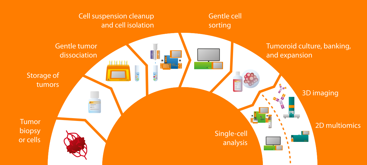
Infographic showing a 3D tumoroid applications workflow from tumor tissue and cell isolation to single-cell analysis and sorting.

Short- and long-term storage of tumors

Keep tumor tissues and cells viable and representative for organoid research.

Short- and long-term storage of tumors

Keep tumor tissues and cells viable and representative for organoid research.

Gentle tumor dissociation

The gentleMACS® Family of tissue dissociators is designed to ensure rapid, standardized, and reliable dissociation of tumor samples. Carefully balanced enzyme kits support exceptional epitope preservation.

Gentle tumor dissociation

The gentleMACS® Family of tissue dissociators is designed to ensure rapid, standardized, and reliable dissociation of tumor samples. Carefully balanced enzyme kits support exceptional epitope preservation.

Cell suspension cleanup and cell isolation

Effective removal of unwanted cell types and debris improves tumoroid establishment and model quality.

Cell suspension cleanup and cell isolation

Effective removal of unwanted cell types and debris improves tumoroid establishment and model quality.

Gentle cell sorting

Sorting dissociated tumor cells prior to tumoroid establishment enables uniform culture seeding and precise control over starting cell populations.

Gentle cell sorting

Sorting dissociated tumor cells prior to tumoroid establishment enables uniform culture seeding and precise control over starting cell populations.

Tumoroid culture, banking, and expansion

Optimized culture conditions enable the generation of consistent 2D and 3D tumor models. TumorMACS™ Media support both formats without the need for transition.

Tumoroid culture, banking, and expansion

Optimized culture conditions enable the generation of consistent 2D and 3D tumor models. TumorMACS™ Media support both formats without the need for transition.

High-resolution 3D tumoroid imaging

Light sheet–based 3D imaging reveals cell-cell interactions and spatial organization in intact tumor organoids. Image up to 48 tumoroids in a single run for high-throughput studies.

High-resolution 3D tumoroid imaging

Light sheet–based 3D imaging reveals cell-cell interactions and spatial organization in intact tumor organoids. Image up to 48 tumoroids in a single run for high-throughput studies.

2D spatial multiomics tumoroid imaging

Multiplex imaging provides essential insights into the cellular and structural characteristics of tumor organoids.

2D spatial multiomics tumoroid imaging

Multiplex imaging provides essential insights into the cellular and structural characteristics of tumor organoids.

Analysis of dissociated tumoroids

Characterize tumoroids by flow cytometry and gently sort cells for downstream single-cell genomics.

Analysis of dissociated tumoroids

Characterize tumoroids by flow cytometry and gently sort cells for downstream single-cell genomics.

1. Short- and long-term storage of tumors

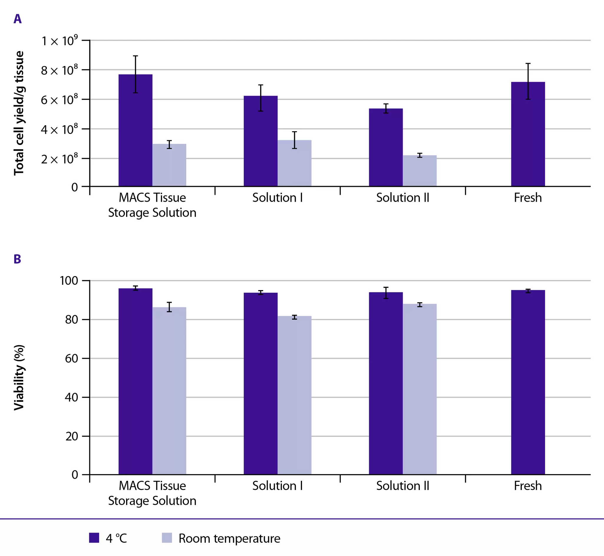
Samples were put in different media, e.g. MACS Tissue Storage Solution and results are shown in barcharts for Total cell yield /g tissue and viability.
Comparison of (A) total cell yield and (B) viability of mouse spleen, stored in different solutions

Transport and storage of tumor tissues and primary cell suspensions are critical for the successful establishment of tumoroids, as delays or poor conditions reduce viability and alter cellular composition. High-quality input reduces clonal bias and improves the formation efficiency of 3D cancer organoids across samples and tumor entities.

Our MACS® Tissue Solution and MACS Cell Storage Solution allow optimized storage of fresh organs, tumor samples, and dissociated cells, while preserving cellular viability and functionality without activating the cells or inducing apoptosis.

Application note

Optimal storage of murine tumors in MACS Tissue Storage Solution

Discover in this application note how our MACS Tissue Storage Solution reliably preserves cell viability, composition, and activation status of cells for at least 48 hours, by keeping stress-related gene expression levels at a minimum when compared to fresh sample
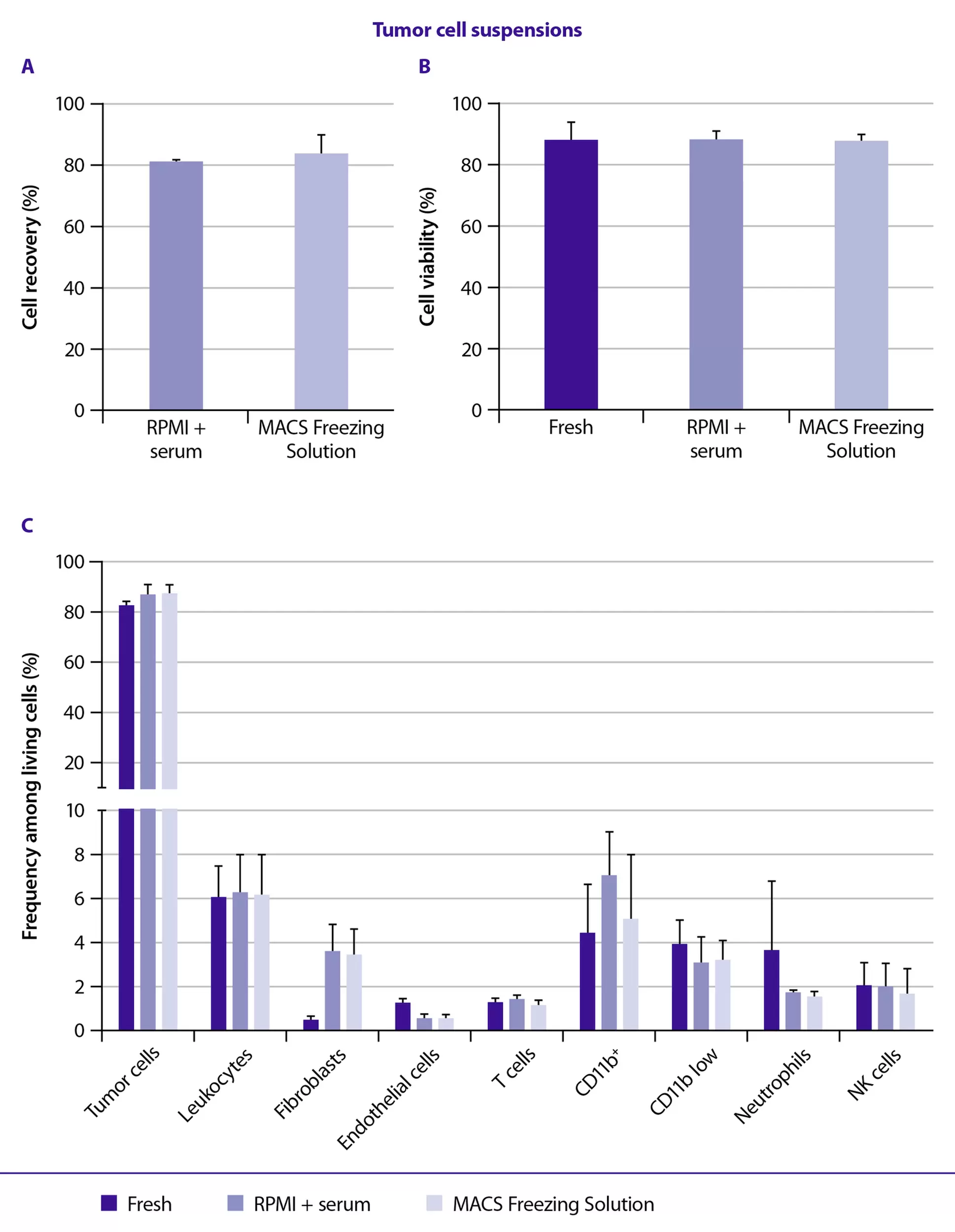
Three barcharts are shown. Tumor sampels were put in differenet cell media e.g. MACS Freezing Solution and it was looked for cell count, viability and Frequency among living cells (%).
Analysis of cryopreserved tumor samples and their cellular composition

Standardized cryopreservation with serum- and animal component–free formulations ensures consistent downstream analysis also after long-term storage. Our MACS Freezing Solution ensures high preservation of viability and recovery of cells and tissues upon thawing.

Application note

Cleaner samples, clearer data

Explore this application note for a case study on human high-grade serous ovarian carcinoma (HGSOC) samples. Biopsies were frozen with the MACS Freezing Solution and stored in liquid nitrogen for 6 to 17 months for later bulk single-cell RNA sequencing (scRNA-seq) analysis.

2. Gentle tumor dissociation

Tumor dissociation is critical for successful tumoroid establishment, as harsh enzymatic, mechanical, or temperature conditions reduce viability, damage surface markers, and bias cell populations.

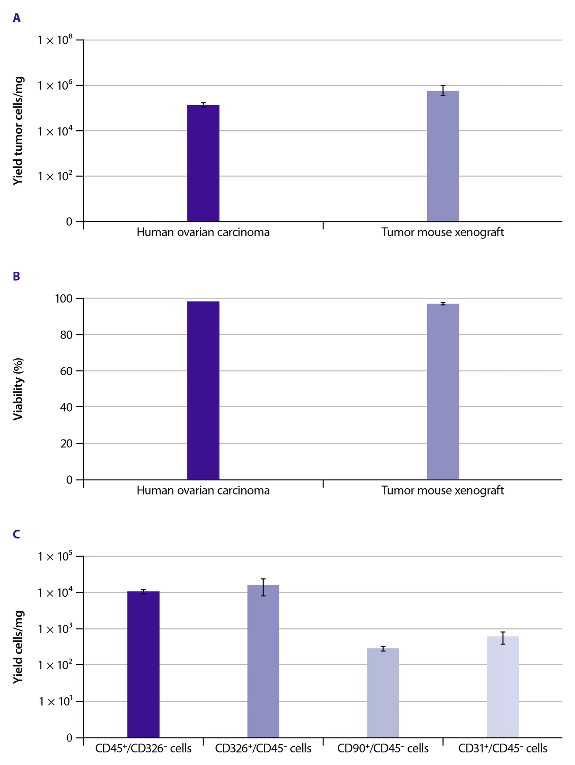
Three barcharts are shown, were the yield, viability and the ration Yield tumor cells per mg are shown. Samples were prepared with the Tumor Dissociation Kit, human.
Analysis of cell yield, viability, and cellular composition after tumor dissociation

Our reliable and automated gentleMACS® Technology combines mechanical disruption and controlled enzymatic digestion to support gentle and efficient release of viable cells from human tumors and xenografts. Using our Tumor Dissociation Kits in combination with predefined, tailored gentleMACS Programs enable optimal preservation of epitopes on single cells, for consistent and reliable cell recovery from different tumor entities.

Human ovarian carcinoma and mouse tumor xenografts were dissociated using the Tumor Dissociation Kit, human, in combination with the gentleMACS Octo Dissociator with Heaters.

References

gentleMACS Dissociators, Tubes, and Reagent Kits – selected references

Investigate a list of selected recent publications using our gentleMACS Technology. See how our products contribute to cutting-edge cancer research by scientists worldwide.

3. Cell suspension cleanup and cell isolation

Human primary tumor specimens contain non-tumor cells, which compromise the success and reproducibility of tumor cell cultures. High levels of dead cells and debris reduce tumoroid formation by impairing cell-cell interactions and culture conditions. Our Tumor Cell Isolation Kit maximizes tumor cell purity, improves establishment rates, and enhances the biological relevance of resulting 3D tumor models.

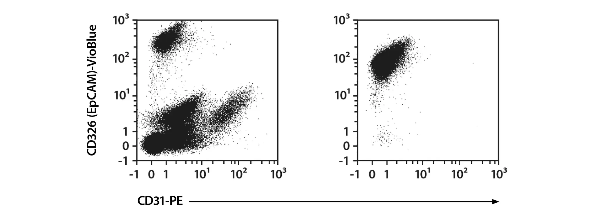
Two plots show flow cytometric characterization of human breast carcinoma cells before and after isolation.
Flow cytometric characterization of human breast carcinoma cells before and after isolation

EpCAM-positive human breast carcinoma cells were isolated from a heterogeneous human cell sample using the Tumor Cell Isolation Kit, human. Tumor cell purity of >95% was achieved in less than 20 minutes. Dot plots show cell composition before (left) and after (right) isolation.

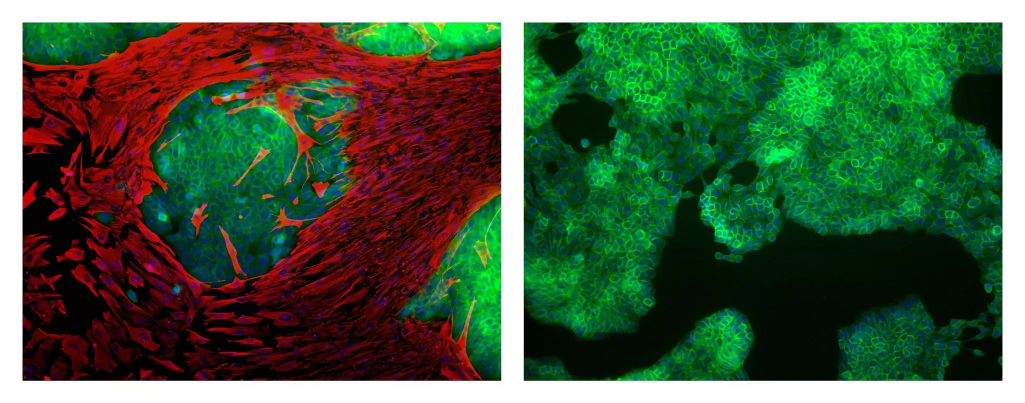
Two immunofluorescence images show bulk tumor cells and isolated tumor cells after three days in culture. While stromal cells outgrew tumor cells in the unseparated fraction, isolated tumor cells maintained high purity over time.
Bulk tumor cells and isolated tumor cells after seven days in culture

Human breast carcinoma cells from a heterogeneous human cell sample were cultured either without separation or following tumor cell isolation. After 7 days of culture, EpCAM (green) and vimentin (red) staining identified tumor cells and stromal cells, respectively. While stromal cells outgrew tumor cells in the unseparated fraction (left), isolated tumor cells maintained high purity over time (right).

Similarly, in patient-derived xenograft models murine cells infiltrate the human tissue, affecting molecular analysis and compromising reliable tumoroid establishment. Our Mouse Cell Depletion Kit efficiently and gently removes mouse cells and cellular debris, preventing cross-species interference and murine overgrowth.

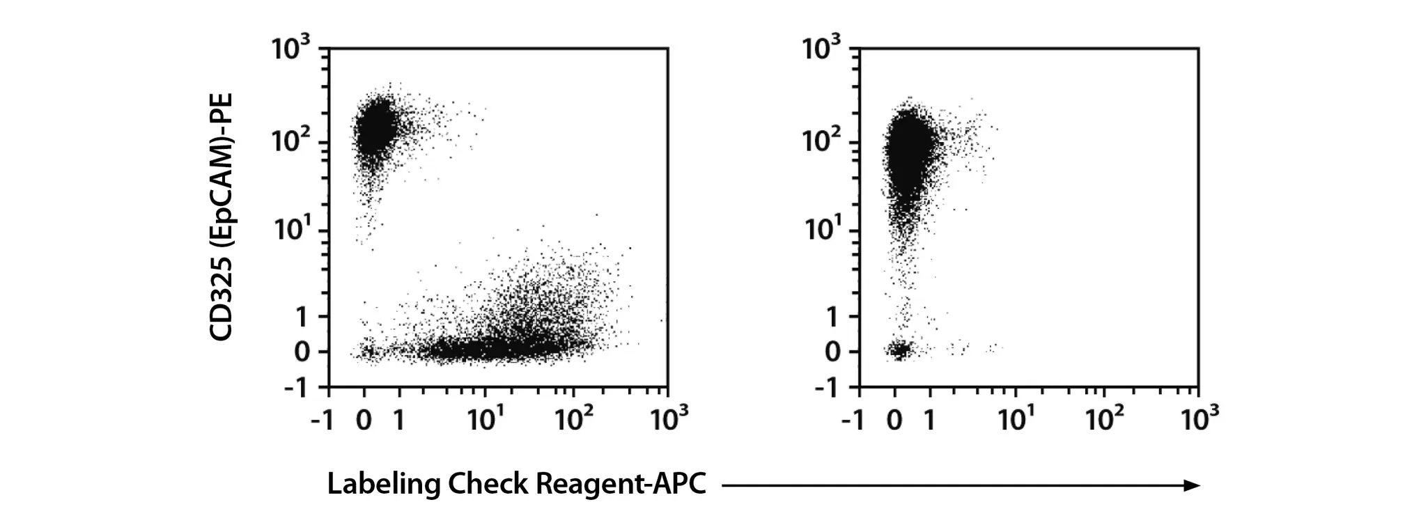
Two dot plots that show comparison of cell populations before and after separation demonstrates efficient enrichment of human carcinoma cells.
Isolation of human tumor cells from xenograft-derived bulk suspension

Human colon carcinoma cells were enriched from a heterogeneous xenograft-derived cell suspension through one-step depletion of mouse cells and debris. Comparison of cell populations before (left) and after (right) separation demonstrates efficient enrichment of human carcinoma cells.

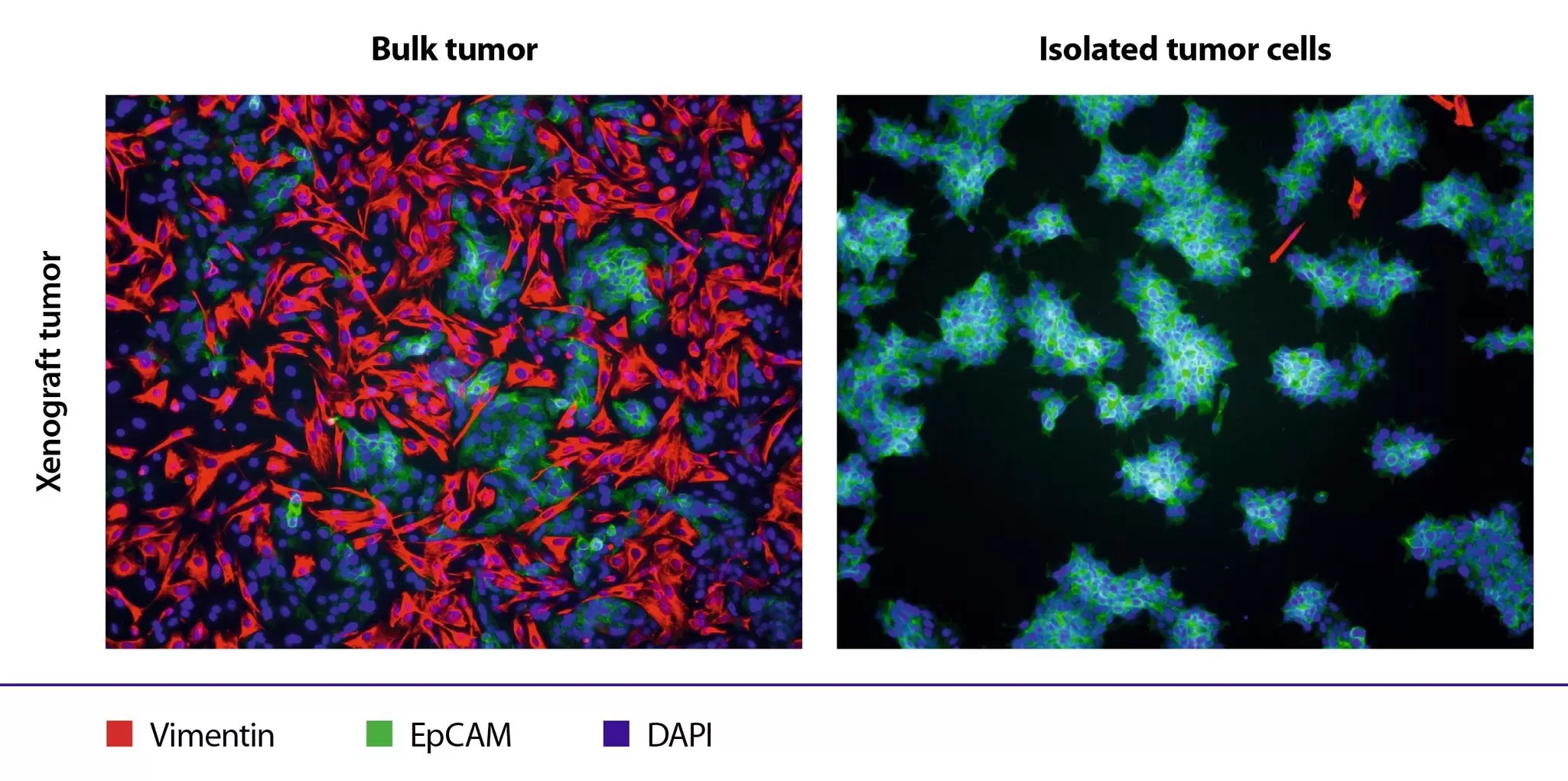
To assess culture purity after mouse cell depletion, bulk and isolated tumor cell fractions were cultured separately. Cells were stained differently.
Magnetic depletion of non-tumor cells to obtain pure tumor cell fractions

To assess culture purity after mouse cell depletion, bulk and isolated tumor cell fractions were cultured separately. EpCAM (green) and murine vimentin (red) staining revealed that the isolated fraction remained highly enriched for EpCAM-positive tumor cells after 7 days in culture (right), while mouse stromal cells expanded in the bulk culture (left).

4. Gentle cell sorting

Sorting dissociated tumor cells prior to tumoroid establishment enables uniform culture seeding and precise control of cellular composition, such as defined tumor-to-fibroblast ratios for modeling tumor microenvironment interactions. Because tumor and stromal cells are often sensitive to shear stress and pressure changes, the gentle microfluidic sorting technology of the MACSQuant® Tyto® Cell Sorter helps preserve viability and functionality. This supports efficient 3D aggregation into stable, physiologically relevant tumoroids that retain cellular heterogeneity.

Enrichment of large and fragile cell types up to 50 μm in diameter is enabled by the MACSQuant Tyto Cartridge LC (Large Cell). Combined with advanced microfluidics and inertial cell focusing, this approach delivers high-performance tumor cell sorting while preserving cell integrity, making it ideally suited for pretumoroid sorting workflows.

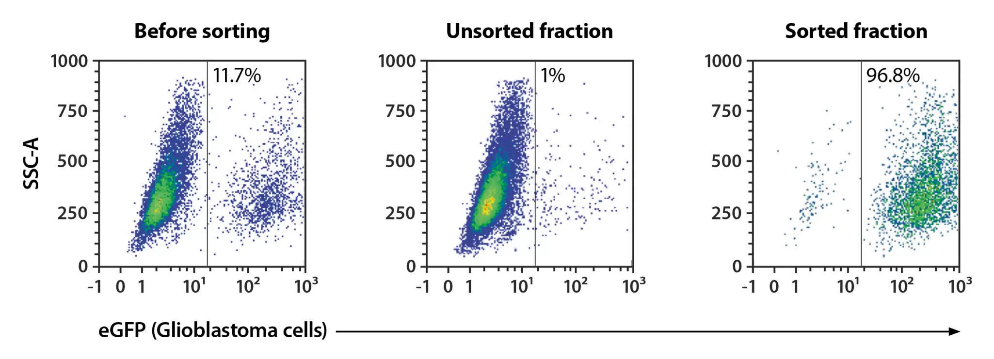
Flow cytometry sorting of primary Glioblastoma (GBM) cells is shown, in three plots: before sorting, unsorted fraction , and the sorthed fraction.
Flow cytometry sorting of large GBM cells

Primary glioblastoma (GBM) tumor cell lines were established in vitro from fresh cavitron ultrasonic aspirator (CUSA) tumor dissociates. Tumor cells were transduced to express eGFP and sorted using the MACSQuant Tyto and the MACSQuant Tyto Cartridge LC. Sorting resulted in >96% tumor cell purity.

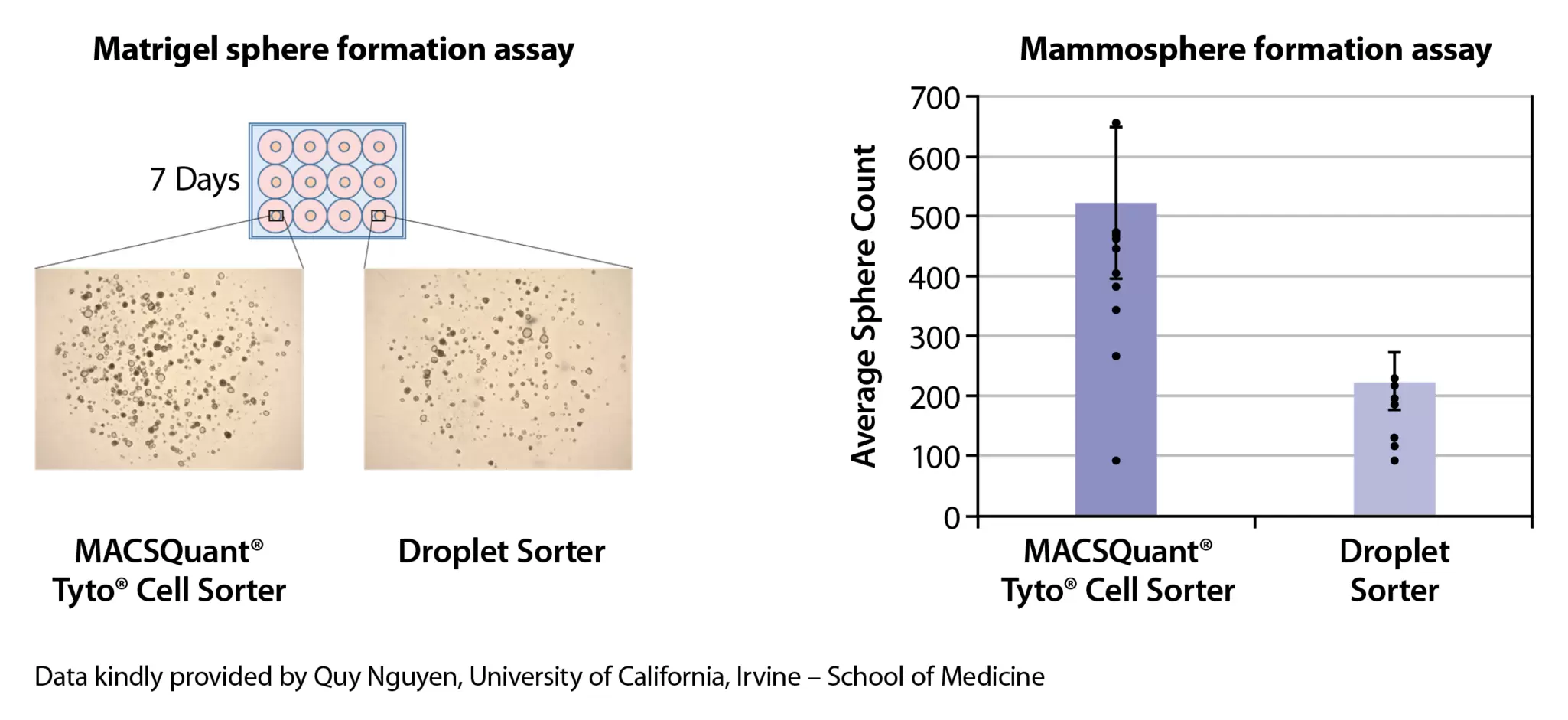
On the right side of this illustration one can see a matrigel sphere formation assay after 7 days of the MACSQuant Tyto Cell Sorter and a droplet sorter. On the right side these instruments were comapred in a mammosphere formation assay. Data kindly provided by Quy Nguyen, University of California.
Enhanced sphere formation after gentle sorting with the MACSQuant Tyto

A Matrigel® sphere formation assay revealed superior sphere-forming capacity of cells sorted with the MACSQuant Tyto compared to a droplet-based sorter. After 7 days, cells sorted with the MACSQuant Tyto formed significantly more spheres, indicating higher viability and functionality.

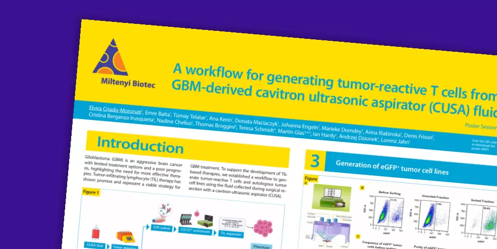
Scientific poster

A workflow for generating tumor-reactive T cells from glioblastoma (GBM)-derived cavitron ultrasonic aspirator (CUSA) fluids

Learn more about how the MACSQuant Tyto facilitated gentle enrichment of large tumor cells in this study.

5. Tumoroid culture, banking, and expansion

Establishing a 3D cancer organoid culture

Tumoroid establishment is the process by which primary tumor cells reorganize within a defined extracellular matrix and growth factor environment to form expandable 3D structures. Efficiency depends on cell viability, preservation of functional states, and controlled seeding conditions.

Our TumorMACS™ Media are based on a standardized serum-free formulation, designed to support tumor heterogeneity and expansion. Our media enable robust proliferation of tumor cells, while maintaining their genomic, transcriptomic, and metabolomic stability.

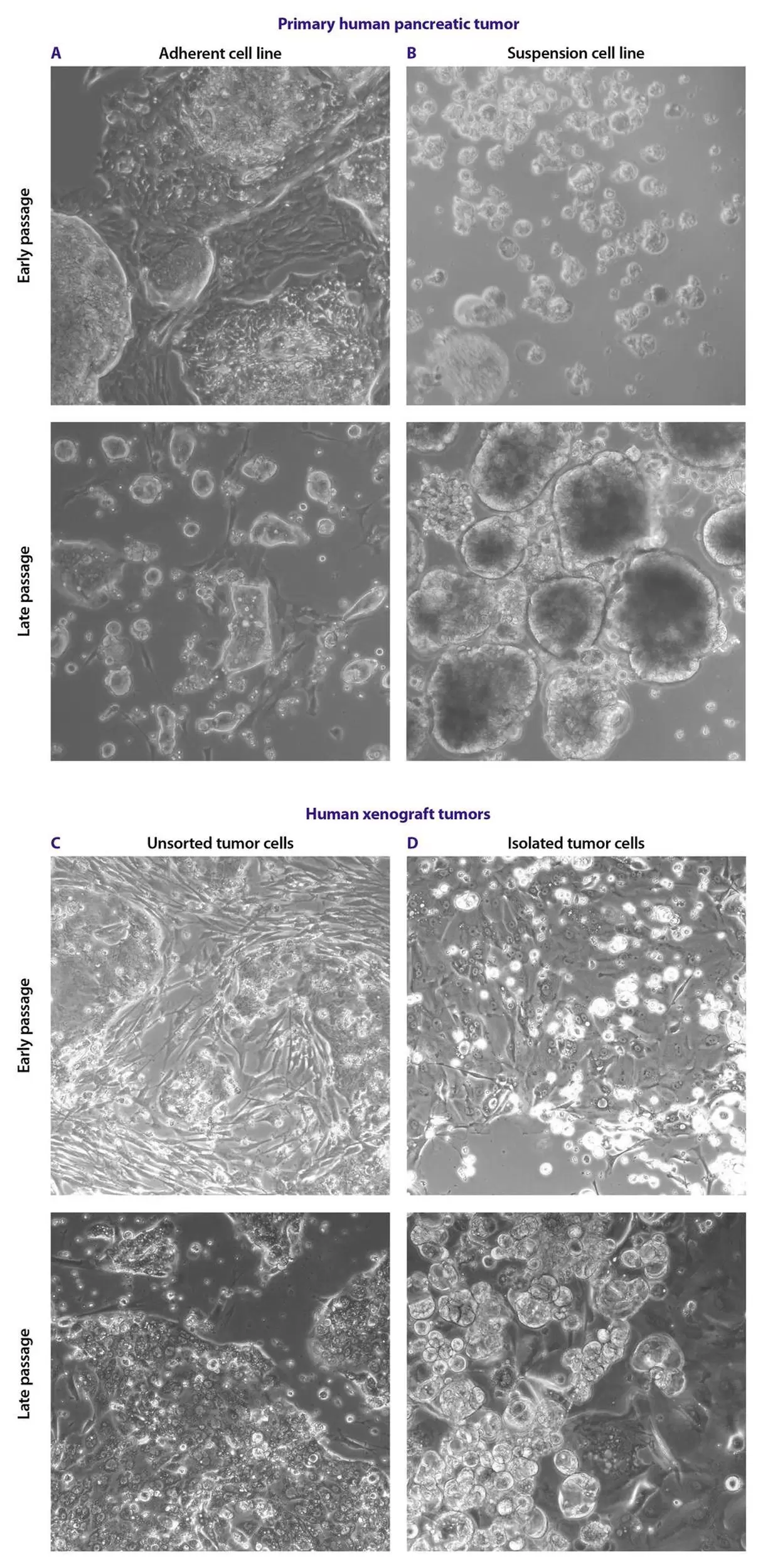
In this illustration one can seee primary human pancreatic and human xenograft samples and that their morphological heterogeneity was preserved not only during initial passages but also in late passages.
TumorMACS Media preserve morphological heterogeneity of primary human tumor cells

Using TumorMACS Media, cells can be cultured directly after tissue dissociation or tumor cell isolation. Both adherent and suspension cells expand efficiently, while primary cell lines retain the morphological heterogeneity of the parent tumor, even at later passages.

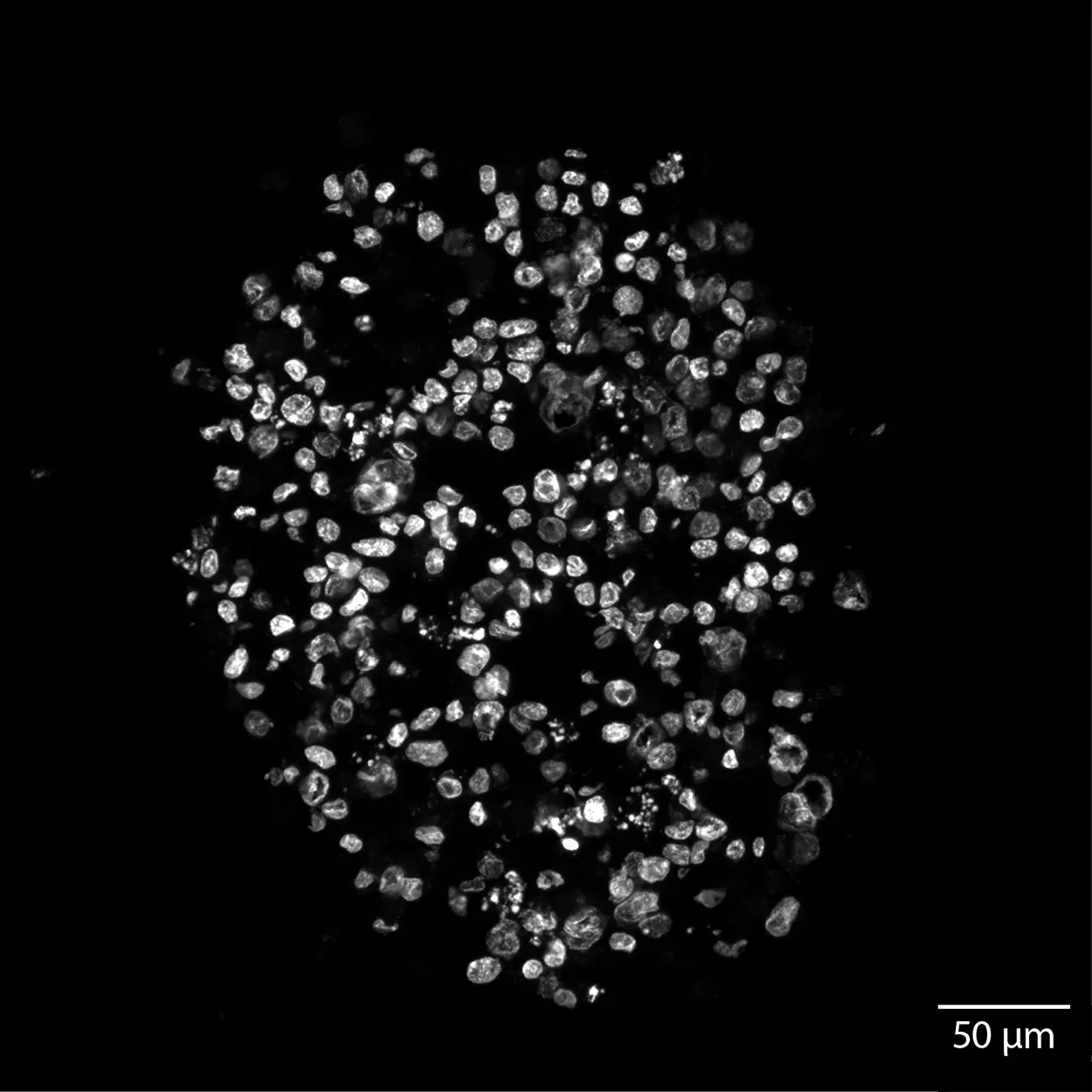
Light sheet microscopy of 3D spheroids cultured in TumorMACS Medium and StemMACS Y27632.
Light sheet microscopy of 3D spheroids cultured in TumorMACS Media and StemMACS Y27632

TumorMACS Media are optimized for both 2D and 3D cultures across multiple cancer types, including pancreatic, renal, colon, ovarian, lung, and breast cancer. Cells can be seamlessly transitioned between 2D and 3D formats without media adaptation. StemMACS™ Y27632 supplement helps preserve the tumorigenic potential during long-term culture.

Isolation and expansion of HSCs icon on purple background

Guide for the initiation of primary tumor cell cultures

Find out more about ROCK inhibitors and how they can significantly enhance survival and plating efficiency by preventing apoptosis that normally occurs when cells lose cell–cell/ECM attachment.

Tumoroid expansion and banking

Tumoroid expansion is a key phase in the tumor-to-model workflow, enabling scalability and long-term use of tumor organoid cultures. Efficient expansion requires optimized conditions that preserve cellular diversity and functional integrity while supporting proliferation. Using our standardized expansion protocol, a stable tumoroid repository can be generated, supporting longitudinal studies and collaborative research.

Tumor organoid banking involves cryopreservation of well-characterized tumoroids for future use without loss of quality. Robust cryopreservation of organoids using the MACS Freezing Solution ensures high viability and preserved function after thawing, enabling reproducible downstream applications (see also workflow section 1: Short- and long-term storage of tumors).

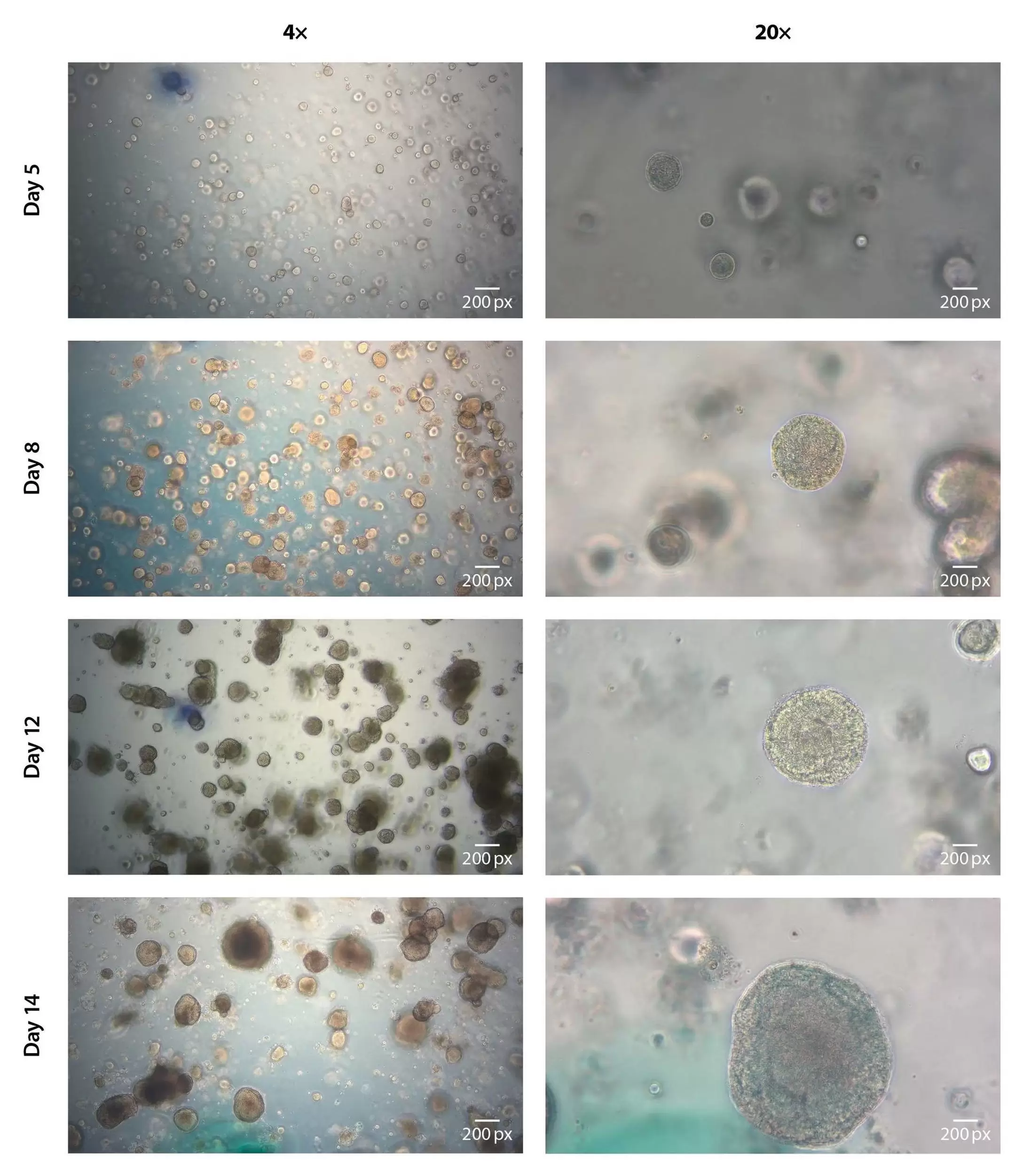
Representative images of primary breast cancer spheroid growth during a passage (days 5, 8, 12, and 14).
Representative images of primary breast cancer spheroid growth during a passage (days 5, 8, 12, and 14)

Brightfield imaging over 14 days demonstrates robust spheroid formation and growth of breast cancer cells in 3D culture cultivated in Breast Cancer TumorMACS Medium supplemented with StemMACS Y27632. Progressive increases in spheroid size and complexity were observed throughout the cultivation period.

6. Analysis of dissociated tumoroids

Flow cytometry enables rapid, quantitative tumoroid characterization, including identity confirmation, phenotypic monitoring, and cellular subpopulation analysis. The MACSQuant Analyzer streamlines workflows through automated labeling, acquisition, analysis, and reporting, while REAfinity® Recombinant Antibodies support reproducible results through recombinant design, uniform isotypes, and lot-to-lot consistency.

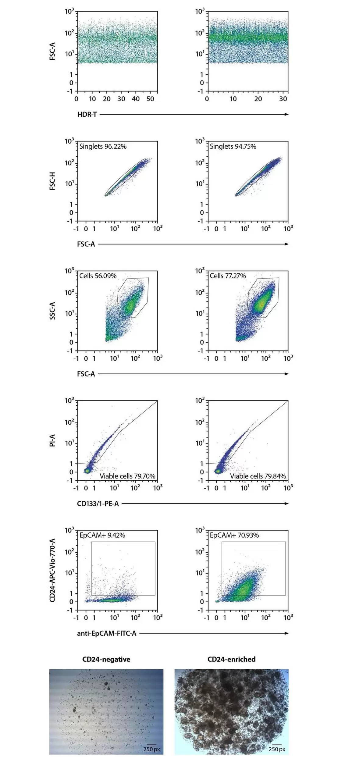
Isolation and flow cytometric analysis of human colon cancer cells from tumoroids. Only CD24-enriched fractions reaggregated into 3D structures, confirming the association of the stemness marker CD24 with tumoroid-forming potential.
Magnetic separation and flow cytometry analysis of CD24-expressing cancer cells from colon organoids

Analysis of cell phenotype and function by flow cytometry

Following dissociation of colon cancer organoids, CD24-positive tumor cells were enriched by magnetic separation and analyzed by flow cytometry. Comparison of cell fractions before and after separation demonstrates efficient enrichment of the CD24-positive population. Isolated cells successfully formed 3D cultures, confirming the organoid-forming potential of this cancer stem cell–associated population.

Gentle sorting for molecular analysis

Preparing tumoroids for downstream single-cell genomics often requires cell sorting, which can affect cell viability and induce stress-related transcriptional changes. The MACSQuant Tyto Cell Sorter uses a gentle microfluidic sorting approach that minimizes shear stress, pressure, and electrical exposure, helping to preserve transcriptomic and genomic states.

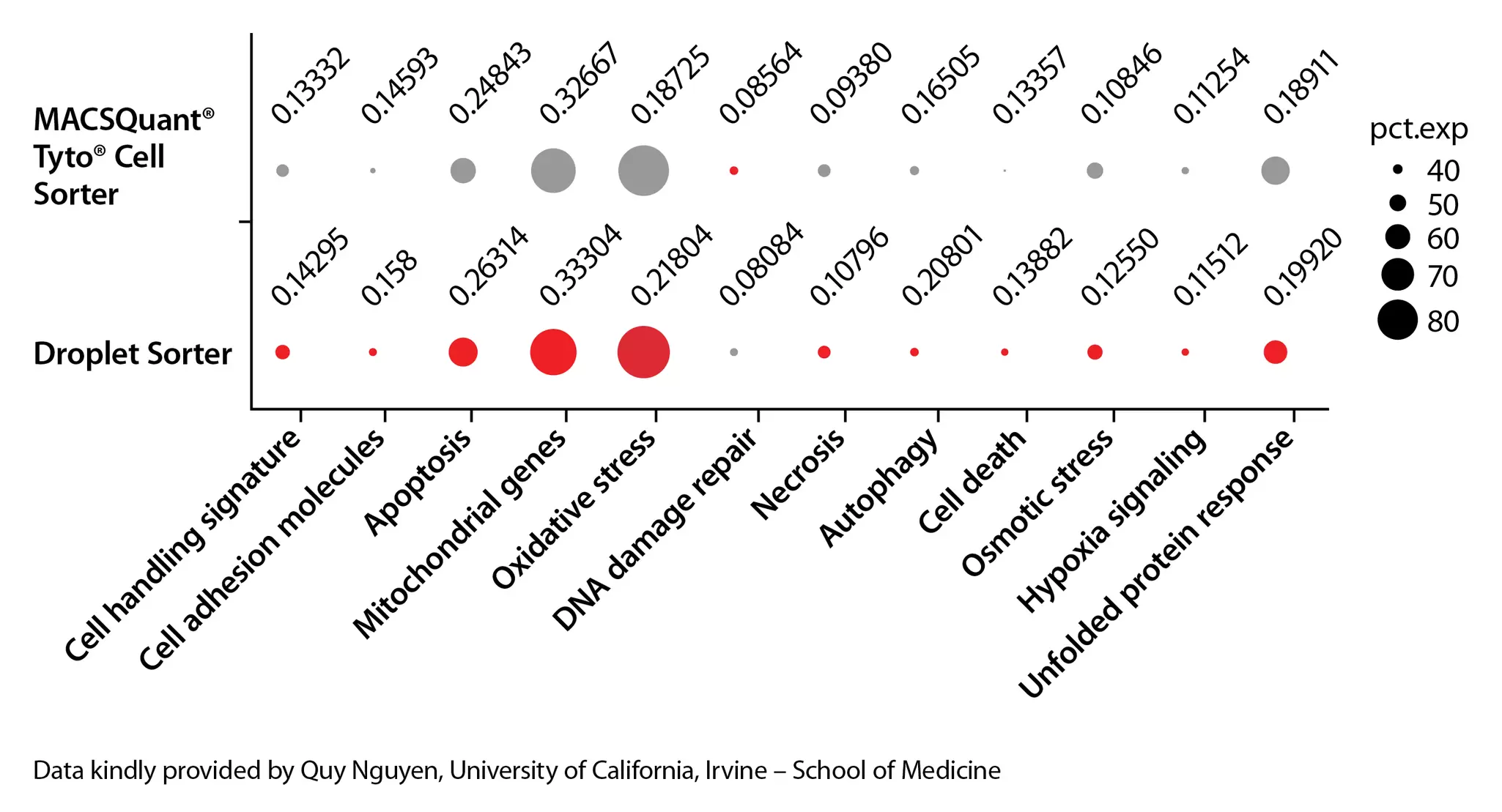
Comparison of gene expression of cells sorted with the MACSQuant Tyto or conventional cell sorter
Reduced stress-related gene induction demonstrated by single-cell RNA sequencing

Cells processed with the MACSQuant Tyto show reduced stress-response signatures and improved transcriptome quality, making the system ideal for tumoroid analysis and single-cell sequencing workflows.

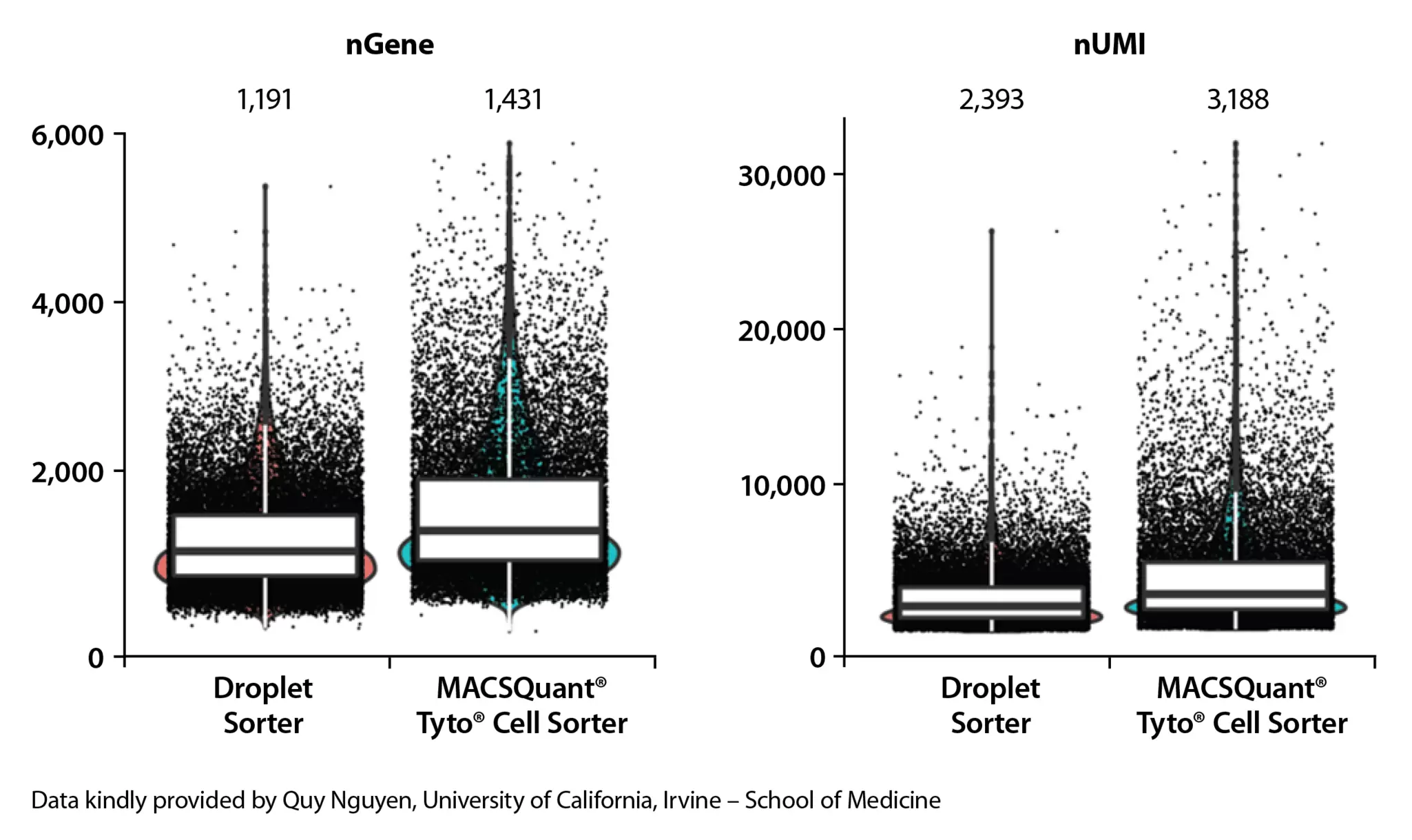
Cell sample quality after scRNA sequencing of samples sorted with the MACSQuant Tyto or a droplet sorter
Increased gene and UMI detection in whole transcriptome sequencing

Compared to a conventional droplet sorter, the MACSQuant Tyto yielded higher numbers of detected genes and unique molecular identifiers (UMIs), indicative of superior sample quality.

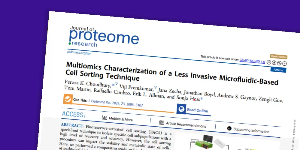
Thumbnail showing the first page of a Journal of Proteome Research article titled “Multiomics Characterization of a Less Invasive Microfluidic-Based Cell Sorting Technique,” featuring the article abstract and diagrams comparing microfluidic chip-based cell sorting with conventional droplet-based sorting methods.
Publication

Multiomics characterization of a less invasive microfluidic-based cell sorting technique

Choudhury F.K., Premkumar V., Zecha J., et al. (2024) J. Proteome Res. 23: 3096-3107.
doi: 10.1021/acs.jproteome.3c00773


Learn how stress-free sorting with the MACSQuant Tyto can benefit your molecular downstream analysis.

7. High-resolution 3D tumoroid imaging

Light sheet–based three-dimensional imaging reveals cell-cell interactions and spatial organization in intact tumoroids, an information often lost in 2D analyses or upon dissociation. Whole-mount immunofluorescence upon tissue clearing enables visualization of organoid architecture, cellular organization, and marker distribution, supporting analysis of heterogeneity, infiltration patterns, and tumor microenvironmental interactions.

The UltraMicroscope Blaze Light Sheet Imaging System combined with MACS UltraMount Sample Holders enables high-throughput cellular imaging of large or multiple tumoroids (up to 48 tumoroids in a single run). Advanced light sheet optics deliver consistent image quality from macro to cellular detail, while validated antibody panels for immuno-oncology ensure robust and reproducible marker detection.

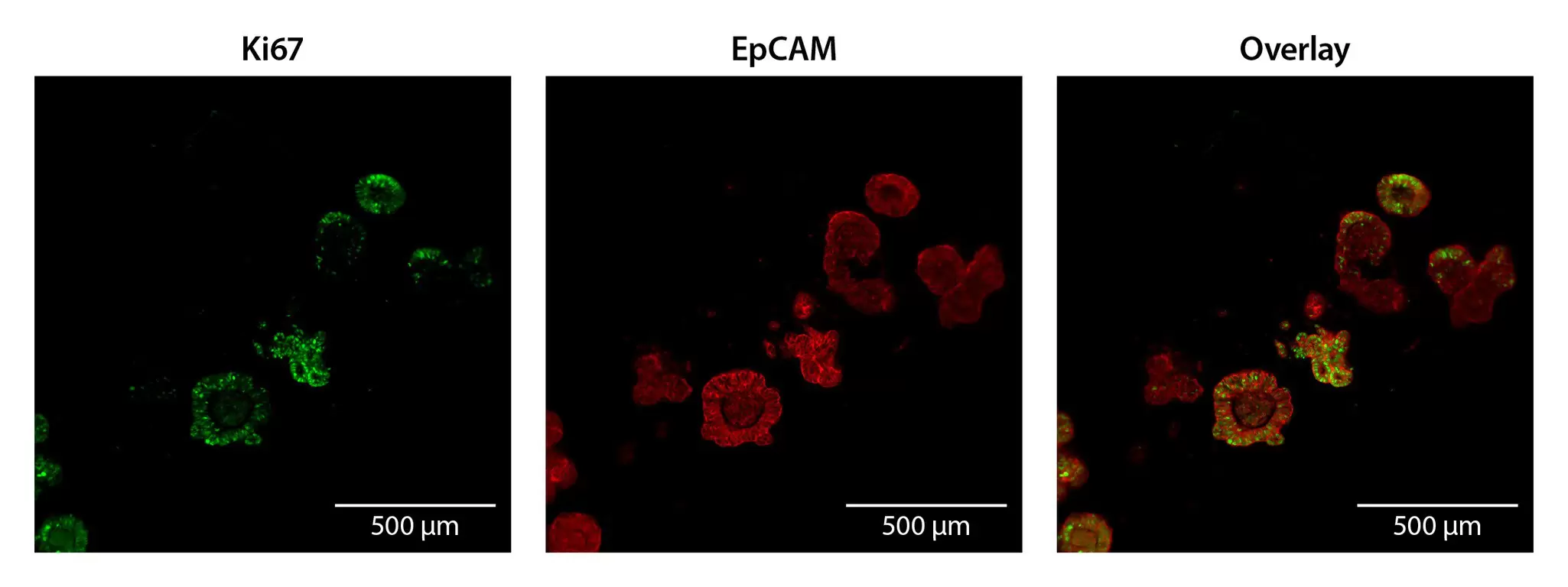
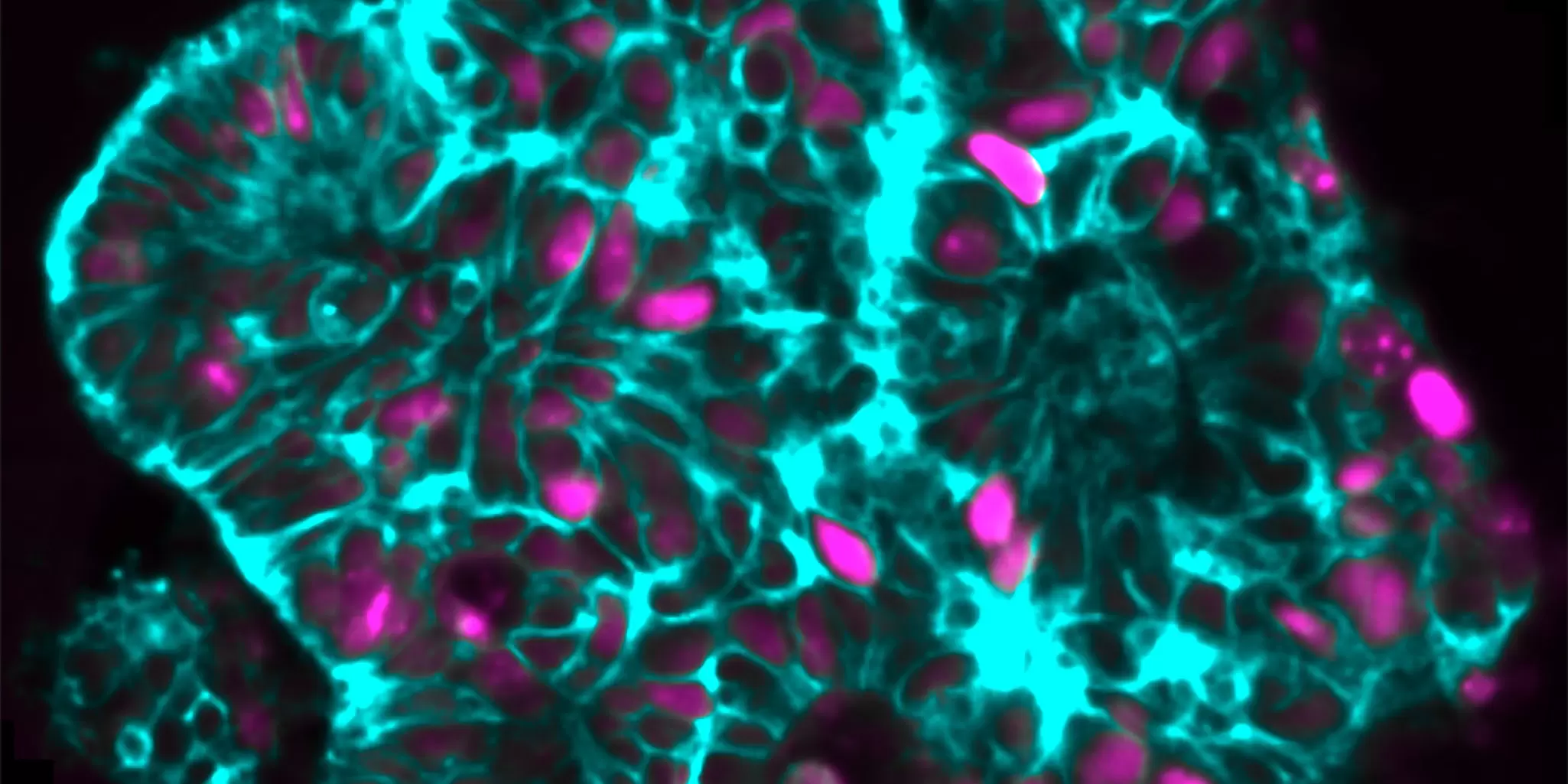
Microscopic image of colon cancer organoids that were grown in Colon TumorMACS media for 20 days, before they were harvested and fixed for subsequent 3D imaging using the UltraMicroscope Blaze™ light sheet instrument.
EpCAM and Ki-67 staining enable 3D visualization of colon cancer organoids

To complement your 3D imaging workflow, relevant sections can be selected and further analyzed using the MACSima® Platform, revealing hundreds of proteins and dozens of RNAs in 2D detail for deep phenotyping of the tumoroid region of interest.

Duration of steps for clearing opaque tumor-derived organoids for 3D imaging using the MACS Clearing Kit.

Tissue typeFixationPermeabilizationImmunostainingDehydrationClearing
Tumoroids (< 1 mm3)20 min6 hours40 hours24 hours6 hours

8. 2D spatial multiomics tumoroid imaging

Multiplex imaging provides essential insights into the cellular and structural characteristics of cancer organoids, offering a straightforward and effective method for examining specific phenotypic surface protein expression and enabling deep phenotyping of the cellular organization.

Our MACSima Platform for streamlined spatial omics workflows is built on cyclic immunofluorescence imaging, enabling the analysis of hundreds of protein markers and dozens of RNAs on a single sample. An extensive antibody portfolio for spatial biology is available, covering more than 600 biomarkers pretested for use on FFPE and frozen tissue sections.

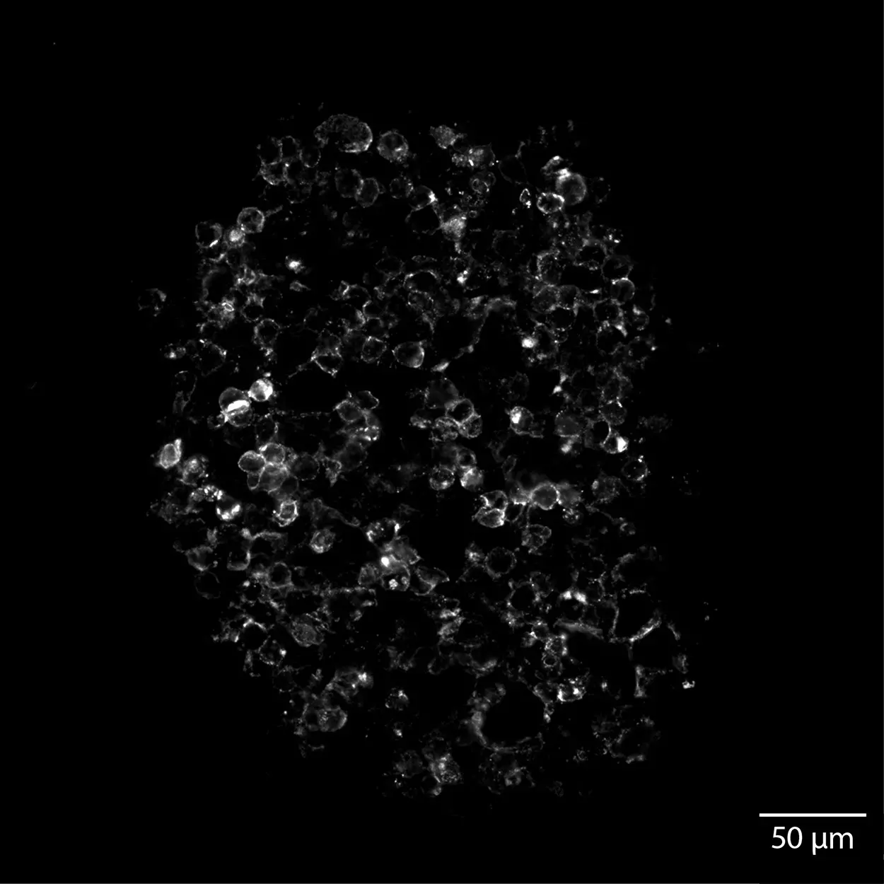
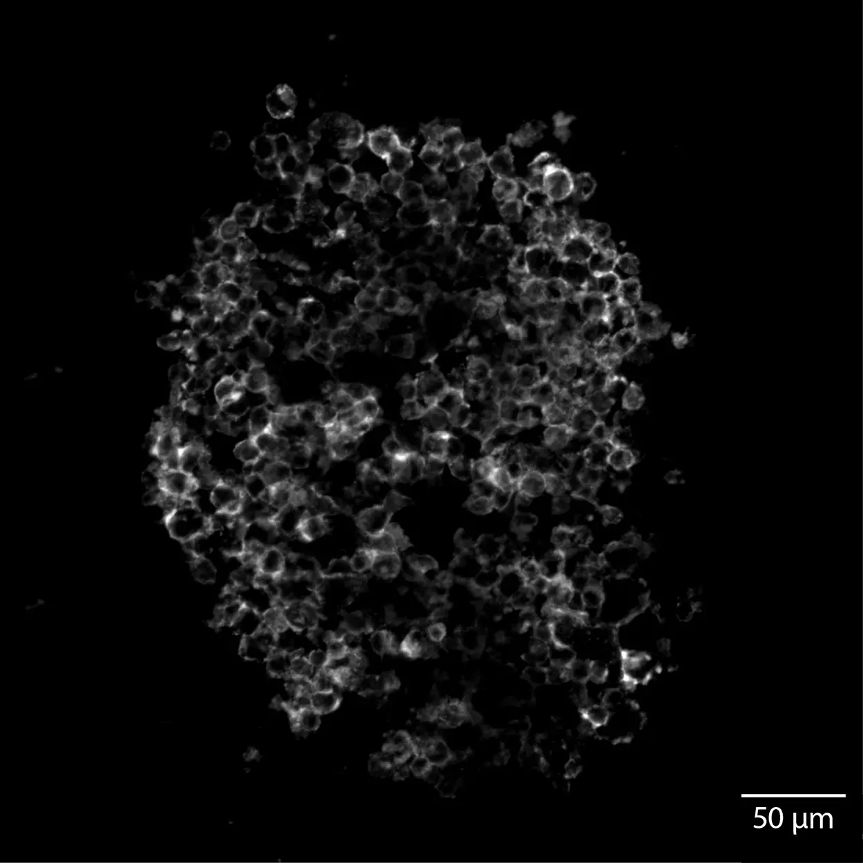
Colon cancer organoids were analyzed by 2D multiomics imaging. Broad expression of epithelial markers, including CD324, CD326, cytokeratins, and integrins, confirmed the epithelial origin of the cells. At the same time, heterogeneous expression of cancer stem cell–associated markers and variable distribution of epithelial proteins revealed the spatial organization and cellular heterogeneity within the 3D cultures.

Heterogeneous expression of cancer stem cell–associated CD44 and CD133, as well as areas with proliferative, Ki-67–expressing cells.

Staining for epithelial markers revealed broad expression patterns, with regional differences characterized by stronger expression of specific epithelial markers in distinct areas.

Staining for integrins and carcinoma-associated markers demonstrated broad, largely homogeneous expression across the 3D cultured cells.

Insights from our R&D experts

Frequently asked questions on tumoroids

What is the difference between tumoroids and cancer organoids?

Tumoroids, also known as cancer organoids or patient-derived organoids (PDOs), are self-organizing 3D models derived from primary tumor tissue that preserve tumor heterogeneity. Tumor spheroids are usually cell line–derived aggregates that lack structured architecture and long-term biological fidelity.

How long does it take to establish patient-derived tumoroids?

Initial tumoroid formation is typically observed within 3–10 days after embedding in a 3D matrix. Stable expansion and passaging generally require 2–4 weeks, depending on tumor entity, tissue quality, and culture conditions. Establishment efficiency varies between cancer types and individual patient samples.

Why do primary tumor cultures fail?

Primary tumor cultures may fail due to low viability after dissociation, overgrowth of stromal cells such as fibroblasts, loss of critical growth signals, excessive enzymatic damage, or suboptimal culture conditions. Variability in tissue handling and processing time also significantly impacts establishment success.

Can tumoroids be used for immunotherapy testing?

Yes. Patient-derived tumoroids can be used for immunotherapy testing when cocultured with immune cells such as T cells, NK cells, or tumor-infiltrating lymphocytes. They enable evaluation of immune-mediated cytotoxicity, checkpoint inhibition responses, and personalized immunotherapy strategies in a physiologically relevant 3D setting.

All relevant products for application on tumoroid research

35 products found

|

You have previously chosen International as your location.
What do you want to do?