Content overview
Genomic analysis of magnetically isolated cells
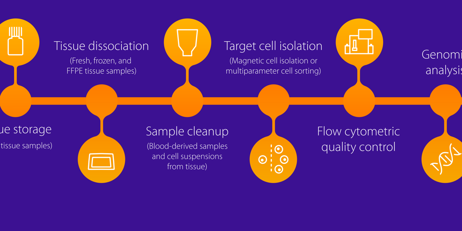
How to isolate target cells using MACS Technology
Content
- Genomic analysis of magnetically isolated cells
- How to isolate target cells using MACS Technology

Isolation of target cells increases the resolution of bulk and single-cell genomic analysis, by excluding the background signals coming from non-target cells, and thereby helps to save time and decrease sequencing costs. MACS® Technology allows the fast and easy immunomagnetic isolation of single cells from suspensions of solid tissue and blood-derived samples. By preserving gene expression during isolation, it supports reliable, high resolution genomic analysis. MACS Technology also includes automated solutions to upscale, standardize, and speed up your magnetic cell separations for reproducible results.
Content overview
Genomic analysis of magnetically isolated cells
How to isolate target cells using MACS Technology
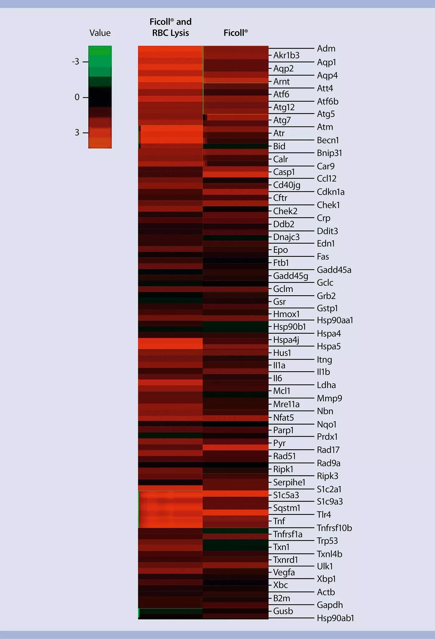
Gentle direct isolation of PBMCs from whole blood using immunomagnetic beads prevents activation of cellular stress responses compared to density gradient centrifugation methods. In addition to that, the automated PBMC isolation procedure is standardized and reproducible, improving purity of the obtained PBMCs population.
Highly pure PBMCs directly isolated from whole blood using the Whole Blood PBMC Isolation Kit, human and Sedimentation Kit 2*
| Sample 1 | Sample 2 | Sample 3 | ||
| Granulocytes | Whole Blood PBMC Isolation Kit | 0,27% | 0,29% | 0,2% |
| Ficoll® | 4,9% | 2,41% | 1,58% | |
| Erythrocytes | Whole Blood PBMC Isolation Kit | 0,12% | 0,06% | 0,06% |
| Ficoll® | 2,59% | 37,81% | 21,94% | |
* Please contact us for product and ordering information

Magnetic isolation improves the resolution of single-cell RNA sequencing of T and B cell receptors, highlighting clonotypes otherwise poorly or altogether unrepresented in the bulk sample.

Magnetic isolation of neurons after dissociation of adult mouse brain substantially increases the resolution of single-cell gene expression profiling, allowing the identification of several neuronal subpopulations which otherwise could not be distinguished.
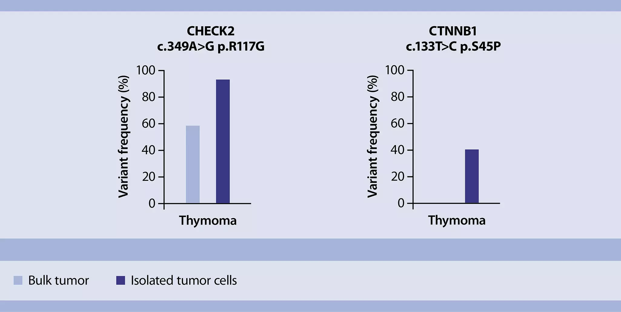
Tumor cell isolation enables accurate detection of single nucleotide polymorphisms (SNP) in solid tumors for the analysis of loss of heterozygosity using next generation sequencing (NGS).
Enrichment of carcinoma cells from FFPE lung adenocarcinoma specimens with low estimated tumor cell content, increases significantly the allele frequency of detected driver mutations compared to macrodissection.
Increased sensitivity of NGS-based mutation analysis of FFPE lung adenocarcinoma specimens by carcinoma cell
| Specimen ID | Pre-processing prior DNA extraction | Concentration of extracted DNA (ng/ul) | DNA input for NGS | Driver mutation identified | Coverage | Allele frequency |
| Specimen 1 (70% tumor cells) | Macrodissection | 48.8 | 40 | EGFR: c.2236_2250del; p.E746_A750del | 1471 | 52.3% |
| Enriched tumor cells | 38.8 | 40 | EGFR: c.2236_2250del; p.E746_A750del | 2522 | 59.9% | |
| Specimen 2 (10% tumor cells) | Macrodissection | 4.78 | 20 | KRAS: c.34G>T; p.G12C | 388 | 11% |
| Enriched tumor cells | 0.46 | 20 | KRAS: c.34G>T; p.G12C | 623 | 28% | |
| Specimen 3 (< 5% tumor cells) | Macrodissection | 5.64 | 40 | KRAS: c.34G>T; p.G12C | 405 | 1% |
| Enriched tumor cells | 2.12 | 40 | KRAS: c.34G>T; p.G12C | 951 | 7% |
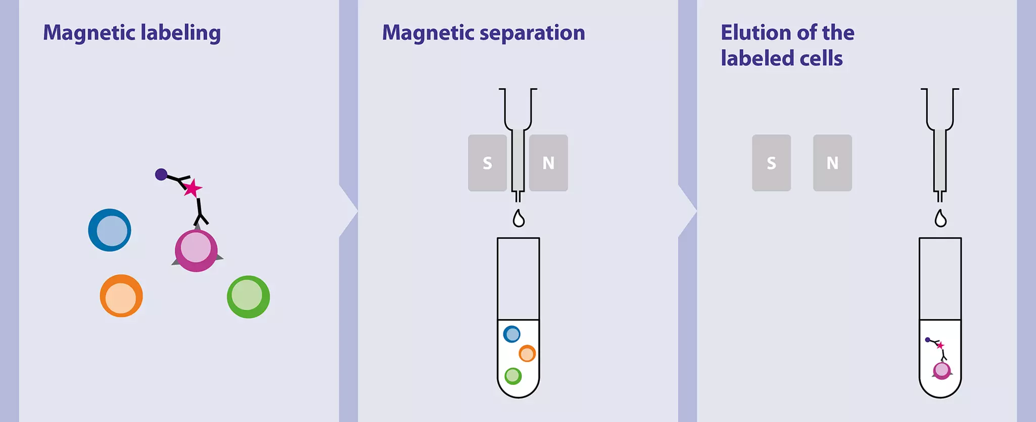
MACS Technology enables the magnetic separation of cell populations by targeting surface antigens with specific antibodies that are conjugated to superparamagnetic beads. Labeled cells are magnetically retained in a separation column, from which they can easily be eluted. The fast and gentle isolation minimizes cellular changes and ensures high viability. Explore the different solutions for target cell isolation with MACS Technology here.

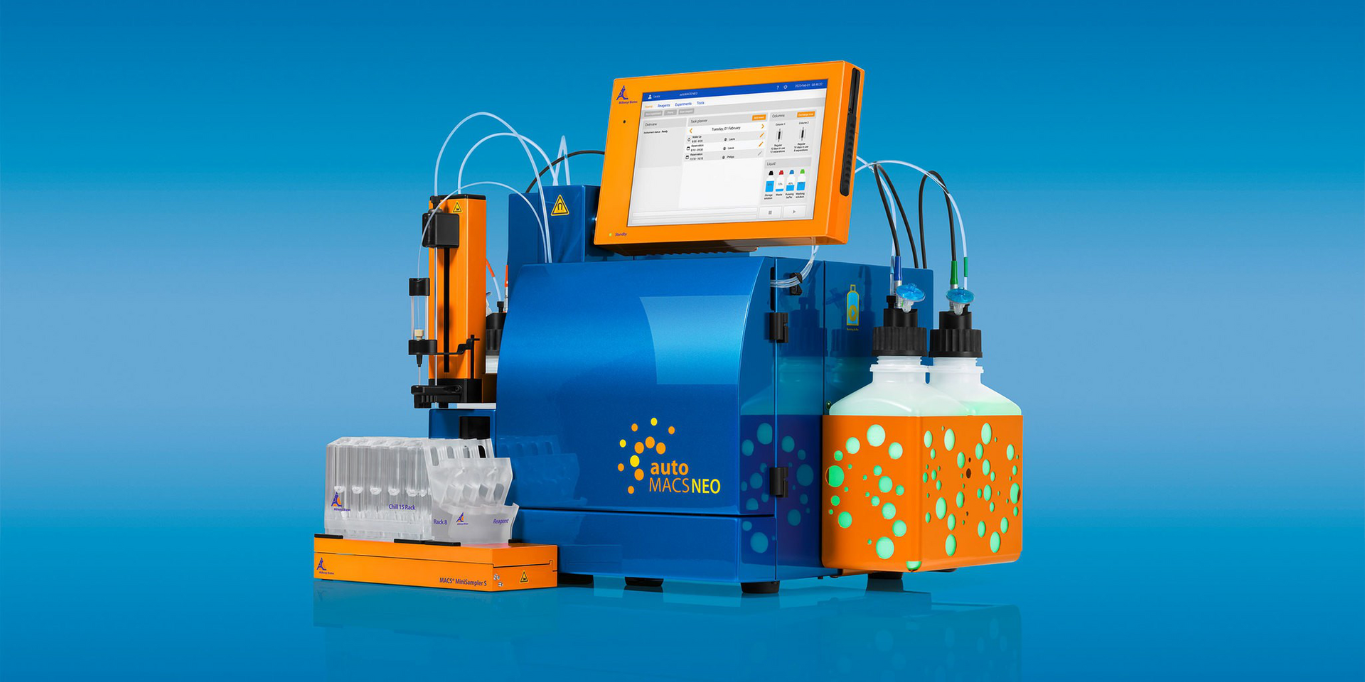
Whether working with only few samples or large sample cohorts, automation helps to streamline cell separation. The MACS Cell Separation instruments allow different levels of automation according to the sample throughputs. In particular, the autoMACS NEO Separator provides full automation for magnetic cell separations using MACS Technology, while minimizing temperature changes and user-dependent variation.
The frequency of certain target cell populations, such as tumor infiltrating leukocytes, can be very low in the starting material, and lead to very long flow sorting times, which add up the higher thenumber of samples to be sorted, increasing the risk for gene expression changes to happen. Pre-enrichment of low abundant cell types with MACS Technology prior flow sorting allows much faster acquisition times and higher quality of the cells for genomic analysis.
Pre-enrichment of target cells reduces the flow acquisition time
| Cell type | Cells to analyze | Events to collect | Flow cytometry time/sample* | Total flow cytometry time** |
| CD4+ T cells | ||||
| Bulk | 5,000 | 7.96×10⁶ | 66.3 min | >10 h |
| Isolated*** | 5,000 | 5.41×10⁴ | 0.5 min | ~11 min |
| CD8+ T cells | ||||
| Bulk | 5000 | 2.80×10⁶ | 23.3 min | >3.5 h |
| Isolated*** | 5000 | 4.37×10⁴ | 0.4 min | ~10 min |
| T cells | ||||
| Bulk | 10,000 | 8.13×10⁵ | 6.8 min | >1 h |
| Isolated*** | 10,000 | 3.24×10⁴ | 0.3 min | <10 min |
| * Flow rate: 2,000 events/s ** Considering 9 samples (3 experimental groups × 3 replicas/group). Includes 45 s automated mixing and rinsing between samples on the MACSQuant® Instrument *** Isolation using CD8 (TIL), CD4 (TIL), or CD4/CD8 (TIL) MicroBeads, respectively | ||||
Isolation of CD4+, CD8+, and pan T cells from different mouse tumor models using mouse CD4 (TIL) MicroBeads, CD8 (TIL) MicroBeads, and CD4/CD8 (TIL) MicroBeads dramatically decreases time of analysis.
You have previously chosen International as your location.
What do you want to do?