Mouse hematopoietic stem cells


1 Introduction to mouse hematopoietic stem cells 2 Mouse HSCs in bone marrow
1 Introduction to mouse hematopoietic stem cells
Hematopoietic stem cells (HSCs) are tissue-specific adult stem cells capable of differentiating into all blood cell types to ensure the homeostasis of blood throughout life.
Research on murine HSCs dates back to the late 1940s/early 1950s when it was discovered that sub-lethally irradiated mice could be rescued from hematopoietic failure by injecting cells from blood-forming organs, such as bone marrow. This finding led to the understanding that cells in bone marrow with repopulation capacity could be further characterized and quantified in transplantation models. In early experiments, decreasing numbers of bone marrow cells were injected into recipient mice to determine the lowest cell number needed to fully reconstitute hematopoiesis. Further experiments that transplanted sub-radioprotective doses of bone marrow cells into mice showed that these mice developed colonies of hematopoietic cells in their spleens. The number of colonies correlated to the number of bone marrow cells originally injected. The advent of fluorescence-activated cell sorting technology and monoclonal antibodies then finally led to the identification of surface mouse HSC markers.
 Related PDFs:
2 Mouse HSCs in bone marrow
The main source to isolate mouse HSCs is bone marrow, where out of all cells, approx. 0.002% of hematopoietic stem cells reside (PMID: 23209149).
2.1 Cell subsets, frequencies, and marker expression
Mouse HSCs are characterized and/or isolated using several surface markers. Firstly, mouse HSCs lack the expression of lineage-specific markers (meaning that they are lin-), and are positive for c-Kit (CD117) and Sca-1, the so-called LSK population. As only approx. 10% of LSK cells are long-term HSCs (PMID: 19023891), additional markers have been identified to further classify alternate HSCs. For improved HSC identification, the marker combinations Lin–Sca1+c-Kit+CD34–Flk2– or Lin–c-Kit+Sca-1+CD48–CD150+ are very often used.
2.2 Miltenyi Biotec application protocols for mouse HSCs from bone marrow
Miltenyi Biotec has created dedicated application protocols to work with and analyze mouse HSCs.
2.3 Sample preparation of bone marrow
Murine HSCs are usually isolated from bone marrow prepared from the tibia of the hind leg.
2.4 Magnetic pre-enrichment of mouse HSCs from bone marrow
Miltenyi Biotec has developed products to preenrich lineage-negative cells containing mouse HSCs from bone marrow cells using MACS® Technology.
For details on MACS Technology, see the MACS Handbook chapter Magnetic cell separation.
 MACS Handbook:
2.4.1 Isolation of mouse HSCs
At a glance: Kits and reagents for the separation of mouse HSCs from bone marrow
| Starting material | Isolation strategy | Comments | Automation | Product |
| Bone marrow | Depletion of non-target cells | Stringent two-step procedure for labeling of lineage-negative cells | Yes | Lineage Cell Depletion Kit, mouse |
| Bone marrow | Depletion of non-target cells | Direct preenrichment of lineage-negative cells Fully automated cell labeling and separation using the autoMACS® Pro Separator possible | Yes | Direct Lineage Cell Depletion Kit, mouse* |
| Bone marrow | Positive selection of target cells | Yes | Anti-Sca-1 MicroBead Kit (Vio® Bright FITC), mouse | |
| Bone marrow | Positive selection of target cells | Yes | CD105 MultiSort Kit (PE), mouse | |
| Bone marrow | Positive selection of target cells | Yes | CD117 MicroBeads, mouse |
*can additionally automate HSC labeling
The Lineage Cell Depletion Kit, mouse, and the Direct Lineage Cell Depletion Kit, mouse, enable fast preenrichment of mouse HSCs from bone marrow by depleting cells expressing lineage markers. Designed to magnetically label mature cells (T cells, B cells, NK cells, dendritic cells, monocytes, granulocytes, and erythroid cells) and their differentiation-committed precursors, the kits generate an enriched population of untouched mouse HSCs and progenitor cells. The Direct Lineage Cell Depletion Kit, mouse, is specially formulated for a quick one-step preenrichment process, whereas the Lineage Cell Depletion Kit, mouse, enables the more stringent depletion of Lin+ cells from bone marrow.
Direct Lineage Cell Depletion Kit, mouse
 View details
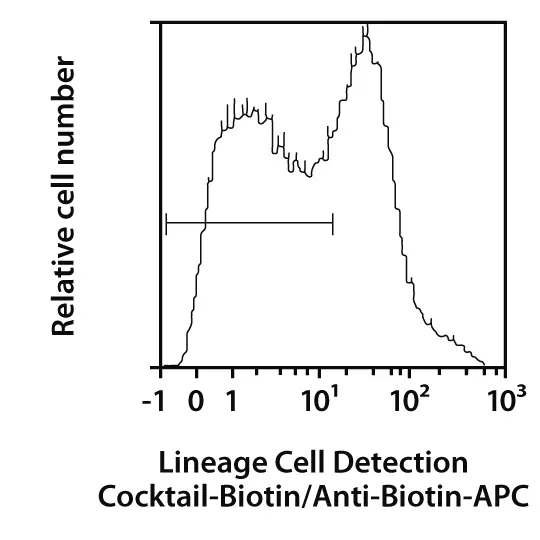
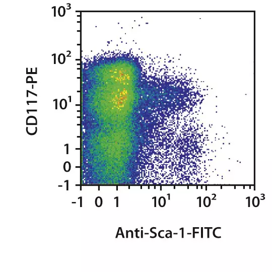
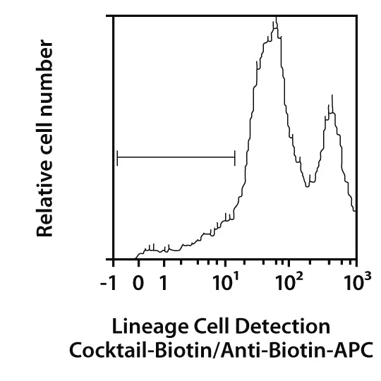
Quick, one-step separation of lineage-negative cells. Untouched lineage-negative cells were isolated from a single-cell suspension of mouse bone marrow using the Direct Lineage Cell Depletion Kit, mouse, and a MidiMACS™ Separator with an LS Column. Cells were fluorescently stained with Hematopoietic Lineage Labeling Cocktail, anti-mouse, Biotin, Anti-Biotin-APC and analyzed by flow cytometry using the MACSQuant® Analyzer. To evaluate the LSK (Lin–Sca-1+c-Kit+) fraction, cells were further stained with CD117-PE (c-Kit) and Anti-Sca-1-FITC. Cell debris and dead cells were excluded from the analysis based on scatter signals and propidium iodide fluorescence.
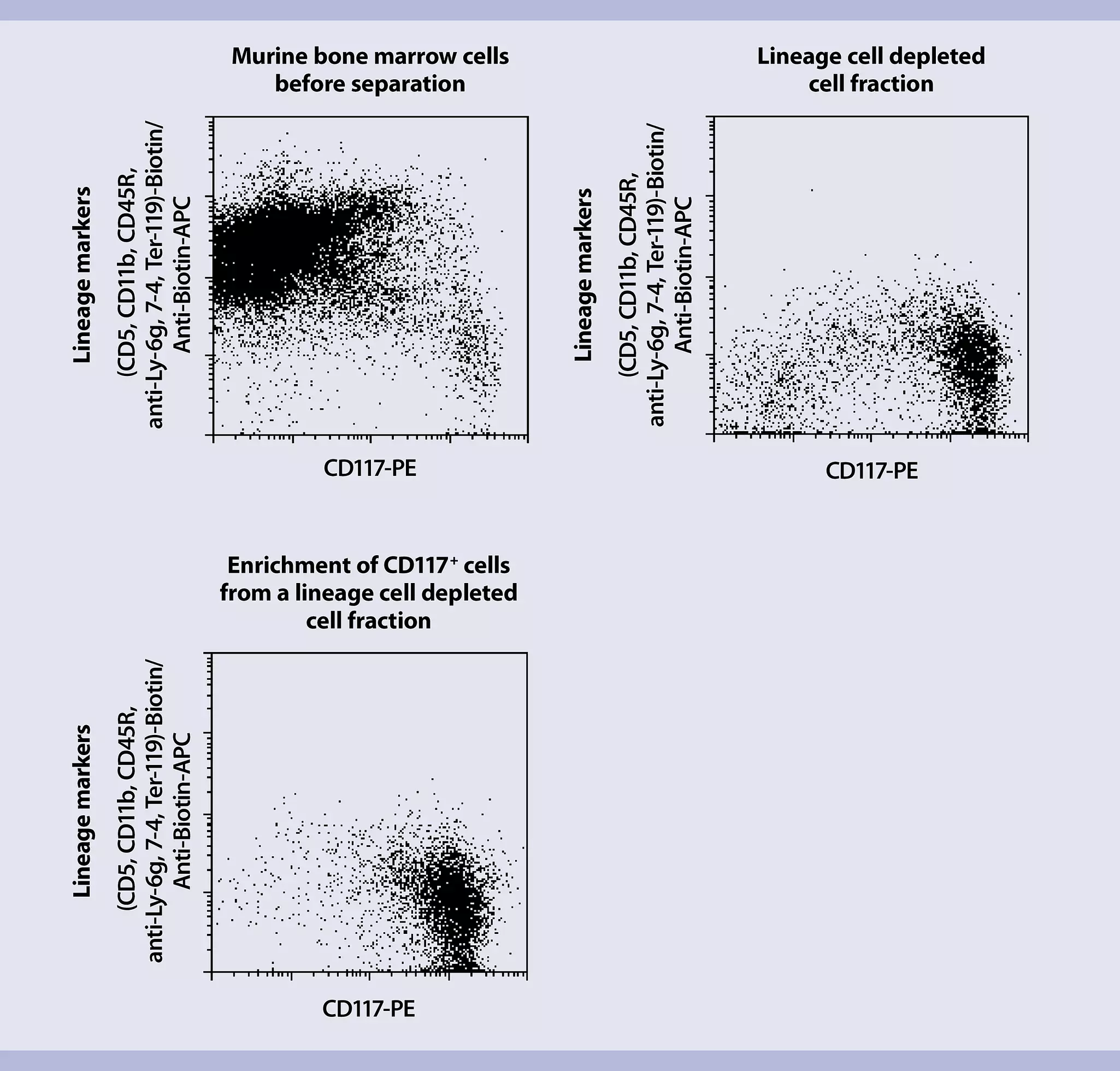
Mouse HSCs can be further separated from lineage negative (Lin–) cells using specific MACS MicroBeads, like the CD117 MicroBeads, mouse, or the Anti-Sca-1 MicroBead Kit (Vio® Bright FITC), mouse.
CD117 MicroBeads isolate murine progenitor cells. CD117 (also known as c-Kit, steel factor receptor, or stem cell factor receptor) encodes a 145 kDa cell surface glycoprotein belonging to the class III receptor tyrosine kinase family. It is expressed on the majority of mouse hematopoietic progenitor cells, including multipotent HSCs and committed myeloid, erythroid, and lymphoid precursor cells. In addition to their hematopoietic cell differentiation potential, CD117+ stem cells from murine bone marrow are reportedly capable of differentiating into smooth muscle cells, myocytes, and endothelial cells in vivo. CD117 is also expressed on a few mature mouse HSCs such as mast cells.
 View details
 Related videos:
2.5 Mouse HSC characterization by flow cytometry
At a glance: Dedicated antibody cocktail for flow cytometry analysis of mouse HSCs
| Reagent used | Markers in cocktail | Comment |
| Hematopoietic Lineage Labeling Cocktail, anti-mouse, Biotin | Monoclonal antibodies against CD5, CD11b, CD45R (B220), Anti-7-4, Anti-Gr-1 (Ly-6G/C), and Anti-Ter-119. | Requires the use of a secondary antibody such as Anti-Biotin-FITC, Anti-Biotin-PE, or Anti-Biotin-APC |
Mouse HSCs are routinely analyzed by flow cytometry which is based on various extracellular and intracellular markers. The Hematopoietic Lineage Labeling Cocktail, anti-mouse, Biotin simplifies the flow cytometric analysis of cells before and after cell separation. All antibodies are optimally titrated to easily add to an aliquot of a cell fraction before analysis.
Miltenyi Biotec also offers a wide range of unique and standard monoclonal antibodies for research of mouse HSCs and progenitor cells, including CD117, Anti-SCA-1, CD150, and CD48. Fluorochrome-conjugated MACS Antibodies are perfectly suited for the identification, enumeration, and characterization of mouse HSCs, which can be combined into panels tailored to specific research needs. An online tool to quickly build an appropriate multicolor flow cytometry panel for each research project is available through the related resources panel to the right.
For details about Miltenyi Biotec antibodies and dyes for cell analysis, see the chapter Flow Cytometry.
Related PDFs:
Related content:
 Related products:
2.6 Cell culture of mouse HSCs
2.6.1 Expansion of mouse HSCs
At a glance: Cytokines for the expansion of mouse HSCs
| Use | Comment | Product |
| Expansion | Premium grade | Mouse SCF |
| Research grade | Mouse FLT-3-Ligand | |
| Premium grade | Mouse IL-3 IS | |
| Premium grade | Mouse IL-6 |
Isolated mouse HSCs can be further expanded in cell culture with HSC-relevant growth factors to obtain a larger number of cells or to elucidate mechanisms and signaling pathways driving self-renewal or differentiation. Early acting cytokines – mouse stem cell factor (SCF), Flt3-Ligand, and interleukin-6 (IL-6) – expand primitive mouse HSCs, although current mouse HSC culture systems allow only limited expansion and maintenance of primitive mouse HSCs that can engraft and completely restore the entire blood system.