3D illustration of regulatory T cells
Logo

TCR Discovery for next-generation cancer immunotherapy

Identifying tumor-reactive T cell receptors (TCRs) is pivotal for developing next-generation TCR T cell therapies and other TCR-based products. Unlike chimeric antigen receptor (CAR)-based products, which are restricted to targets on the surface of cells, these emerging therapies recognize intracellular antigens presented by major histocompatibility complex (MHC) molecules on antigen-presenting cells. This broadens the range of antigens beyond those accessible to CAR T cells and offers new opportunities against solid tumors.

This application page focuses on the identification of alpha beta (αβ) TCRs, but the process can be reproduced with gamma delta (γδ) TCRs. A range of cells can then be engineered with the tumor-reactive TCR (e.g., T cells, NK cells, or γδT cells). Our flexible and established workflow empowers researchers to efficiently pinpoint the key TCRs that drive potent immune responses against cancer.

Content overview

Content

  • Application overview
  • How to perform TCR discovery
  • 1. T cell and monocyte isolation
  • 2. Antigen-specific T cell stimulation
  • 3. Selection of antigen-specific T cell
  • 4. Flow cytometric characterization of antigen-specific T cells
  • 5. Tumor-reactive TCR sequencing
  • 6. Engineering TCR T cells
A flexible workflow for antigen-specific TCR identification

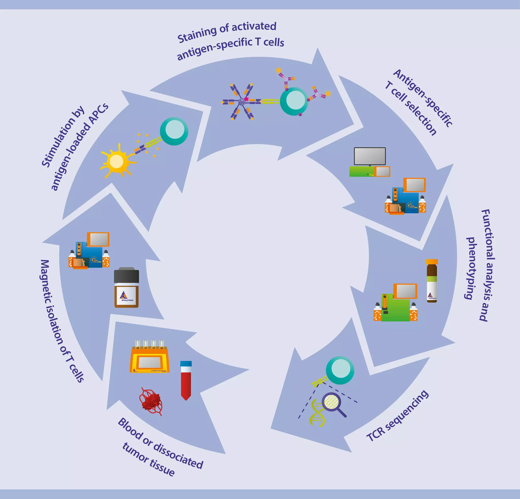
The workflow for antigen-specific TCR identification, from starting material to TCR sequencing.
Workflow for antigen-specific TCR identification.

A therapeutically effective TCR recognizes and eliminates cancer cells. Finding that TCR involves selecting tumor-reactive T cells and leveraging their antigen specificity to determine the optimal TCR sequence.

Our comprehensive preclinical workflow begins with the efficient isolation of T cells from dissociated tumor tissues or healthy donor peripheral blood while ensuring high purity and viability. These T cells are subjected to antigen-specific stimulation through a targeted coculture with mature monocyte-derived dendritic cells (Mo-DCs) loaded with standardized peptides. This step robustly activates tumor-reactive T cells. They are subsequently enriched, either directly with antigen-loaded multimers or indirectly based on their expression of activation markers or cytokine secretion. Finally, precise TCR sequencing is performed to decode the TCR repertoire, enabling researchers to identify the most promising TCR candidates for therapeutic development.

Webinar

Working with antigen-specific T cells

Learn about Miltenyi Biotec’s unique solutions for the isolation, stimulation, pre-enrichment, and analysis of tumor-reactive T cells, leading to the identification of your TCR of interest.

How to perform TCR discovery

We offer solutions designed for consistency and efficiency to make the discovery of your TCR of interest easy. These include solutions for antigen-specific T cell stimulation through coculture with differentiated Mo-DCs loaded with standardized peptides of interest, sensitive selection of the antigen-specific T cells, and precise TCR sequencing.

What is the role of T cells in developing cancer immunotherapies?

While the final cell type used in TCR-based cell therapies can be T cells, NK cells, or others, the process of identifying the therapeutic TCR almost always starts with T cells. Here's why:

  • Natural TCRs reside on T cells: TCRs are the natural antigen recognition molecules of T cells. Therefore, the initial discovery and characterization of TCRs with the desired target specificity are typically done using T cells.
  • T cell-based discovery methods: The techniques used to identify and isolate TCRs are generally designed to work with T cells.
  • Understanding TCR function in T cells: A significant amount of knowledge about TCR signaling, antigen recognition, and downstream effector functions comes from studying T cells, which is essential in selecting and engineering TCRs.

Once a candidate TCR is identified using T cells, its genetic code can be introduced into other cell types to create an engineered TCR therapy. The TCR provides the antigen-targeting ability, while the base cell (e.g., NK cell) contributes its effector mechanisms.

1. T cell and monocyte isolation

Efficient isolation of T cells is the cornerstone of robust TCR discovery for cancer research. Start from either dissociated tumor tissues or healthy donor peripheral blood to obtain highly pure and viable isolated T cells. These are perfect for the accurate identification of tumor-reactive T cells. Monocytes can also be isolated from the same donor and used to stimulate the antigen-specific T cells in the following steps.

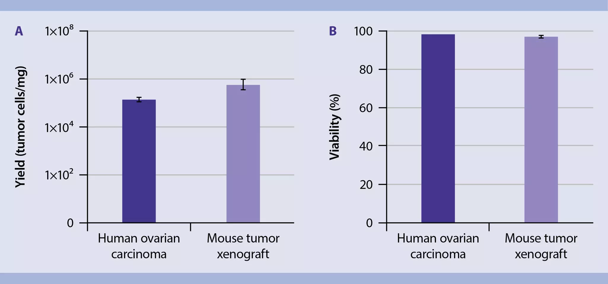
Bar chart showing high yield and viability of tumor cells from dissociated ovarian and xenograft tumors.
Efficient dissociation of ovarian carcinoma and tumor xenografts yields high counts of viable tumor cells.

Enzymatic tumor dissociation for efficient recovery of tumor-reactive T cells

If you have access to patient material, use the Tumor Dissociation Kit for fast, standardized, and automated processing of tumor samples. Together with the gentleMACS™ Octo Dissociator with Heaters and C Tubes, it improves reproducibility and helps you to recover key populations of tumor-reactive T cells, independently from the operator.

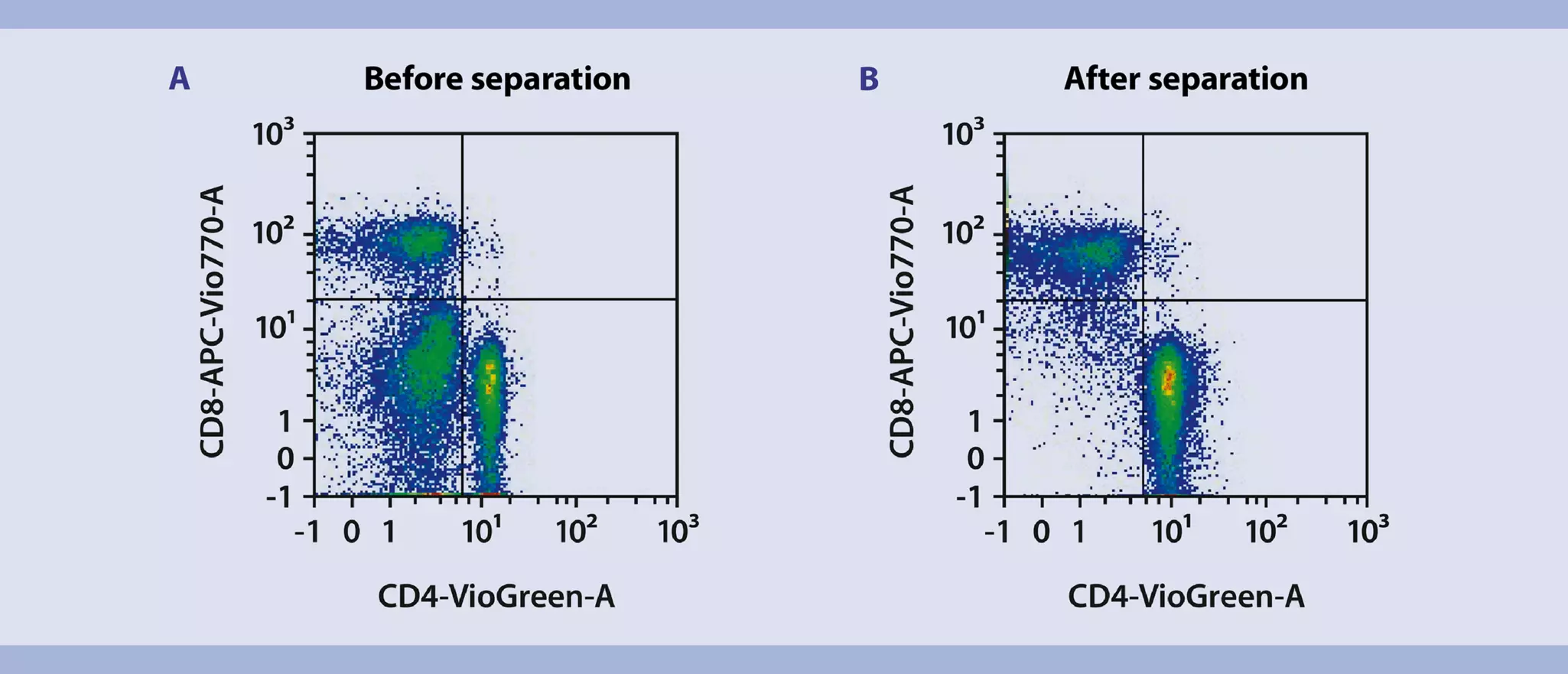
Purity of CD4+ and CD8+ T cells before and after enrichment with MACS Technology analyzed by flow cytometry using the MACSQuant Analyzer
Efficient enrichment of CD4+ and CD8+ T cells via positive magnetic selection.

Magnetic isolation of tumor-reactive T cells from dissociated tissues or PBMCs

Positive selection from peripheral blood mononuclear cells (PBMCs) or dissociated tissues using MACS® MicroBead Kits enables straightforward isolation of either or both CD4+ and CD8+ T cells. Simultaneous enrichment of CD4+ and CD8+ T cells makes for a homogeneous starting cell population, allowing for robust, standardized, and reproducible results.

If subsequent magnetic sorting steps are required, untouched cell isolation using Pan T Cell Isolation Kits allows for the isolation of unlabeled T cells via depletion of non-target cells.

Illustration of a scatter chart showing purity and recovery of monocyte-derived dendritic cells isolated with CD14 microbeads.
High-purity isolation of monocytes from PBMCs via magnetic separation.

Isolation of highly pure, debris-free monocytes from PBMCs

Monocytes can also be isolated from PBMCs using CD14 MicroBeads UltraPure. Their high specificity ensures a reduced amount of debris. Conversely, this purity increases the efficacy for downstream applications, such as ex vivo differentiation into Mo-DCs.

Bar charts showing high purity, recovery, and cell counts of T cells and monocytes isolated directly from whole blood.
Purity, recovery, and yield of CD4+, CD8+ and CD14+ cells isolated from a leukapheresis product.

Magnetic isolation of T cells and monocytes directly from donor blood

Using StraightFrom® Technology rapidly enriches T cells and monocytes directly from the same healthy donor's blood (buffy coat, Leukocyte reduction System Cones or LRSC, and Leukopak®) without the need to separate PBMCs first. These isolated cells can be cocultivated to stimulate antigen-specific T cells.

  • No density gradient centrifugation, no erythrocyte lysis.
  • Preservation of cell functionality for full downstream compatibility.
  • Automated and standardized isolation of different cell types with the autoMACS NEO Separator.
  • Semi-automated isolation of multiple samples or large volume samples with MultiMACS™ Cell24 Separator Plus.
  • StraightFrom Technology can also be used to isolate PBMCs directly without a density gradient.

Learn more about StraightFrom Technology here.

2. Antigen-specific T cell stimulation

In this step, antigen-specific in vitro stimulation of T cells is performed to enrich target T cells for their subsequent high-purity sorting. Our PepTivator® Peptide Pools or MACSpep Single Peptides can be loaded on differentiated antigen-presenting cells (APCs) to enable the efficient activation and expansion of antigen-specific CD4+ and CD8+ T cells. PepTivator Peptide Pools are available in RUO or MACS GMP-Grade for smooth translational applications.

 PepTivator Peptide PoolsMACSpep Single Peptides
Main applications
  • Peptide pool-based activation of antigen-specific T cells (CD4+ and CD8+)
  • Peptide-based enrichment and analysis of T cell pools using rapid ARTE protocol
  • Epitope-specific activation of antigen-specific T cells
  • Peptide-loading of MHC MACSimers for analysis of antigen-specific T cells by peptide-MHC multimer staining
Peptide structure
  • Pool of 15-mer peptides sequences with 11-amino-acid overlaps
  • Covering the sequence of a complete T cell antigen
  • One specific peptide sequence, restricted to bind either a MHC class I or MHC class II molecule
  • MHC I binders are 8- to 12-mer in length; MHC II binders are 13- to 25-mer in length
T cell expansion properties
  • T cell expansion against multiple peptide-MHC combinations
  • Tracking of multiple populations of antigen-specific T cells with different TCR-specificities
  • T cell expansion against one peptide-MHC combination
  • Tracking of one population of epitope-specific T cells

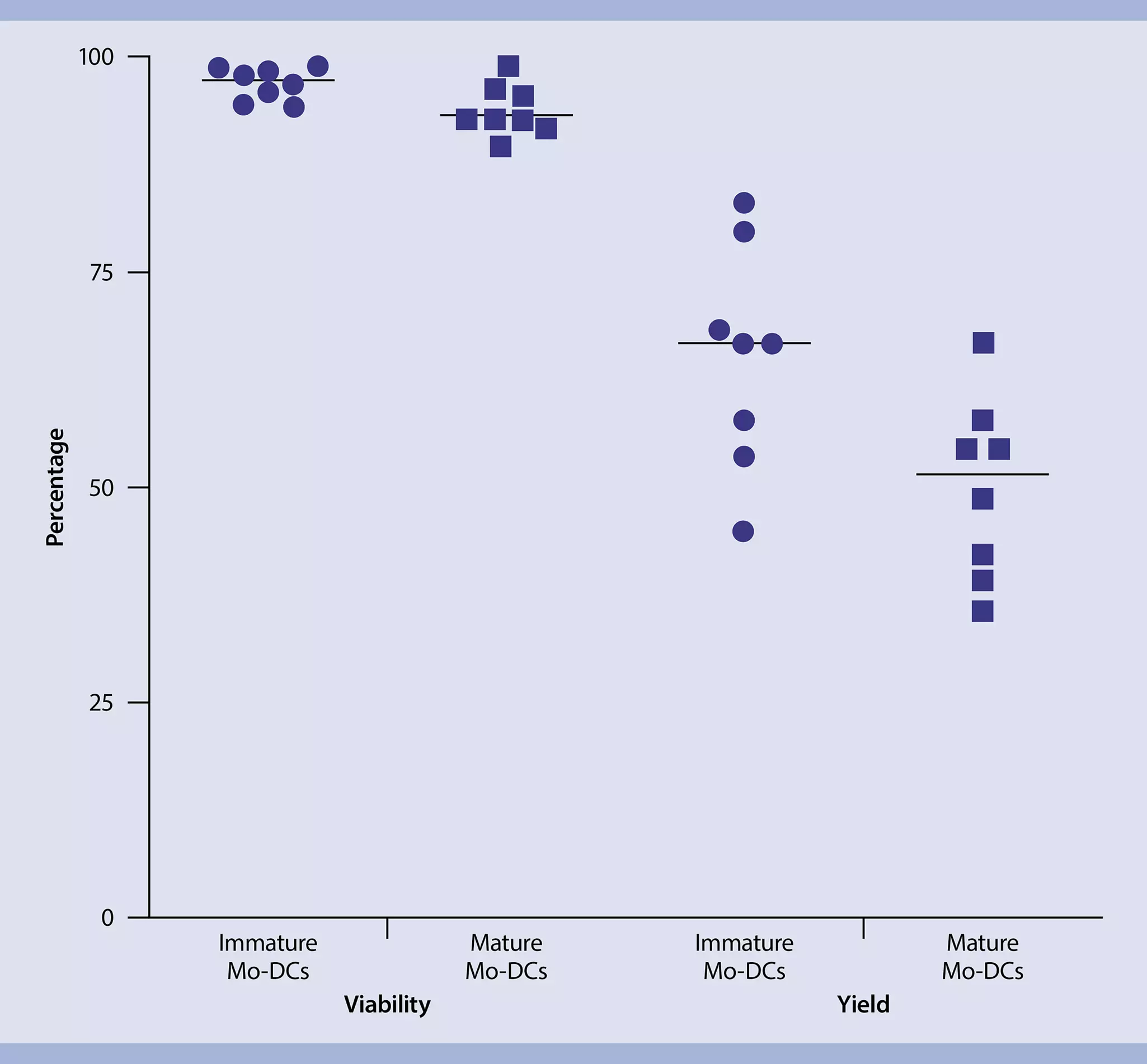
Scatter chart showing high viability (>90%) and yield of immature and mature monocyte-derived dendritic cells.
High numbers of viable immature and mature Mo-DCs are generated from isolated monocytes.

Monocyte-derived dendritic cell generation

Isolated monocytes can be differentiated into dendritic cells (Mo-DCs) to serve as antigen-presenting cells (APCs).4,5 The mature Mo-DCs are loaded with the peptide of interest (e.g., PepTivator Peptide Pools or MACSpep Single Peptides) and cocultivated with the isolated T cells for the next step: stimulation of the antigen-specific T cells.5,6

Cultivate the monocytes in medium supplemented with IL-4 and GM-CSF for seven days and obtain viable Mo-DCs. Learn more in our detailed application protocol below or explore the data.

Icon application on an orange background
Step-by-step protocol

Generation of Mo-DCs

This application protocol describes a reliable and reproducible protocol for the generation of Mo-DCs. Using high-quality reagents that ensure cell viability, the resulting Mo-DCs can be used for downstream phenotypic and functional characterization.

Bar chart comparing the effective activation of CD4 and CD8 T cells using peptide stimulation versus no stimulation.
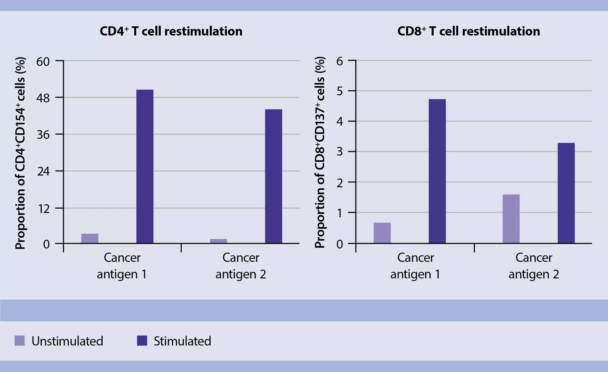
Stimulation by PepTivator Peptide Pools effectively activates antigen-specific T cells.

Antigen-specific T cell stimulation via peptides combined with APCs

Identifying a specific peptide (or a specific neoantigen)7 and triggering a cognate response from antigen-stimulated T cells is challenging. The different PepTivator Peptide Pools give you the flexibility to work with a wide range of antigens, including neoantigens such as RAS mutation collection (e.g., PepTivator RAS G12C). At the same time, it ensures reactivity to one of its peptide fractions, facilitating antigen-specific TCR identification.

After narrowing down your selection to one specific T cell epitope, you can use MACSpep single peptide sequences for the expansion of T cells against that single peptide sequence (e.g., MACSpep KRAS G12C). For the fast, easy, and sensitive detection of peptide-reactive T cells via the analysis of surface markers and intracellular cytokines, use our Antigen-specific T Cell Analysis Kits, anti-human, REAfinity®.

3. Selection of antigen-specific T cell

Isolation of pure and viable antigen-specific T cells is challenging due to their low frequency within complex cell populations. Once stimulated with the peptide of interest (see previous step), the antigen-specific T cells proliferate, enriching their proportion within the cell population. They can be detected indirectly by analyzing their expression of activation markers (e.g., CD137, CD154), their cytokine secretion, or directly through interaction with MHC multimer reagents coupled with the peptide of interest.

Our MACS MicroBeads, antibodies, detection kits, and MHC MACSimers ensure that tumor-reactive T cells can be specifically isolated via magnetic selection or flow cytometry sorting.

Dot plots before and after sorting of CMV pp65-specific T cells by cell sorter or magnetic separation.
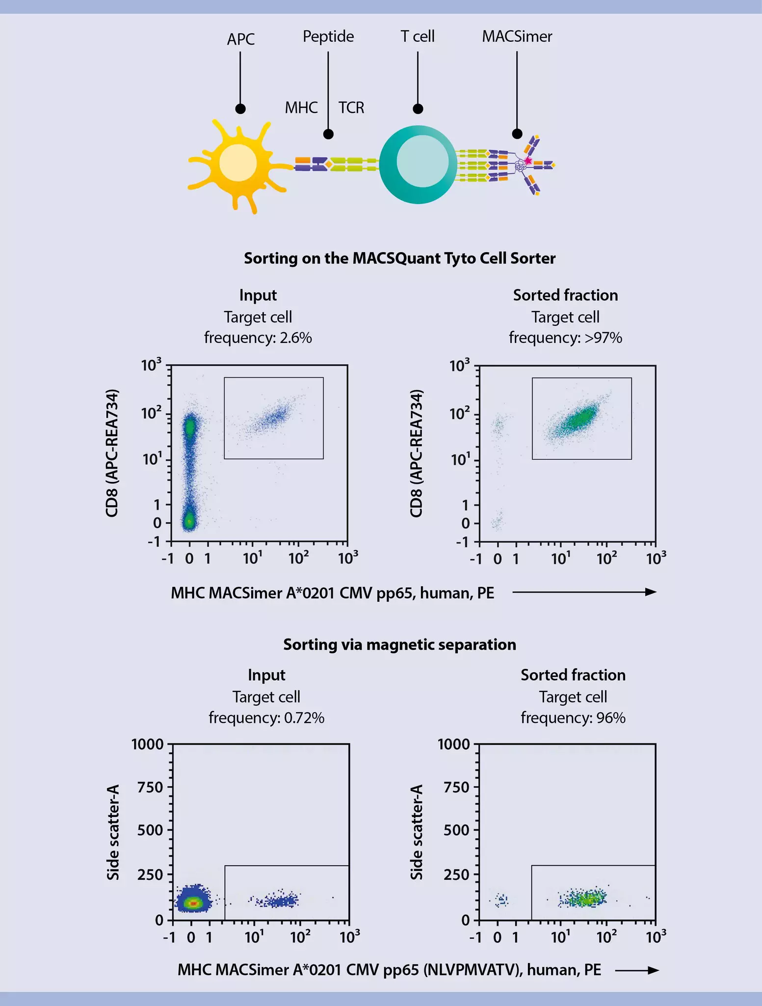
Sorting high-purity CMV pp65-specific T cells using the MACSQuant Tyto Cell Sorter and MHC MACSimers.

Direct isolation of antigen-specific T cells via their TCR using MHC MACSimers

Antigen-specific T cells express highly unique TCRs on their surface to detect unique MHC molecules loaded with specific T cell antigens. This biological process of TCR-mediated antigen recognition via peptide-MHC complexes can be mimicked using fluorescently tagged MHC MACSimers loaded with a peptide of interest (e.g., standardized MACSpep single peptides). You can then efficiently isolate the bound antigen-specific T cells by flow sorting with the MACSQuant® Tyto® Cell Sorter or magnetically using anti-PE MicroBeads.

Learn more about the MHC MACSimer technology here.

Webinar

Successful discovery, analysis, and quality control of Ag-specific T cells targeting solid cancers

This webinar delves into strategies for successful TCR discovery and quality control assay development for TCR-based therapies targeting solid cancers. Marc Schuster and Marek Wieczorek team up to illustrate the identification of tumor-specific TCRs. They also provide concrete examples and advice on developing robust assays for in-process controls and release testing, including the use of MHC multimers in flow cytometry. Sign in to Miltenyi University to view the slides from the video and explore more learning opportunities. miltenyibiotec.com/university

Indirect isolation of antigen-specific T cells via activation markers

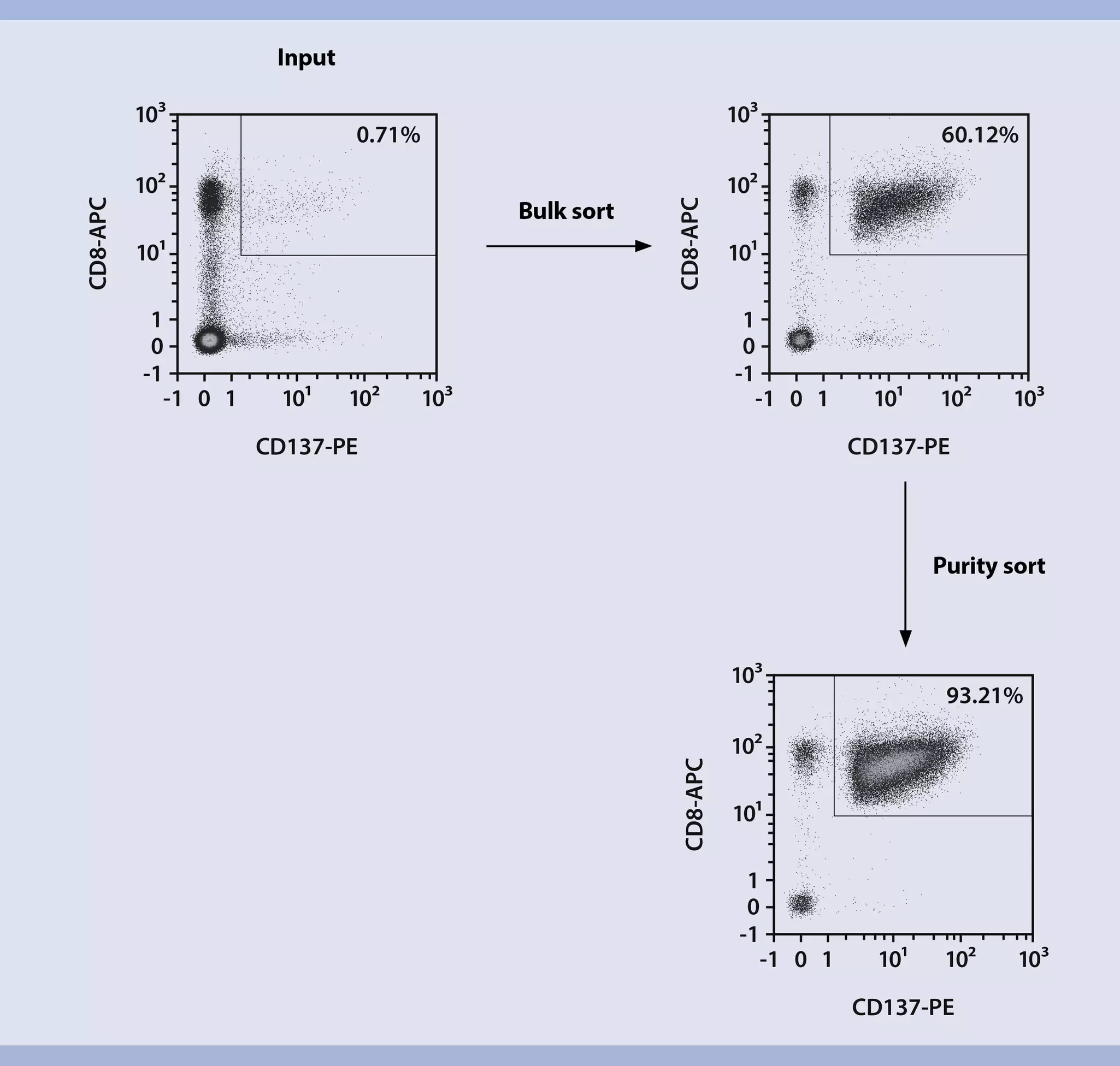
Within hours after activation, CD154 (CD40L) is transiently up-regulated on activated CD4+ T cells. This makes it an ideal marker of the activated antigen-specific T cells. CD137 is a suitable marker of activated human CD8+ T cells, as it is not expressed on resting cells and is reliably induced after 24 hours of stimulation. Following antigen-specific stimulation, staining for these markers allows isolating the activated antigen-specific T cells via flow cytometry using the MACSQuant Tyto Cell Sorter.

To standardize your process, they can also be isolated via magnetic enrichment using anti-CD137 or anti-CD154 MicroBeads in combination with the autoMACS NEO Separator.

Dot plots demonstrating a two-step sorting strategy to efficiently separate antigen-specific T cells.
Surface markers enable the efficient isolation of activated antigen-specific T cells on the MACSQuant Tyto Cell Sorter.

The MACSQuant Tyto Cell Sorter ensures gentle sorting to preserve cell viability and phenotype. Its disposable sterile cartridges provide maximal safety. Revolutionize cell sorting and enrich antigen-specific T cells from <0.05% to >90% purity in a single run.

Webinar

Cell sorting on the MACSQuant Tyto

Find out more about the easy cell sorting procedure on the MACSQuant Tyto: It's simple, it's gentle, it's sterile. This video will give you an overview of the straightforward operator steps from sample loading to retrieval.

Indirect isolation of antigen-specific T cells via pro-inflammatory cytokine secretion

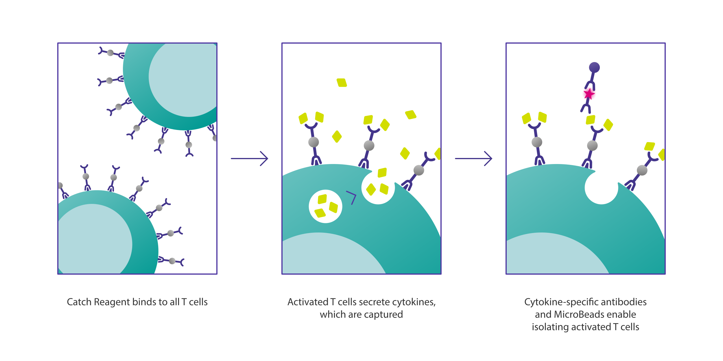
Following antigen-specific stimulation, the activated antigen-specific T cells secrete specific pro-inflammatory cytokines, such as interferon γ (IFN-γ) or TNF-α. MACS Cytokine Secretion Assays are unique tools for enriching cytokine-secreting T cells with a sensitivity of one in a million cells. Assays are available for human cells secreting TNF-α, GM-CSF, IFN-α, IFN-γ, or IL-2, IL-4, IL-5, IL-10, IL-12, IL-13, IL-17, IL-22. Refer to the individual product pages below for performance data.

Illustration of how activated T cells can be efficiently isolated based on their secretion of cytokines.
Principle of Miltenyi Biotec's Cytokine Secretion Assay – Cell Enrichment and Detection Kits. Antigen-specific T cells are detected and isolated based on their secretion of cytokines.

4. Flow cytometric characterization of antigen-specific T cells

Flow cytometric analysis after antigen-specific T cell isolation is critical for TCR identification. Make sure your rare antigen-specific T cells are pure, functional, and phenotypically relevant by using standardized reagents, optimized and validated flow panels, and detailed application notes.

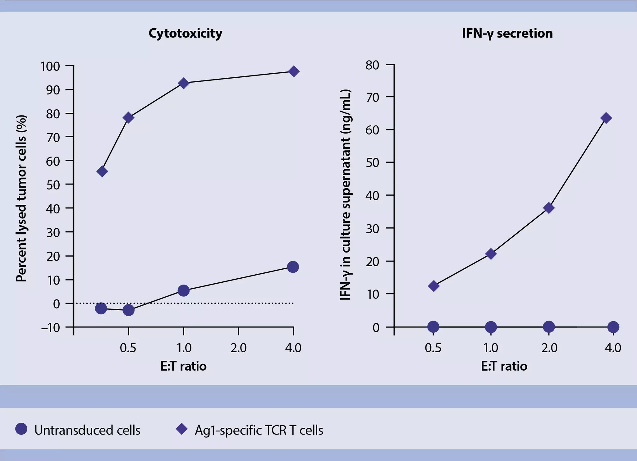
Scatter charts comparing the cytotoxicity and IFN-γ secretion of untransduced versus Ag1-specific TCR T cells.
Cytotoxicity and IFN-γ secretion of antigen-specific T cells compared to untransduced T cells.

  • REAfinity Recombinant Antibodies provide superior lot-to-lot consistency and eliminate background signal.
  • MACSplex Cytokine Kits enable multiplexed analysis of secreted cytokines in cell culture supernatant.
  • Rapid Cytokine Inspector Kit combines surface marker and intracellular cytokine staining for multiparameter analysis of activated human T cells.
  • MACSimers perfectly integrate into multicolor flow cytometry panels for T cell characterization, e.g., for assessing cell maturation.
  • MACSQuant Analyzers are fully automated benchtop flow cytometers for highly sensitive, multiparameter flow analyses.
  • Build your own T cell flow panel with our Flow Panel Builder.

Optimized multicolor flow cytometry panels

Phenotypic characterization of antigen-specific T cells by flow cytometry can be performed using optimized panels of fluorochrome-conjugated REAfinity Recombinant Antibodies. Our Miltenyi Biotec tested panels (MBTPs) are developed and validated by Miltenyi Biotec’s research and development experts, as well as by customers and collaborators.

Discover our relevant flow panels for antigen-specific T cells:

MBTP 28Immunophenotyping of virus-specific T cells from human blood using flow cytometry
MBTP 31Immunophenotyping of human T cells via transcription factor profiling using flow cytometry
MBTP 33Immunophenotyping of activated T cells within antigen-stimulated human PBMCs using flow cytometry
MBTP 29Immunophenotyping of antigen-specific CD4+ T cells from human PBMCs using flow cytometry

5. Tumor-reactive TCR sequencing

Following the isolation and characterization of tumor-reactive T cells, the next critical phase is TCR sequencing. This step involves decoding the unique TCR alpha and beta chain sequences, allowing researchers to identify the specific receptors responsible for anti-tumor activity. By characterizing the TCR repertoire, scientists gain insights into the molecular diversity and clonality of the immune response. This is essential for selecting the most promising candidates for therapeutic development and lays the groundwork for designing targeted TCR T cell therapies.

6. Engineering TCR T cells

Once you have identified a tumor-reactive TCR specific to your target of interest, it is time to generate TCR T cells. You'll need to isolate pure T cells, activate, transduce, and expand them to move forward on developing next-generation cell therapies against cancer. Miltenyi Biotec offers solutions to support your preclinical research, as well as a portfolio of instruments, consumables, and applications for the subsequent clinical manufacturing of your cell product.

Scientist preparing a sample for flow cytometry analysis on a MACSQuant Analyzer.

TCR engineering for research settings

Discover Miltenyi Biotec tools and reagents to streamline your research, boost confidence in results, and ease your transition to clinical settings.

Learn more

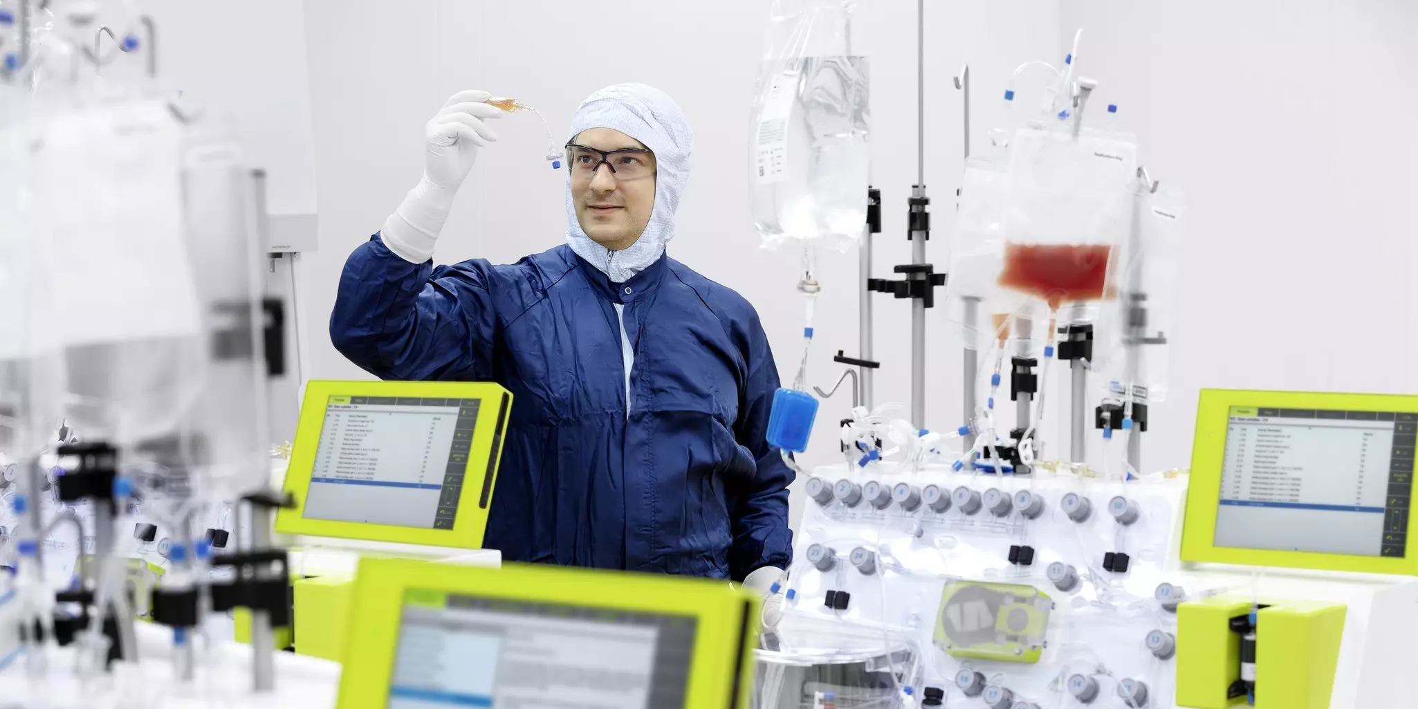
Operator in a cleanroom supervising cell manufacturing on several CliniMACS Prodigy Instruments.

TCR engineering for cell manufacturing

Transform your manufacturing workflows with our CliniMACS® Instruments for automated processing of autologous and allogeneic cell products.

Automate today