CD8 MicroBeads were developed for the positive selection or depletion of human cytotoxic CD8+ T cells from different cell sources. The most commonly used cell sources are peripheral blood and cord blood. In addition, cytotoxic T cells have also been isolated from lymph nodes, thymus, skin biopsies1, and spleen.


Related products for CD8 MicroBeads, human

CD8 MicroBeads were developed for the positive selection or depletion of human cytotoxic CD8+ T cells from different cell sources. The most commonly used cell sources are peripheral blood and cord blood. In addition, cytotoxic T cells have also been isolated from lymph nodes, thymus, skin biopsies1, and spleen.
The CD8 antigen forms a complex together with the T cell receptor and acts as an accessory molecule in the recognition of MHC class I/peptide complexes by the TCR heterodimer on CD8+ cytotoxic T cells. The CD8 molecule consists of either an α/β heterodimer or an α/α homodimer. It is expressed strongly on cytotoxic T cells and dimly on a subset of NK cells. CD8 is found on most thymocytes and on about one third of all peripheral blood T cells. CD8+ cytotoxic T cells play an important role in the killing of virus-infected cells and tumor cells.
Isolation or depletion of CD8+ cytotoxic T cells is performed in many different research fields such as infectious diseases, autoimmune diseases, allergy, and asthma, as well as tumor immunology.
Cytotoxic T cells isolated by MACS® Technology remain viable and functional. Therefore, they can be used for further functional studies, such as proliferation assays2,9 and cytotoxicity assays2,3, but also for the analysis of in vitro cytokine production4. CD8 MicroBeads have also been used for the depletion of CD8+ T cells from human PBMCs for immune reconstitution experiments in SCID mice.5
In combination with CD4 MultiSort MicroBeads, CD8 MicroBeads have been used for the isolation of CD4+CD8+ double-positive thymocytes.8
Other examples include various studies on viral infections, e.g., in vitro infections of CD8+ cells with HIV6, or monitoring of immune abnormalities in children of HIV-infected mothers7.
For positive selection: MS, LS, XS, or autoMACS® Columns. For depletion: LD, D, or autoMACS Columns.
Separation of CD8+ cells from PBMCs using CD8 MicroBeads, an LS Column, and a MidiMACS™ Separator.
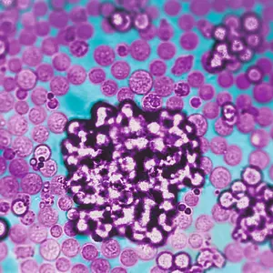
Scanning electron micrograph of CD8+ T cells isolated using CD8 MicroBeads (courtesy of Prof. Peter Groscurth, Institute of Anatomy, University of Zürich, Switzerland).
- Akdis, M. et al. (1999) Skin homing (cutaneous lymphocyte-associated antigen-positive) CD8+ T cells respond to superantigen and contribute to eosinophilia and IgE production in atopic dermatitis. J Immunol 163: 466-475
- Walker, W. and Gallagher, G. (1994) The in vivo production of specific human antibodies by vaccination of human-PBL-SCID mice. Immunology 83: 163-170
- Turner, J. and Dockrell, H. M. (1996) Stimulation of human peripheral blood mononuclear cells with live Mycobacterium bovis BCG activates cytolytic CD8+ T cells in vitro. Immunology 87: 339-342
- Kutlesa, S. et al. (2002) Developmentally regulated interactions of human thymocytes with different laminin isoforms. Immunology 105: 407-418
- Al-Shanti, N. et al. (2004) Investigation of alpha nascent polypeptide-associated complex functions in a human CD8+ T cell ex vivo expansion model using antisense oligonucleotides. Immunology 112: 397-403
- Akdis, A. C. et al. (1995) Cytokine immunotrapping: an assay to study the kinetics of production and consumption or degradation of human interferon-gamma. J. Immunol. Methods 182: 251-261
- Parra, E. et al. (1997) The role of B7-1 and LFA-3 in costimulation of CD8+ T cells. J Immunol 158: 637-642
- Nielsen, S. D. et al. (2001) Impaired progenitor cell function in HIV-negative infants of HIV-positive mothers results in decreased thymic output and low CD4 counts. Blood 98: 398-404
- Ennen, J. et al. (1994) CD8+ T lymphocytes of African green monkeys secrete an immunodeficiency virus-suppressing lymphokine. Proc. Natl. Acad. Sci. U.S.A. 91: 7207-7211