Logo

hPSC-derived cerebral organoids for human brain development research

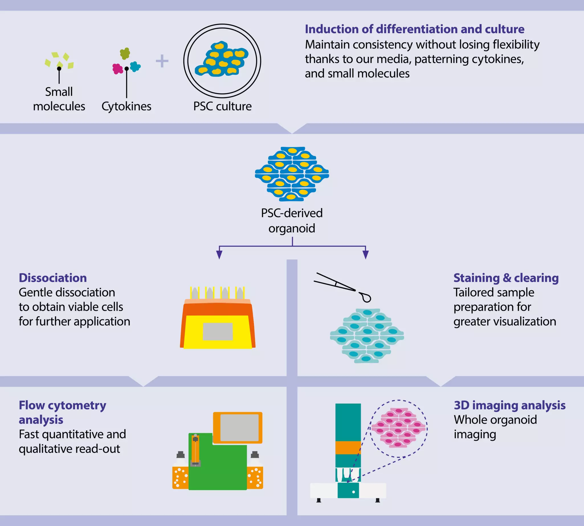
Cerebral organoids (brain organoids) derived from human pluripotent stem cells (hPSCs), either embryonic or induced PSCs, have emerged as novel model systems that can be used to investigate human brain development and disorders. Browse through our cerebral organoid dissociation options for single-cell analysis or our cutting-edge solutions for whole organoid visualization.

Content overview

Content

  • Application overview
  • 1. Culture of hPSC-derived cerebral organoids
  • 2. Dissociation of hPSC-derived cerebral organoids
  • 3. Analysis by flow cytometry of hPSC-derived cerebral organoids
  • 4. Staining and clearing of hPSC-derived cerebral organoids
  • 5. Analysis by 3D imaging of hPSC-derived brain organoids
  • Step-by-step protocols

1. Culture of hPSC-derived cerebral organoids

To obtain viable and reliable cerebral organoids it is pivotal to use high-quality PSC lines as starting material. Our cell culture media, StemMACS™ iPS-Brew XF, human and StemMACS PSC-Brew XF, human, have been specifically designed to provide your cells with the best-quality nutrients to sustain robust growth under feeder-free conditions and to maintain full pluripotent potential and the ability to differentiate into the three embryonic germ layers.

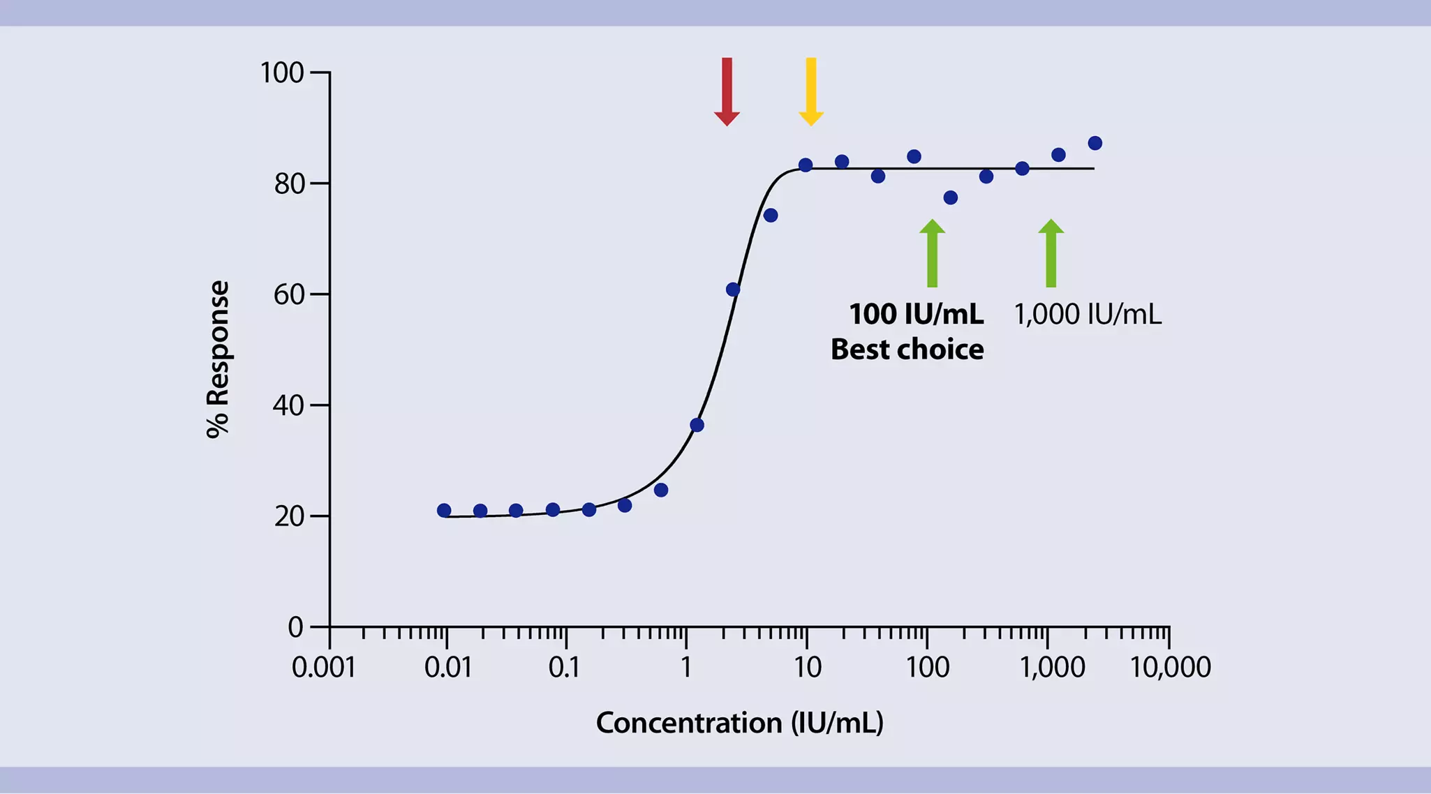
Illustration of line chart showing effective cytokine usage with optimal unit dosing indicates cytokine concentrations that result in maximal cellular response with MACS® Premium-Grade Cytokines
Efficient cytokine usage with specific unit dosing

Importance of premium-grade cytokines

Generation of hPSC-derived cerebral organoids is achieved by timely addition of patterning factors. For this sensitive phase, administration of precise amounts of active cytokine or small molecules is pivotal, both for success of the differentiation itself but also to ensure experimental reproducibility. MACS® Premium-Grade Cytokines come with specific biological activity for each lot so you can precisely apply cytokines according to their biological activity. In this way, you can generate reproducible cell culture conditions and aim for natural cytokine concentrations while avoiding possibly harmful oversaturation, artificial experimental results, and at the same time reduce costs.

2. Dissociation of hPSC-derived cerebral organoids

Dissociation of hPSC-derived cerebral organoids into single cells might be necessary to analyze particular cellular subpopulations within the organoid. These sensitive structures require specific reagents and gentle mechanical dissociation in order to preserve cell viability and functionality. We provide a well-orchestrated portfolio of products that allow you to dissociate mature cerebral organoids gently and obtain highly viable cell populations for further isolation, adherent culture, analysis by flow cytometry, or immunofluorescence staining.

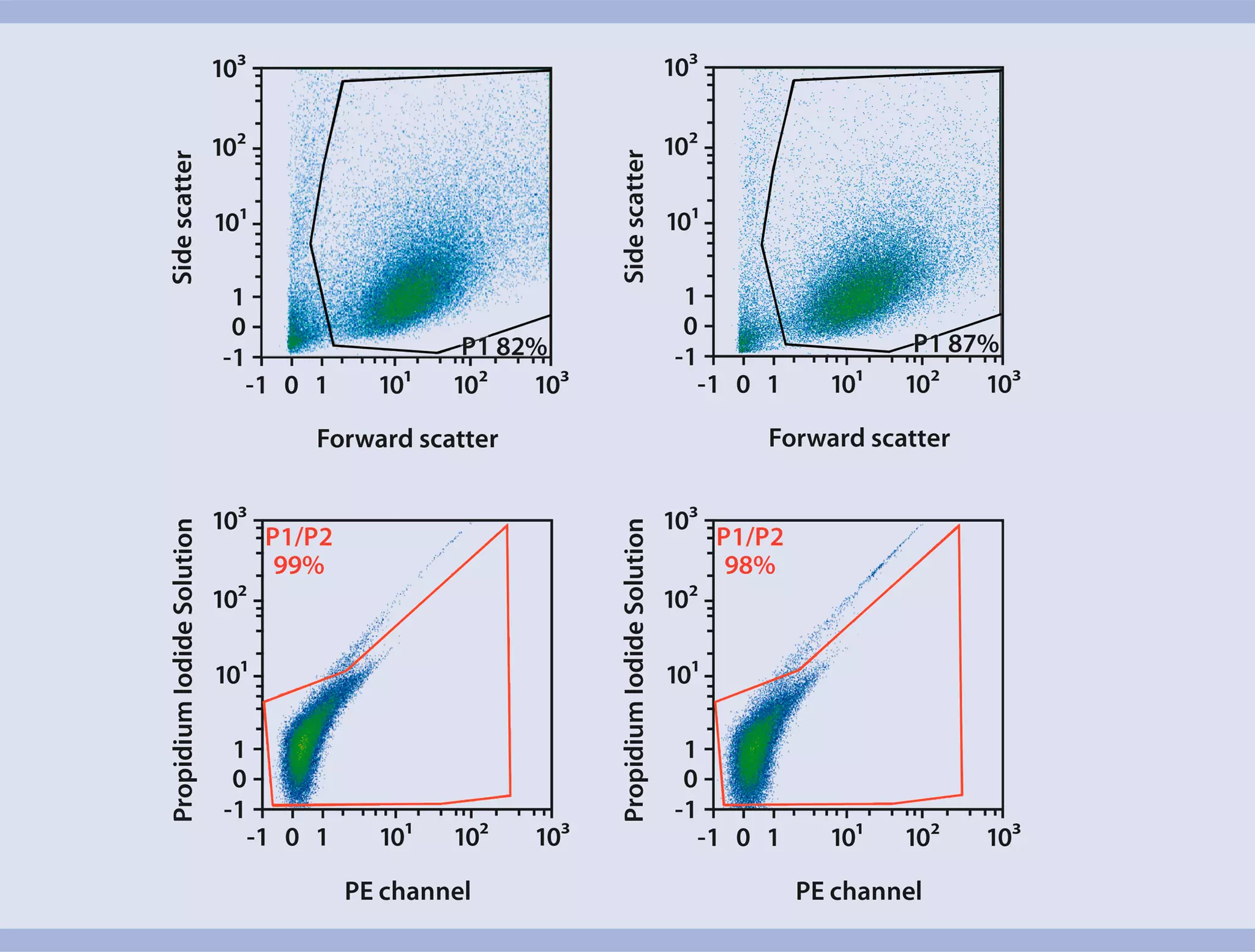
Flow cytometric analysis of showing a large number of viable single cells after dissociation using Neural Tissue Dissociation Kits with a gentle two-step enzymatic dissociation procedure
Flow cytometric analysis of single cells after dissociation using Neural Tissue Dissociation Kits

Neural Tissue Dissociation Kit

With the Neural Tissue Dissociation Kit, based on a gentle two-step enzymatic dissociation procedure, you can obtain large numbers of viable cells in less than an hour. Preserve key epitopes by choosing the Neural Tissue Dissociation Kit (P) with papain or the Neural Tissue Dissociation Kit (T) with trypsin. For maximal cell viability, we recommend the use of the Neural Tissue Dissociation Kit together with the gentleMACS™ Octo Dissociator with Heaters. After dissociation, cells can be maintained in culture with MACS Neuro Medium supplemented by MACS NeuroBrew®-21.

3. Analysis by flow cytometry of hPSC-derived cerebral organoids

Flow cytometry is a fast, qualitative, and quantitative analysis method that can be applied after organoid dissociation to confirm the presence of cells of interest or analyze the expression of specific markers.

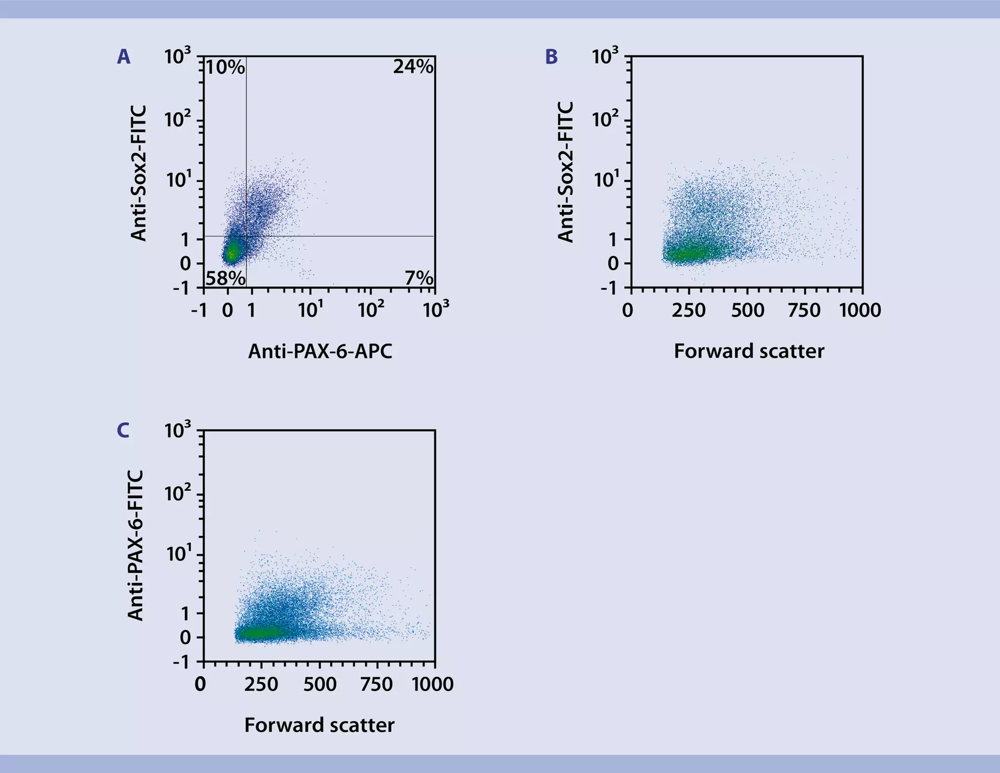
Flow cytometric analysis of PSC-derived brain organoids after dissociation into single cells using expression of the neural progenitor markers Sox2 and Pax6
Example of flow analysis of PSC-derived brain organoids dissociated into single cells

Observe superior consistency

hPSC-derived cerebral organoids are complex structures that can be analyzed by evaluating many different markers. Our REAfinity® Antibodies have been recombinantly engineered to gain high specificity without the need of blocking the Fc receptor. Browse through our large portfolio of recombinant antibodies optimized for flow cytometry that provide superior lot-to-lot consistency and purity compared to mouse or rat hybridoma-derived, monoclonal antibodies.

4. Staining and clearing of hPSC-derived cerebral organoids

hPSC-derived cerebral organoids model rudimental brains that include multiple cellular populations, providing the opportunity to study connectivity and cellular interaction of neural cells. With our complete 3D imaging workflow analyzing the structure and composition of whole cerebral organoids has never been easier. This workflow relies on 3D-immunofluorescence (3D-IF) validated antibodies, a ready-to-use tissue clearing kit, and cutting-edge imaging techniques.

Video

Validated antibodies provide conclusive 3D imaging data

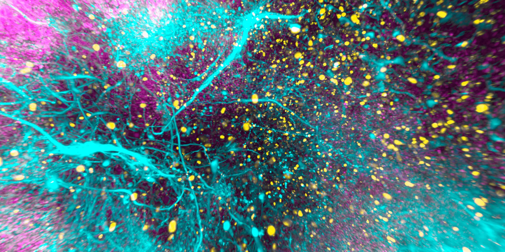
The video shows an hPSC-derived cerebral organoid labeled with Miltenyi Biotec recombinant antibodies (Ki-67 Antibody, Vio R667, REA183 (yellow); SOX2 Antibody, Vio B515, REA320 (magenta); and β-Tubulin 3 Antibody, Vio G570, REA1152 (cyan)), and optically cleared using the MACS Clearing Kit. Imaged with an UltraMicroscope Light Sheet Instrument and processed as a 3D-rendered maximum intensity projection.

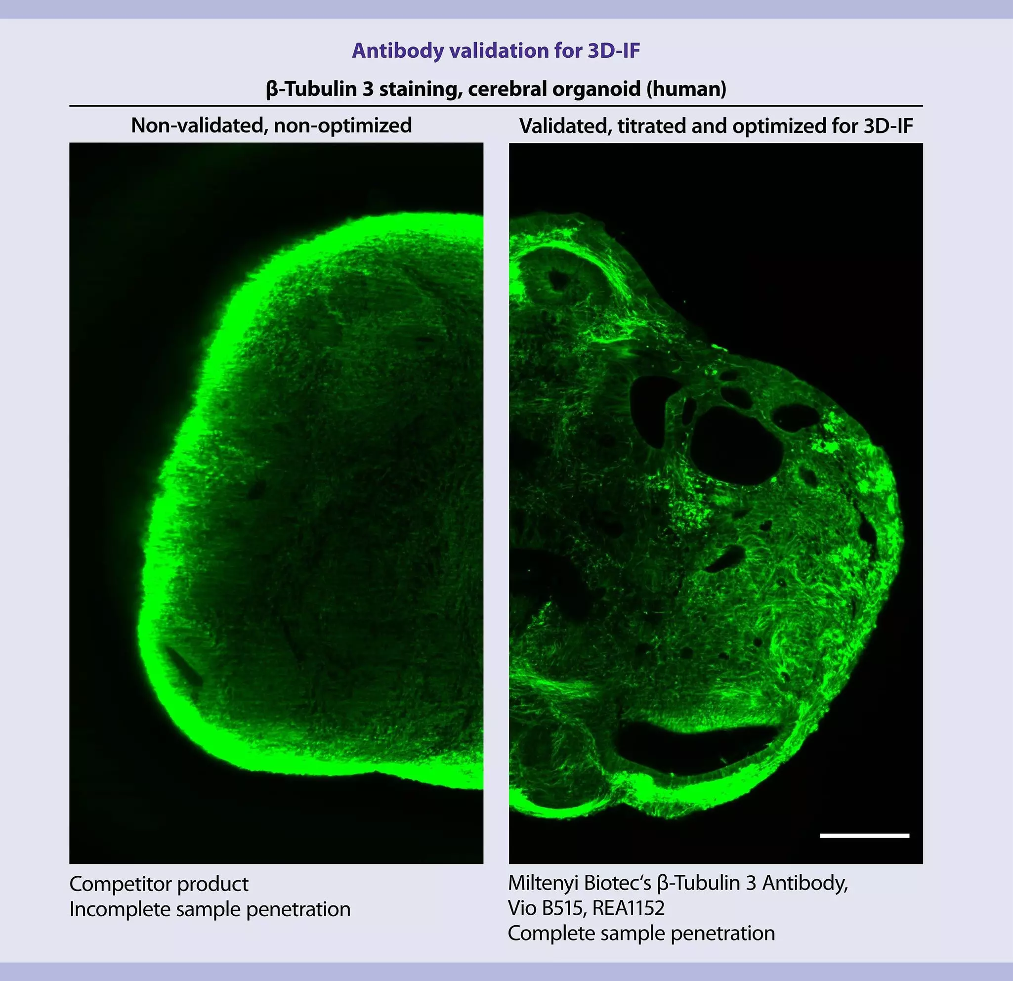
 Antibody validation for 3D-IF
Antibody validation for 3D-Immunofluorescence

Optimal performance for 3D imaging

Our 3D-IF antibodies are already validated for 3D imaging of large samples such as organoids. Tailor-made to work with our MACS Clearing Kit and directly conjugated to bright and photostable Vio® Dyes, our recombinantly engineered REAfinity Antibodies offer you all the advantages of direct 3D-IF microscopy: reduced staining time, great signal-to-noise ratio, and minimal background interference.

Immunofluorescence light sheet microscopy of cerebral organoid using the UltraMicroscope II

Antibodies cleared for 3D-immunofluorescence microscopy

No matter if you explore your samples using a light sheet or confocal microscope, our antibodies will get you superior results every time.

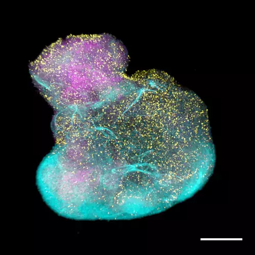
A cerebral organoid generated from human iPS cells

Clearing without integrity loss

The MACS Clearing Kit enables clearing of up to 60 hPSC-derived cerebral organoids of approximately 1 mm3. By minimizing light scattering of opaque tissue, it allows you to sharply image structures without compromising their integrity. To make things even easier, we have generated a specific easy-to-follow tissue staining and clearing protocol that guides you through this hassle-free process step by step.

Duration of steps for clearing opaque hPSC-derived cerebral organoids for 3D imaging using the MACS Clearing Kit.

Tissue typeFixationPermeabilizationImmunostainingDehydrationClearing
Cerebral organoids
(1 mm3)*
20 min6 hours40 hours24 hours6 hours

Duration of immunostaining step is based on the use of directly conjugated antibodies.

5. Analysis by 3D imaging of hPSC-derived brain organoids

Light sheet microscopy enables the visualization of different cell populations without disrupting the macro-architecture of the hPSC-derived cerebral organoid.

Discover our light sheet microscope family for 3D high-resolution imaging. The fully automated UltraMicroscope Blaze™ enables overnight imaging of multiple samples in a single imaging experiment, without manual intervention or monitoring. The batch measurement mode ensures that 3D data will be available for in-depth analysis the next morning. Our pioneering UltraMicroscope technology combined with cutting-edge light sheet optics ensures excellent data quality from macro to subcellular imaging.

MACS® UltraMount 48 organoids holder imaged in 3D with the UltraMicroscope Blaze™

3D image of 48 cerebral brain organoids captured using the UltraMicroscope Blaze Light Sheet System and a MACS UltraMount sample holder.

Step-by-step protocols for isolation and analysis of cerebral organoids

Get acquainted with our protocols for your research on PSC-derived cerebral organoids.