Dissociation, culture, and analysis of cerebral organoids
This application protocol describes the step-by-step dissociation of PSC-derived cerebral organoids into single cells, using the Neural Tissue Dissociation Kit (P) or (T) in combination with the gentleMACS® Dissociator. After dissociation, single cells can be either analyzed by flow cytometry using the MACSQuant® Analyzer 10 or can be cultivated for immunostaining and microscopic analysis.
Protocol
Dissociation and analysis of organoids
1 h
Dissociation of cerebral organoids
1 h
Staining of single cells for flow cytometry
6 days
Cultivation of dissociated cells
2 days
Immunofluorescent staining of dissociated cells
Protocol

This protocol includes detailed instructions for preparing necessary reagents and materials and then carrying out each step.
- Use at least 14 organoids (30 days old) per tube for organoid dissociation.
- Up to 400 mg (approximately 100 pieces) organoids can be dissociated.
Preparation of the enzyme mixes using the Neural Tissue Dissociation Kit (T) or (P)
Enzyme T and Enzyme P of the Neural Tissue Dissociation Kit are ready-to-use. Prepare aliquots of appropriate volume to avoid repeated freeze-thaw-cycles. Store aliquots at –20 °C. This solution is stable for 6 months.
Resuspend the lyophylized Enzyme T of the Neural Tissue Dissociation Kit (T) in 3 mL of Buffer X. Close the lid and invert the vial. Wait for 5–10 minutes. Do not pipette up and down. Prepare aliquots to avoid repeated freeze-thaw-cycles. Store aliquots at –20 °C. This solution is stable for 6 months after reconstitution.
For both kits, resuspend the lyophilized powder in the vial labeled Enzyme A with 1 mL sterile, deionized water. Do not vortex. This solution should be aliquoted and stored at –20 °C for later use. Avoid repeated freeze-thaw-cycles.
Table 1: Enzyme mixes of the Neural Tissue Dissociation Kits
| Enzyme mix 1 | Enzyme mix 2 | |||
| Neural Tissue Dissociation Kit (P) | Enzyme P | Buffer X | Buffer Y | Enzyme A |
| 50 µL | 1900 µL | 20 µL | 10 µL | |
| Neural Tissue Dissociation Kit (T) | Enzyme T | Buffer X | Buffer Y | Enzyme A |
| 60 µL | 1890 µL | 20 µL | 10 µL | |
Preparation of stop solution
Prepare a solution of Dulbecco′s Modified Eagle′s Medium (DMEM) containing 20% fetal calf serum.
Preparations for staining for flow cytometry
Staining buffer
Prepare a solution containing phosphate-buffered saline (PBS), pH 7.2, 0.5% bovine serum albumin (BSA), and 2 mM EDTA by diluting MACS® BSA Stock Solution 1:20 with autoMACS® Rinsing Solution. Keep buffer cold (2−8 °C).
Fixation/Permeabilization Solution
To achieve the appropriate working concentration for safe fixation and permeabilization of cells, Fixation/Permeabilization Solution 1 (component of FoxP3 Staining Buffer Set) must be diluted 1:4 with the Fixation/Permeabilization Solution 2 (component of FoxP3 Staining Buffer Set) (i.e. for 106 cells use 0.25 mL of Fixation/Permeabilization Solution 1 plus 0.75 mL of Fixation/Permeabilization Solution 2).
Permeabilization Buffer
To achieve the appropriate working concentration for safe permeabilization of cells, 10× Permeabilization Buffer (component of FoxP3 Staining Buffer Set) must be diluted 1:10 with deionized or distilled water before use (i.e. 1 mL of 10× Permeabilization Buffer plus 9 mL of deionized/distilled water).
Preparations for cultivation
Poly-L-lysine coating of Imaging Plate CG 1.0 (24 well)
Add 500 µL poly-L-lysine per well and incubate the plate at 37 °C overnight. Discard poly-L-lysine directly before plating of cells.
Culture medium
Prepare the culture medium by adding 2% MACS® NeuroBrew®-21, 1% penicillin/streptomycin, and 0.5 mM L-glutamine to MACS Neuro Medium.
Preparations for immunofluorescent staining and microscopic analysis
Blocking solution
Prepare a solution containing 1% Triton X 100 and 10% fetal calf serum in D-PBS without calcium and magnesium.
- Use at least 14 organoids per gentleMACS™ C Tube.
- Organoids should be at least 25–30 days old.
- Generation of cerebral organoids can be done using standard protocols.
- Gently remove organoids from the dish or bioreactor using wide or cut pipette tips and transfer them into a falcon tube.
- To remove residual medium, wash the organoids with a sufficient amount of D-PBS without Ca2+ and Mg2+.
- Transfer 1950 μL of enzyme mix 1 (refer to table in section “Reagent and instrument preparation”) for up to 400 mg (approx. 100 pieces) organoids into a gentleMACS C Tube and pre-heat at 37 °C for 10–15 minutes.
- Transfer the organoids into the C Tube containing the pre-heated enzyme mix 1.
- Tightly close the C Tube and attach it upside down onto the sleeve of the dissociator.
▲ Note: It has to be ensured that the sample material is located in the area of
the rotator/stator.
▲ Note: Organoids tend to stick at the wall of the gentle MACS Tube. To avoid this, slightly sway the tube while turning it gently upside down. - Run the gentleMACS Program m_brain_01.
- Incubate the sample for 15 minutes at 37 °C under slow, continuous rotation using the MACSmix™ Tube Rotator.
- Attach the C Tube upside down onto the sleeve of the gentleMACS Dissociator.
- Run the gentleMACS Program m_brain_02.
- Transfer 30 μL of enzyme mix 2 (refer to table in section “Reagent and instrument preparation”) into the C Tube. Invert gently to mix. Do not vortex.
- Incubate the sample for 10 minutes at 37 °C under slow, continuous rotation using the MACSmix Tube Rotator.
- Attach the C Tube upside down onto the sleeve of the gentleMACS Dissociator.
- Run the gentleMACS Program m_brain_03.
- After termination of the program, detach the C Tube from the gentleMACS Dissociator.
- Add 10 mL of stop solution per C Tube.
- Mix the sample and apply the cell suspension onto a MACS SmartStrainer (70 μm) placed on a 50 mL tube.
▲ Note: Moisten the MACS SmartStrainer with buffer before use.
▲ Note: When upscaling the reagent volume and total volumes, increase also the number of MACS SmartStrainers (70 μm). One MACS SmartStrainer (70 μm) can be used for up to 2 mL.
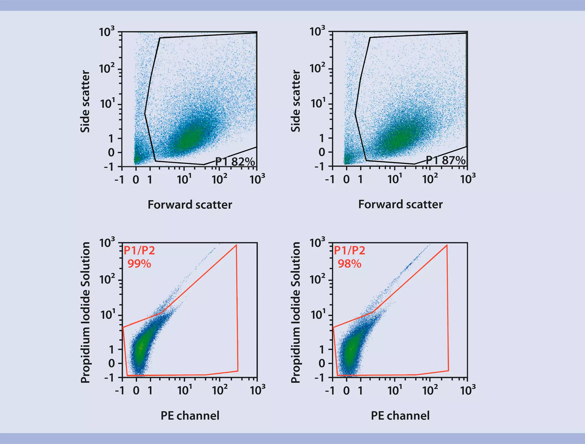
▲ Note: Cells with a diameter >70 μm may be lost. To obtain these cells within the flow through, use a cell strainer with an appropriate mesh size. - Determine the cell number using the MACSQuant Analyzer.
- Proceed with downstream analysis.
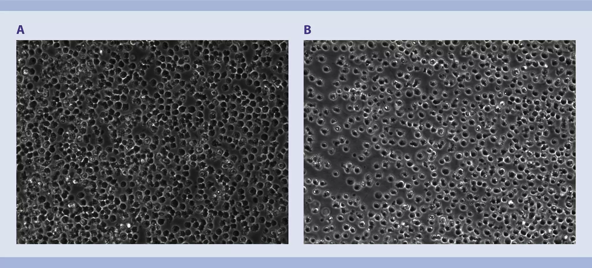
 View details
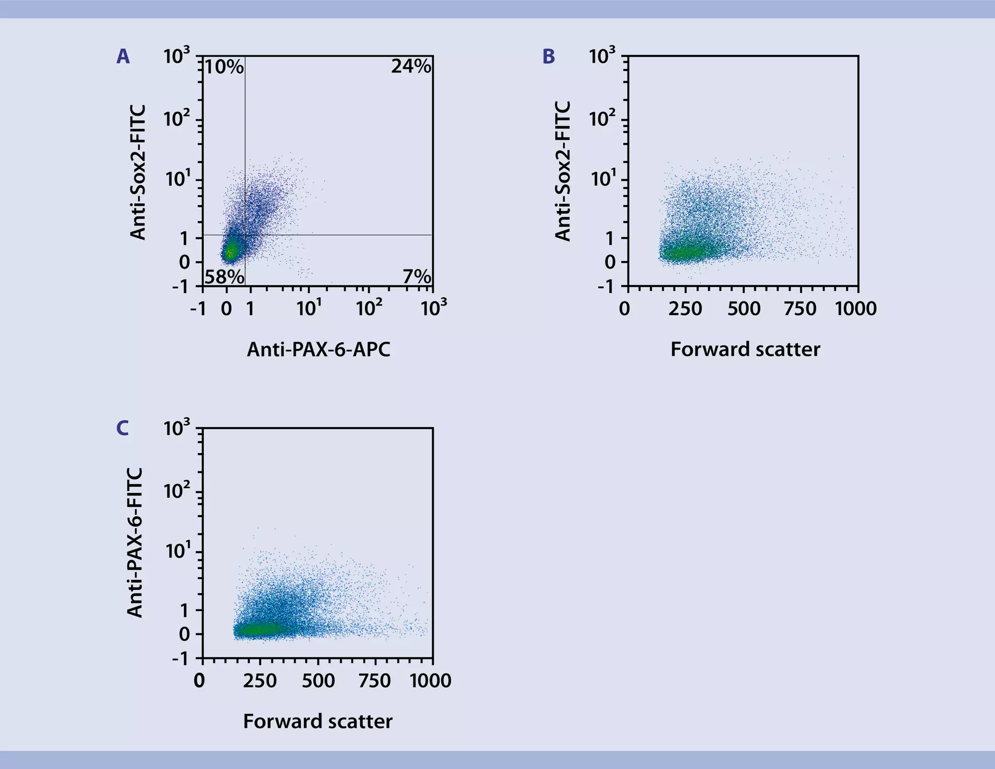
- The recommended antibody dilution of PAX-6 Antibody-APC and Sox2 Antibody-FITC for labeling of cells and subsequent analysis by flow cytometry is 1:11 for up to 106 cells/100 µL.
- Volumes given below are for up to 106 nucleated cells. When working with fewer than 106 cells, use the same volumes as indicated. When working with higher cell numbers, scale up all reagent volumes and total volumes accordingly.
- Always prepare Fixation/Permeabilization Solution freshly as described in the data sheet of the FoxP3 Staining Buffer Set or in section “Reagent and instrument preparation”.
- Wash up to 106 cells by adding 1–2 mL of buffer and centrifuge at 300×g for 10 minutes. Aspirate supernatant completely.
- (Optional) Stain cell surface antigens with appropriate antibodies according to the manufacturer's recommendations. Wash cells by adding 1–2 mL of buffer and centrifuge at 300×g for 10 minutes. Aspirate supernatant completely.
- Resuspend up to 106 cells in 1 mL of cold, freshly prepared Fixation/Permeabilization Solution.
- Mix well and incubate for 30 minutes in the dark in the refrigerator (2−8 °C).
- Wash cells by adding 1 mL of cold buffer per 106 cells and centrifuge at 300×g for 5 minutes at 4 °C. Aspirate supernatant completely.
- Wash cells by adding 1 mL of cold 1× Permeabilization Buffer per 106 cells and centrifuge at 300×g for 5 minutes at 4 °C. Aspirate supernatant completely.
- Resuspend up to 106 nucleated cells in 81.82 μL of cold 1× Permeabilization Buffer.
- Add 9.09 μL of Sox2 Antibody-FITC and 9.09 µL of PAX-6 Antibody-APC.
- Mix well and incubate for 30 minutes in the dark in the refrigerator (2−8 °C).
- Wash cells by adding 1 mL of cold 1× Permeabilization Buffer per 10 cells and centrifuge at 300×g for 5 minutes at 4 °C. Aspirate supernatant completely.
- Resuspend cell pellet in a suitable amount of buffer for analysis by flow cytometry or fluorescence microscopy. Store cells at 2–8 °C in the dark until analysis. Mix well before flow cytometric acquisition.
▲ Note: Do not use propidium iodide (PI) or 7-AAD staining.
- Prepare the culture plate and the medium as described in section “Reagent and instrument preparation”.
- Transfer the desired number of cells (5×10⁴ cells per well for a 24-well plate) in a falcon tube and centrifuge at 300×g for 5 minutes. Aspirate supernatant completely.
- Resuspend cells in 50 µL culture medium per well (1,2 mL for a complete 24-well plate).
- Plate 5×10⁴ cells in 50 µL medium in the middle of each well. Incubate plate at 37 °C for 30 minutes to let cells sink to the bottom and attach.
- Wash the cells once by adding 450 µL of medium per well. Aspirate the medium and add 500 µL fresh medium. Incubate the cells at 37 °C and 5% CO2.
- Exchange half of the medium every 2–3 days.
- After 6 days of culture cells can be analyzed.
▲ Note: Depending on the application cells can be cultivated for >6 days.
- Immunofluorescent staining will be performed in the 24-well Imaging Plate the cells were cultured in.
- Prepare the blocking solution as described in section “Reagent and instrument preparation”.
- Discard old medium and wash cells twice with 500 µL of D-PBS without Ca2+ and Mg2+ per well.
- Add 500 µL 4% PFA per well and incubate for 20 minutes at room temperature.
- Aspirate 4% PFA and wash cells three times with 500 µL D-PBS without Ca2+ and Mg2+ per well. Aspirate supernatant.
- Add 200 µL of blocking solution per well and incubate for 1 hour at room temperature. Aspirate supernatant.
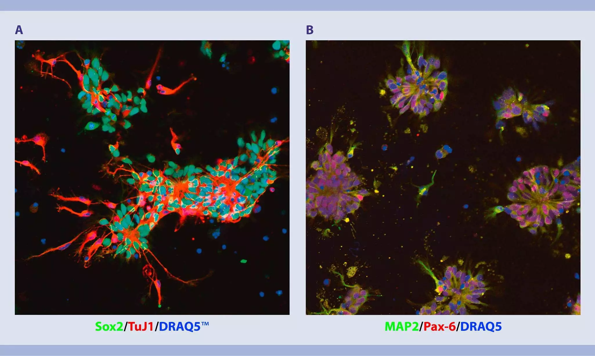
- For Sox2 and TuJ1 stainings add 160 µL of blocking solution, 20 µL Sox2 antibody, and 20 µL TuJ1 antibody per well. For PAX-6 and MAP2 stainings add 179 µL of blocking solution, 20 µL PAX-6 antibody, and 1 µL MAP2 antibody per well.
- Incubate plate overnight in the refrigerator (2–8 °C).
- Remove staining solution.
- Wash cells three times with 500 µL of D-PBS without Ca2+ and Mg2+ per well. Aspirate supernatant completely.
- For nuclear and secondary antibody staining of Sox2, add 99.8 µL blocking solution, 0.1 µL Goat anti-Human IgG (H+L) Cross-Adsorbed Secondary Antibody, Alexa Fluor 488, and 0.1 µL DRAQ5. For nuclear and secondary antibody stainings of PAX-6 and MAP2, add 99.7 µL blocking solution, 0.1 µL Goat anti-Human IgG (H+L) Cross-Adsorbed Secondary Antibody, Alexa Fluor 546, 0.1 µL Goat anti-Mouse IgG (H+L) Cross-Adsorbed Secondary Antibody, Alexa Fluor 488, and 0.1 µL DRAQ5.
- Incubate plate for 1 hour at room temperature in the dark.
- Aspirate staining solution and wash cells three times with 500 µL D-PBS without Ca2+ and Mg2+ per well.
- For imaging add 500 µL of D-PBS without Ca2+ and Mg2+ per well and investigate cells using an appropriate microscope.