Diagnostic research of multiple myeloma*
* for research purposes only
Increase the efficiency of plasma cell isolation in your research of malignant cells in multiple myeloma. Our solutions offer you advantages in the enrichment process for cytogenic studies as well as flow analysis of malignant plasma cells.
Plasma cell isolation
Cell isolation solutions for cytogenetic analysis and efficient enrichment of CD138+ plasma cells is a prerequisite for valid cytogenetic analysis of bone marrow samples. Conventional methods for the isolation of CD138+ plasma cells requires laborious density gradient centrifugation and generation of mononuclear cells prior to any plasma cell purification. MACSprep™ Multiple Myeloma CD138 MicroBeads, human allow the isolation of CD138+ plasma cells directly from bone marrow or peripheral blood without the need for gradient centrifugation or erythrocyte lysis. The minimal manipulation of sample ensures high integrity of CD138+ plasma cells, suitable for further cellular analysis (flow cytometry), cytogenetic analysis (FISH), or molecular analysis (sequencing, PCR, microarray).
Find more information on MACSprep products.
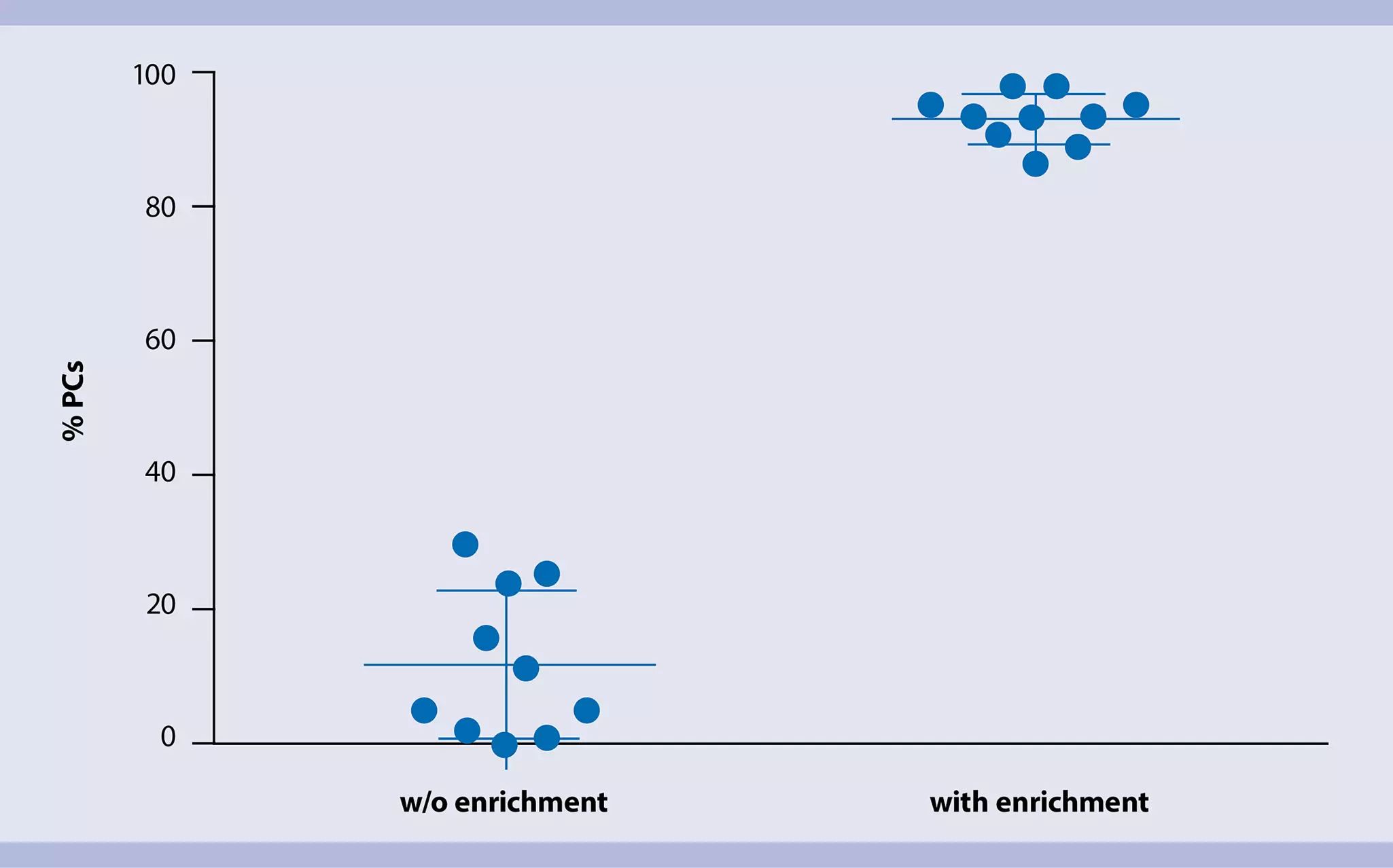
Isolation of plasma cells using MACSprep Multiple Myeloma CD138 MicroBeads, human
Frequency of CD138+ plasma cells prior to and after automated enrichment from bone marrow using MACSprep Multiple Myeloma CD138 MicroBeads, human in combination with the autoMACS Separator. Plasma cells were analyzed by flow cytometry. Each data point represents one individual bone marrow sample. Using this method, the frequency of plasma cells can be significantly increased compared to cell isolation without enrichment. Automated cell isolation on the autoMACS Separator minimizes hands-on time, ensures consistency, and delivers highly pure cells. By this anylsis of rare patient materials is ensured and quality of data obtained is enriched.

Video
Direct cell isolation from bone marrow or blood with the autoMACS® NEOSeparator
With the autoMACS® NEO Separator, CD138+ plasma cells can be automatically isolated from bone marrow or peripheral blood samples. Take this short instrument tour and see how your target cells are being separated without the need for density gradient centrifugation or red blood cell lysis.

Cytogenetic and flow analysis of malignant plasma cells
Most multiple myeloma studies perform fluorescent in situ hybridization (FISH) analysis with isolated CD138+ plasma cells. More studies are incorporating new molecular methods to diagnose multiple myeloma, such as next generation sequencing (NGS) and single nucleotide polymorphism (SNP) array. These assays require DNA isolation from CD138+ plasma cells, as well as analysis of phenotype of malignant plasma cells, which can be determined by flow cytometry.
Cytogenetic studies
FISH is a standard method for identifying genomic aberrations in multiple myeloma. It can also be performed at the interphase of cells, iFISH. In multiple myeloma applications, iFISH is primarily performed. To analyze the cells at interphase directly, isolation of CD138+ cells is necessary to show the aberrant chromosome/genes are from plasma cells
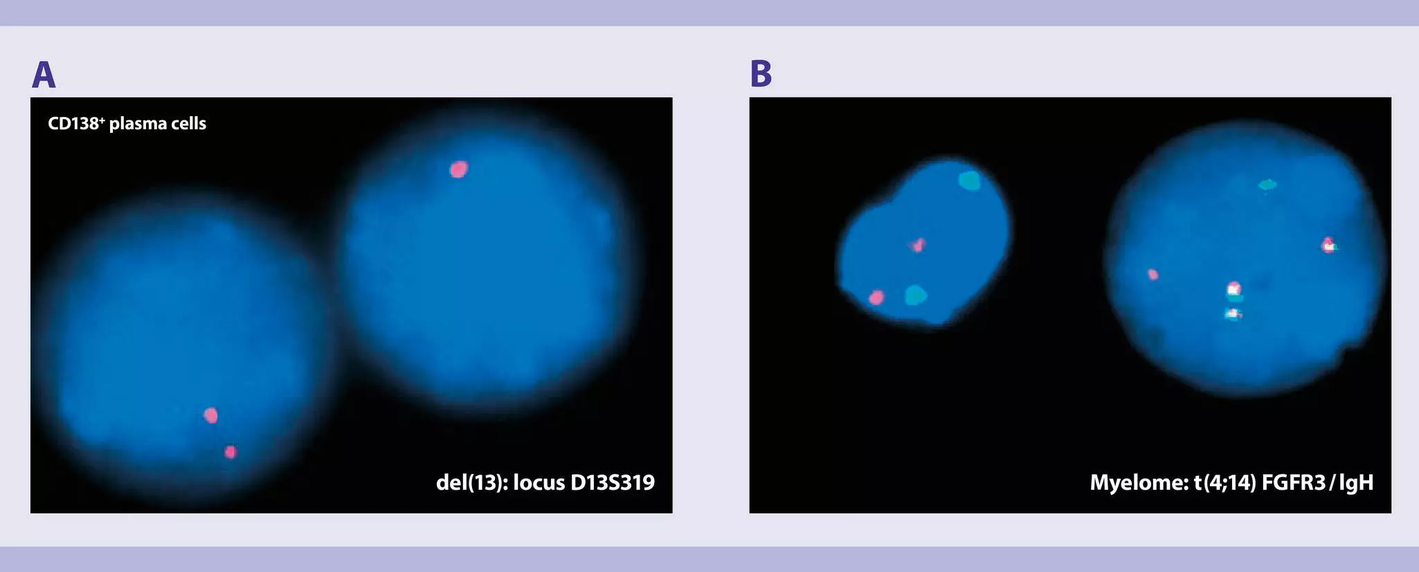
CD138+ Plasma cell FISH analysis
Using our Whole Blood CD138 MicroBeads, we have developed an SOP for an automated, reliable, and standardized method, which allows the processing of multiple samples in a single day, while maintaining sample integrity and increasing the sensitivity of FISH analysis and whole genome arrays.
This technology proved superior to a different automated technology, Including less sample manipulation due to the avoidance of RBC lysis, and higher recovery and purity. Moreover there is no need for density gradient centrifugation. The detection rate of chromosomal abnormalities per sample in multiple myeloma and plasma cell dyscrasia significantly improves when analysis is performed on purified populations of high-quality CD138+ plasma cells.
References
A. Palumbo et al.; J Clin Oncol; Revised International Staging System for Multiple Myeloma: A Report From International Myeloma Working Group
P. Martin et al.; Modern Pathology, hMLH1 and MGMT inactivation as a mechanism of tumorigenesis in monoclonal gammopathies
Multiparameter flow analysis of malignant plasma cells
To gain more information on the isolated malignant plasma cells flow analysis can be performed. Our flow reagents ensure that the data you generate on your flow cytometer is reliable and consistent:
Build your own B cell flow panel with our Flow Panel Builder
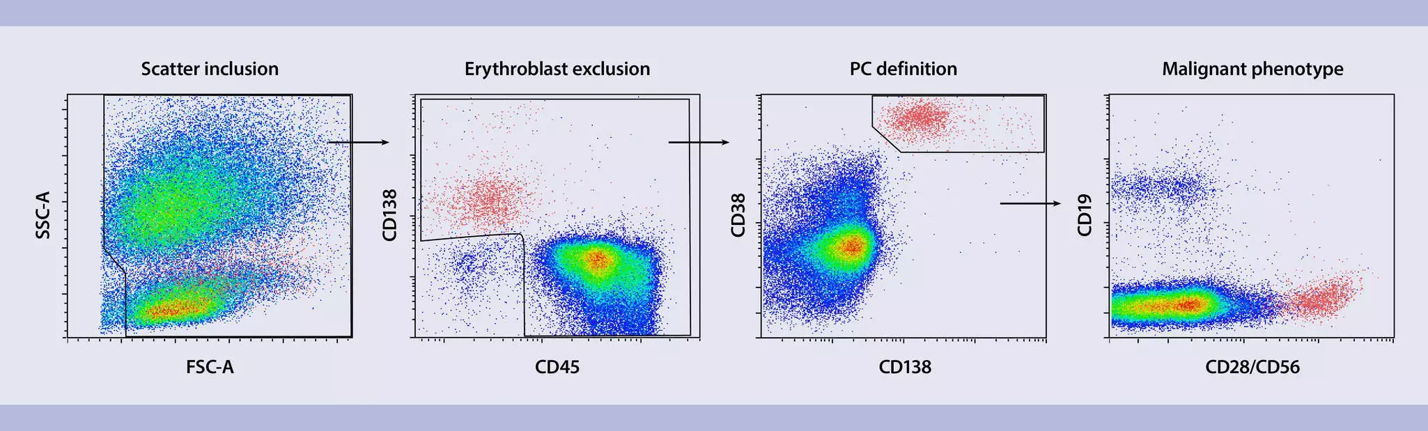
Gating strategy to define malignant plasma cells by flow cytometry
For this application we have designed a specific panel that can help you with the analysis of malignant plasma cells.
| Antigen/parameter | Fluorochrome |
| CD138 | APC |
| CD38 | sFITC |
| CD235a (Glycophorin A) | PE-Vio® 700 |
| CD45 | VioGreen™ |
| CD19 | VioBlue® |
| CD28/CD56 | APC-Vio 770 |
| Viability | Propidium iodide |
Step-by-step protocols for B cell research
Get acquainted with our protocols and antibody panels for the isolation and analysis of human and mouse B cells.
Search:
6 results