Expansion of isolated B cells with the B Cell Expansion Kit, human
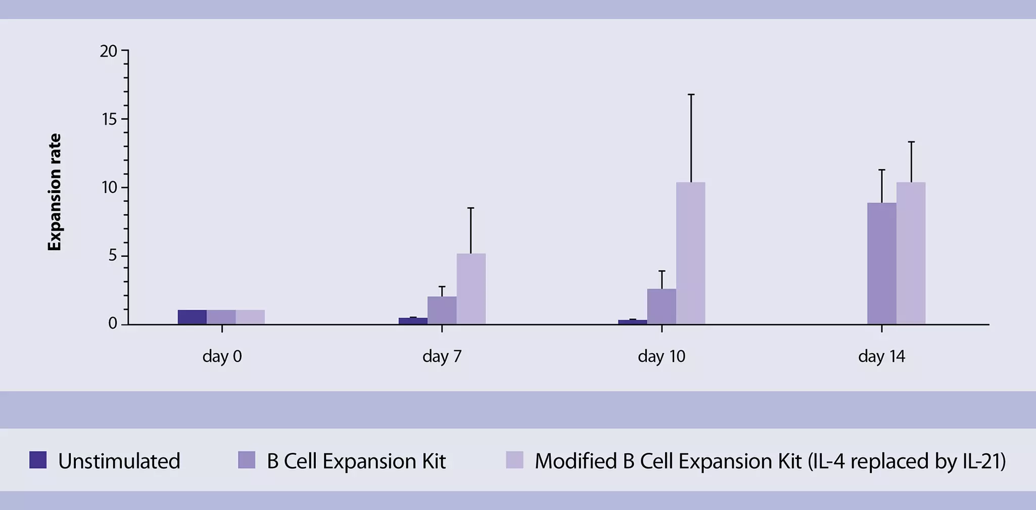
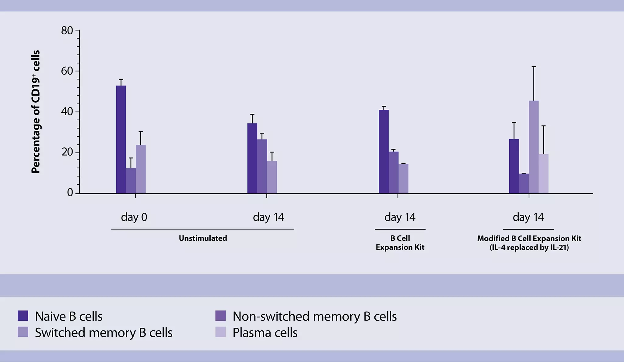
This application protocol describes the in vitro expansion of primary human B cells. Many downstream applications require expansion of B cells due to their low frequencies in peripheral blood. In vitro cultivation of B cells can be time-consuming and can often result in dead cells and low expansion rates. The B Cell Expansion Kit, human has a defined formulation that enables standardized expansion of isolated B cells. Expanded cells are fully functional and ready for downstream applications. The expansion rate reaches up to a ten-fold increase after 14 days of in vitro culture. The phenotype and characteristics of the expanded B cells are also comparable to the starting material, therefore preserving all B cell subsets.
Protocol
For the isolation of human PBMCs, refer to the special protocol "Isolation of mononuclear cells from human peripheral blood by density gradient centrifugation".
Reagent preparation
Preparation of PEB buffer (for staining of isolated B cells)
PEB buffer: Prepare a solution containing phosphate-buffered saline (PBS), pH 7.2, 0.5% bovine serum albumin (BSA), and 2 mM EDTA by diluting MACS® BSA Stock Solution (# 130-091-376) 1:20 with autoMACS® Rinsing Solution (# 130-091-222). Keep buffer cold (2–8 °C). Always use freshly prepared buffer. Do not use autoMACS Running Buffer or MACSQuant® Running Buffer as they contain a small amount of sodium azide that could affect the results.
For B cell isolation various reagents and kits are available according to the kind of isolation.
- Untouched isolation of B cells from PBMCs by depleting all unwanted cells.
- Pan B Cell Isolation Kit, human (# 130-101-638)
- Memory B Cell Isolation Kit, human (# 130-093-546)
- B cell isolation directly from blood products using StraightFrom® MicroBeads. This allows magnetic cell separation directly from human blood products, such as whole blood, buffy coat, leukoreduction system chambers (LRSCs), and Leukopaks.
- StraightFrom® Whole Blood CD19 MicroBeads, human (# 130-090-880)
- Label-free B cell isolation. REAlease® Technology allows for magnetic cell isolation with subsequent removal of any beads and labels from the cells of interest. Clever utilization of recombinant antibody fragments allows for cell surface labeling in just a few steps.
- REAlease® CD19 MicroBead Kit, human (# 130-117-034)
- B cell isolation. For the protocol B cells were separated using MACS® MicroBeads. For details refer to the respective data sheet.
- CD19 MicroBeads, human (# 130-050-301)
Download the quick guide and find a comprehensive overview of all B cell markers and products for convenient isolation and analysis.
Labelling of B cells
- To obtain the results shown in this protocol a total of 1×10⁶ cells has been stained in a total volume of 100 µL.
- Centrifuge cell suspension at 300×g for 10 minutes. Aspirate supernatant completely.
- Prepare a staining cocktail with all fluorochrome-conjugated antibodies according to the respective data sheets.
- Add staining cocktail to each tube containing cell suspension.
- Mix well and incubate for 10 minutes in the dark in the refrigerator (2−8 °C).
▲ Note: Higher temperatures and/or longer incubation times may lead to non-specific cell labeling. Working on ice requires increased incubation times. - Wash cells in each tube by adding 1 mL of PEB buffer and centrifuge at 300×g for 10 minutes. Aspirate supernatant completely.
- Resuspend cells in an appropriate volume of PEB buffer (1× PBS, 0.5% BSA, 2 mM EDTA) and proceed to flow cytometric analysis.
Flow cytometric analysis
- Prior to acquisition of samples in a flow cytometer, e.g., the MACSQuant® Analyzer 10, make sure lasers are properly calibrated (by using, e.g., the MACSQuant Calibration Beads) and compensation of the spillover of fluorochrome emissions has been established in advance using cells or the MACS® Comp Bead Kit, anti-REA. Prior toacquisition adjust also forward scatter (FSC)/side scatter (SSC) parameters to properly locate all leukocyte populations in the plots.
- Exclude doublets by using FSC-H/FSC-A parameters.
- Use gated single cells to exclude erythrocytes and debris using SSC/FSC parameters.
- Use the SSC/FSC gate to exclude dead cells from the analysis using the 7-AAD Staining Solution.
- Use gated viable cells to identify B cell subsets.

 View details

Reconstitution of lyophilized reagents
- Reconstitute lyophilized Human CD40-Ligand (component of the B Cell Expansion Kit, human) with deionized sterile-filtered water to a final concentration of 0.5 mg/mL in a volume of 200 μL (for 100 μg size) or 1000 μL (for 500 μg size) and store at –20 °C.
- Reconstitute Human IL-4 with deionized sterile-filtered water to a final concentration of 2.5×10⁵ IU/mL, e.g., if the lot has a biological activity of 8×10⁶ IU/mg, ise 160 μL water for reconstitution of 5 μg.
- Mix components by pipetting up and down until resuspended. To avoid repeated freeze-thaw cycles, reconstituted reagents should be aliquoted and stored at –20 °C or below until use.
B cell expansion
This protocol is optimized for the activation and expansion of B cells using the Human CD40-Ligand Multimer Kit (component of the B Cell Expansion Kit, human). B cells can either be expanded in low density (0.15×10⁶ cells/mL) or high density (1×10⁶ cells/mL) cell culture.
- Volumes given below are for 10 mL of final expansion medium. When working with larger volumes, scale up accordingly.
- The final expansion medium should be prepared directly before the stimulation. Long-term storage is not recommended.
▲ Note: In case of differentiation and expansion of B cells towards a matured, switched status, use Human IL-21, premium grade at a concentration of 0.7 U/mL instead of human IL-4.
- To increase the biological activity, Human CD40-Ligand and Cross-Linking Antibody (component of the B Cell Expansion Kit, human) must be pre-incubated. Mix 40 μL reconstituted Human CD40-Ligand with 40 μL Cross-Linking Antibody. Incubate for 30 minutes at room temperature (19–25 °C).
- Prepare 10 mL StemMACS™ HSC Expansion Media XF (component of the B Cell Expansion Kit, human) with
2 µL reconstituted Human IL-4, and 5% AB serum. Add the multimerized Human CD40-Ligand (80 μL) to the prepared medium to get the final expansion medium. - Resuspend B cells at a density of 1×10⁶ cells/mL or 0.15×10⁶ cells/mL of the final expansion medium. Culture in an appropriate plate (for details refer to table 1).
- Incubate at 37 °C, 5% CO2.
- On days 7 and 10, exchange half of the medium with fresh medium supplemented with 5% AB serum and
50 units of IL-4/mL (0.2 μL Human IL-4 per mL medium from step 2). Harvested supernatants can be used for downstream analysis. - On day 14, harvest B cells and proceed to downstream applications.
Table 1: Culture plate sizes and required medium volumes for different cell sizes.
| Total cell number (high density) | Total cell number (low density) | Final medium volume | Culture plate |
| 0.5×10⁶ | 0.075×10⁶ | 0.5 mL | 48 well |
| 1.0×10⁶ | 0.15×10⁶ | 1 mL | 24 well |
| 2.0×10⁶ | 0.3×10⁶ | 2 mL | 12 well |
| 4.0×10⁶ | 0.6×10⁶ | 4 mL | 6 well |