Advanced neuroscience research solutions

Neurological disorders are amongst the most prevalent diseases and represent a burden for patients, their families, and our society. For more than three decades, Miltenyi Biotec has worked on developing cutting-edge technologies to support neuroscientists in paving the way towards the most challenging tasks: understanding how the brain works and developing potential therapies.
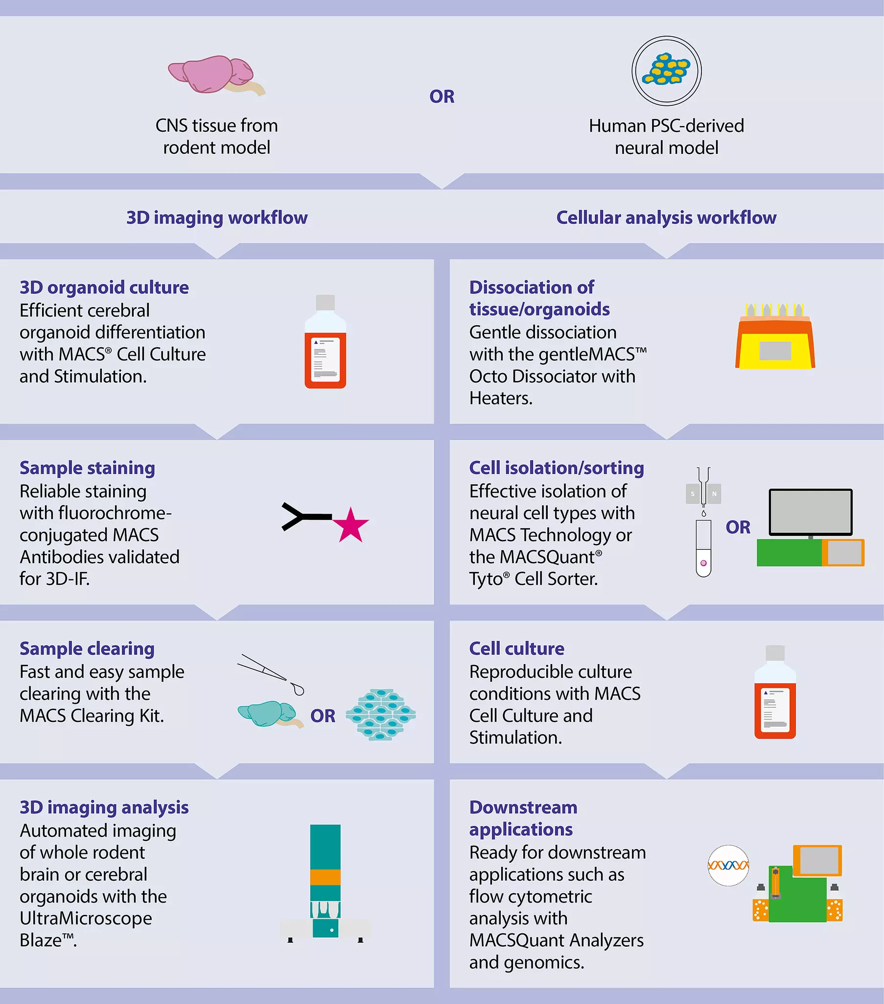
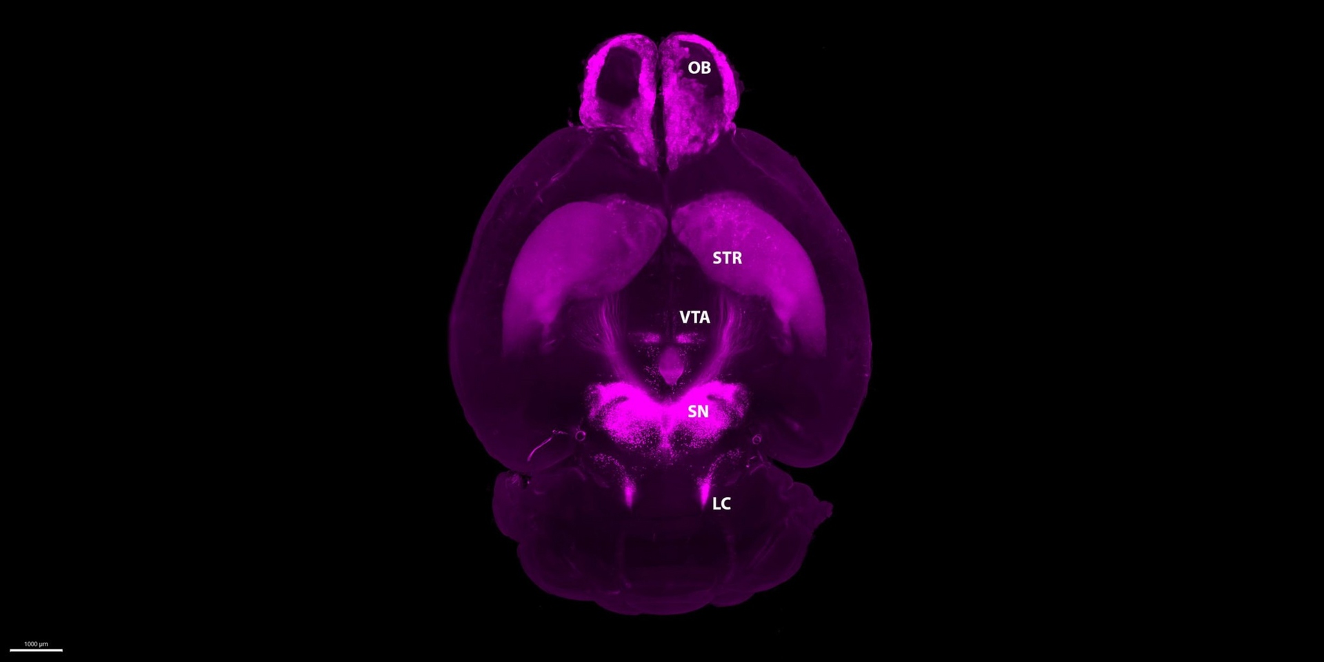
We offer comprehensive product portfolios that address common challenges and facilitate day-to-day research efforts, while ensuring maximal reproducibility. Whether your research relies on rodent or human-derived neural models, we got you covered! Our integrated workflows allow you not only to analyze the cellular components of the rodent and human brain or hPSC-derived cerebral organoids (brain organoids), but also to study their complex neural networks using advanced 3D light sheet microscopy.
Neuroscience models
Choose one of the following research fields to discover detailed scientific data and application protocols for your neuroscience workflow.
Rodent models
Whether your research focuses on adult or neonatal rodent brain, our solutions for tissue dissociation are designed to maintain the delicate nature of synapses, axons, and dendrites.
The neural cell suspension can be further isolated with our MACS® Technology or the MACSQuant® Tyto® Cell Sorter, which will ensure high cell purity.
Isolated cells are suitable for, e.g., single-cell sequencing, cultivation with our serum-free medium, and flow cytometric or immunostaining analysis with our recombinant antibodies.
Human-derived neural models
Our workflows for human-derived neural models (primary, immortalized, and hPS cell lines) include the use of cytokines with lot-specific activity and neural media to ensure the right culture environment.
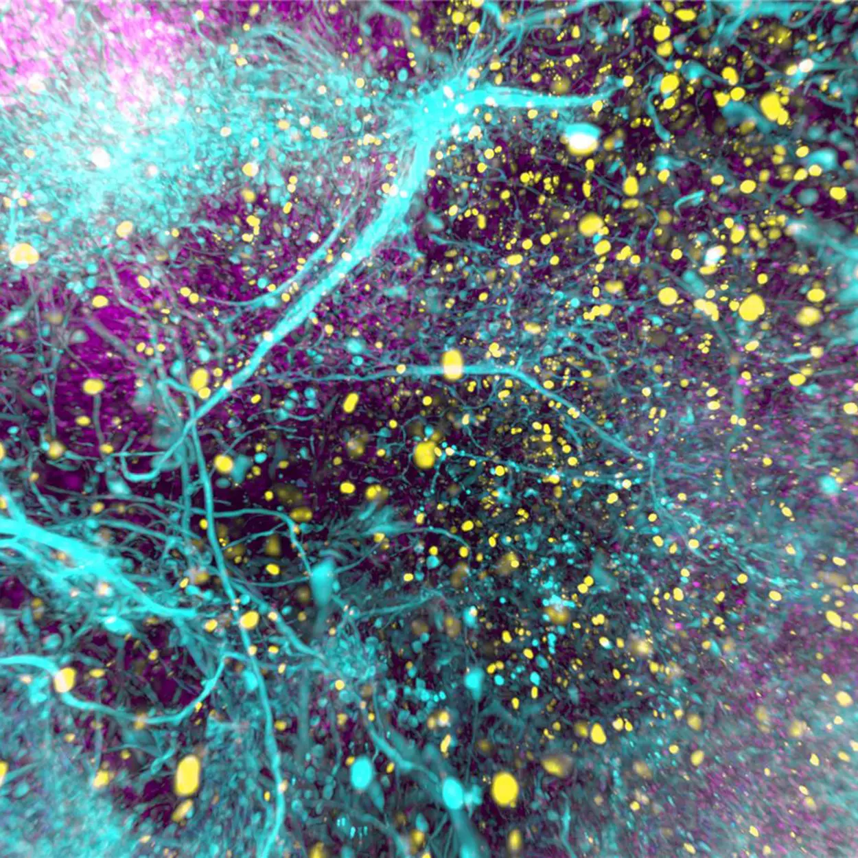
In addition, our specialized portfolio offers solutions from neural cell isolation and flow cytometric analysis, to cutting-edge 3D light sheet microscopy of hPSC-derived cerebral organoids.
Translational research – manufacturing of dopaminergic progenitors
If you focus on developing cell therapies, discover our midbrain dopaminergic progenitor workflow integrated in the CliniMACS Prodigy® Adherent Cell Culture process.
Our neuroscience workflows in action
Dr. Ali Ertürk speaking on advantages of 3D imaging workflow
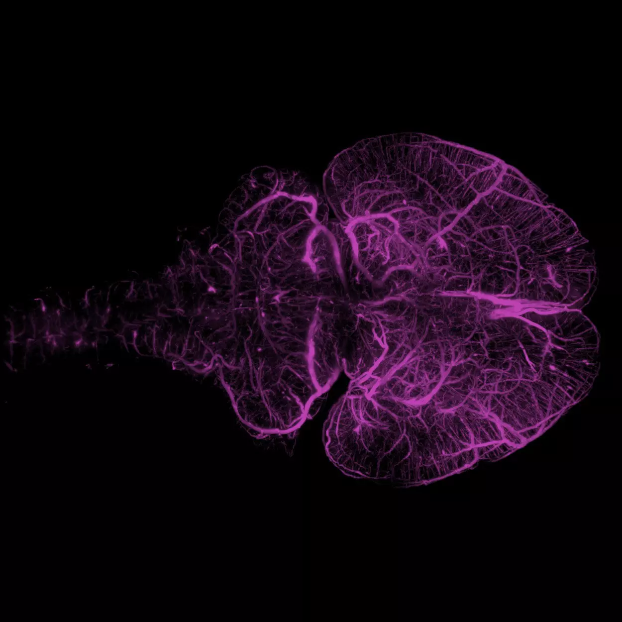
In this interview, Dr. Ali Ertürk, Director of iTERM, Helmholtz Zentrum München, Germany shows how our light sheet microscope, the UltraMicroscope Blaze™, enabled him and his team to visualize a whole cleared mouse brain in 3D and analyze the complete vasculature in detail – from the carotid arteries through to small capillaries in the cortex.

Overcoming limitations in Alzheimer's disease research
Check out Prof. Renzo Mancuso’s and Prof. Bart de Strooper’s work on Alzheimer’s disease and how they overcame the challenges for gentle and sterile microglia sorting using our MACSQuant Tyto Cell Sorter.
Effective sorting of iPSC-derived dopaminergic neurons
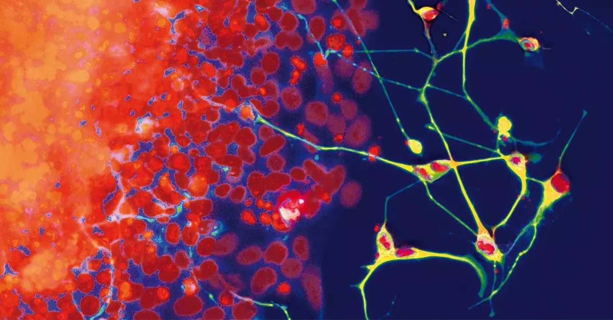
Read about Prof. Jun Takahashi’s renowned laboratory at CiRA (Kyoto, Japan) using the MACSQuant Tyto Cell Sorter for GMP-compliant sorting of iPSC-derived dopaminergic progenitors in a trial for Parkinson’s disease.

Step-by-step protocols for neural cell research
Get acquainted with our protocols and antibody panels for the isolation, cultivation, and staining of neural cells.