
Dopaminergic neurons – study in 3D

The dopaminergic system is essential in neuromodulation of processes such as motor control, reward processing, motivation, and cognition. Dopamine is a neurotransmitter synthesized in both the central and peripheral nervous systems, acting through distinct pathways and brain regions. Dysregulation of dopaminergic signaling is a hallmark of several neurological and psychiatric disorders, such as Parkinson’s disease, Alzheimer’s disease, schizophrenia, and addiction.1, 8

To have a better understanding of dopaminergic neurons and the diseases related to its dysregulation, 3D imaging methods like light sheet microscopy are essential, as they preserve 3D context and reveal dopaminergic pathways in entire mouse brains or even entire mouse bodies.

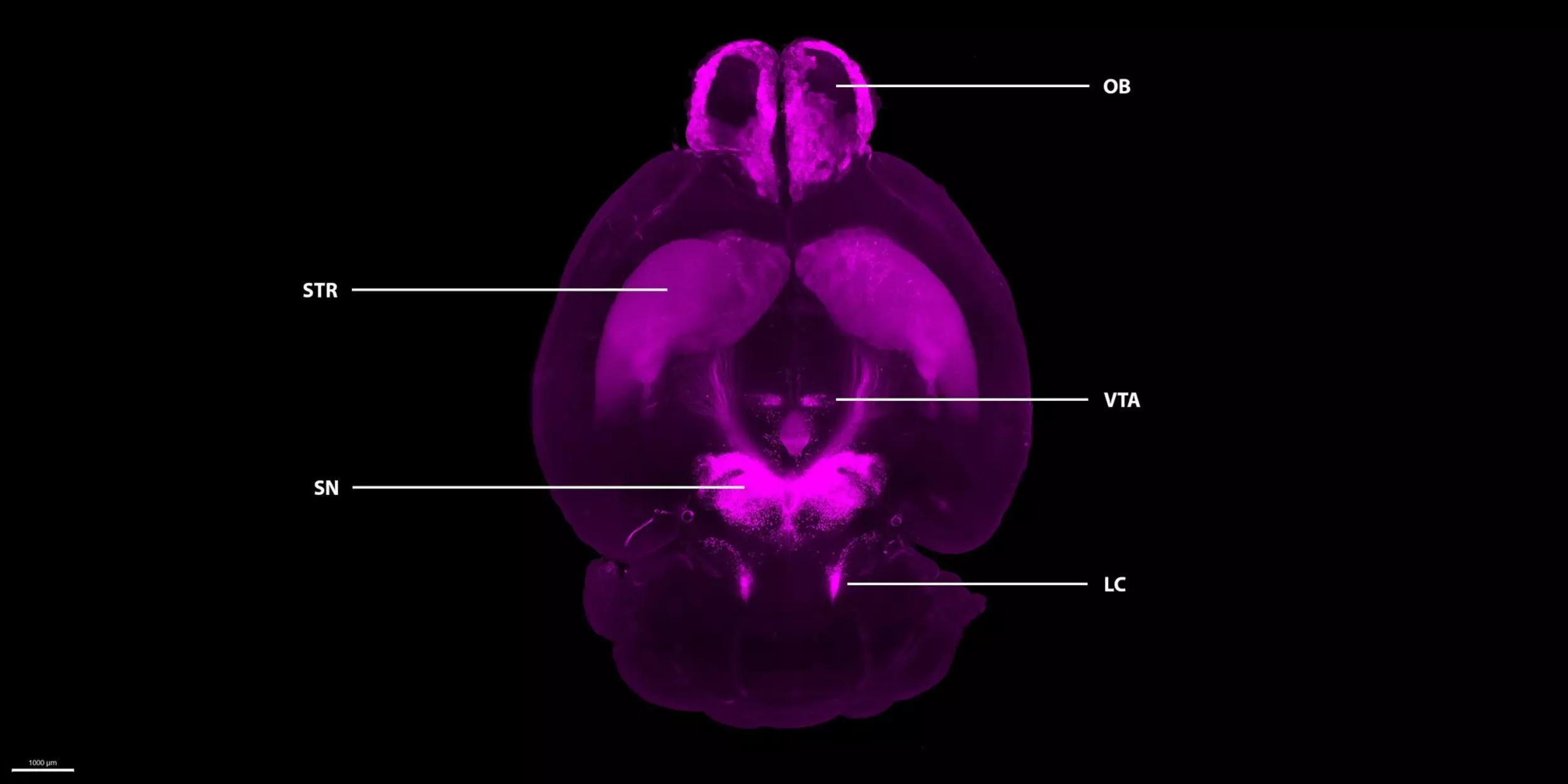
Explore the figure below to learn more about different brain regions' roles

Below you see the 3D image of an entire mouse brain. The brain tissue was cleared using the MACS® Deep Clearing Kit, labeled with Tyrosine Hydroxylase Antibody, Vio® G570, and imaged on the UltraMicroscope™ Platform. Tyrosine hydroxylase is the rate-limiting enzyme in dopamine, epinephrine, and norepinephrine synthesis, and is widely used as a marker for the neurons expressing these neuromodulators.7

Olfactory bulb (OB)

Dopaminergic neurons in the OB support olfactory sensation, and are affected in both Parkinson’s and Alzheimer’s diseases.2

A close up 3D immunofluorescence image of a murine olfactory bulb

Olfactory bulb (OB)

Dopaminergic neurons in the OB support olfactory sensation, and are affected in both Parkinson’s and Alzheimer’s diseases.2

Striatum (STR)

The STR receives dopaminergic inputs from neurons in the substantia nigra pars compacta (SNc). In Parkinson's disease, the dopaminergic neurons of the SNc degenerate, causing a dramatic drop of dopamine release into the STR, and leading to the characteristic motor symptoms. 3

A close up 3D immunofluorescence image of a murine  striatum

Striatum (STR)

The STR receives dopaminergic inputs from neurons in the substantia nigra pars compacta (SNc). In Parkinson's disease, the dopaminergic neurons of the SNc degenerate, causing a dramatic drop of dopamine release into the STR, and leading to the characteristic motor symptoms. 3

Ventral tegmental area (VTA)

The VTA is a source of dopamine for the brain’s reward and motivation circuits. Disruptions here are linked to addiction and depression.4

A close up 3D immunofluorescence image of a murine  ventral tegmental area

Ventral tegmental area (VTA)

The VTA is a source of dopamine for the brain’s reward and motivation circuits. Disruptions here are linked to addiction and depression.4

Substantia nigra (SN)

The SN is the brain’s dopamine engine for movement control. In Parkinson’s, its neurons progressively degenerate, making it the disease’s hallmark site and a critical target for therapies.5

A close up 3D immunofluorescence image of a murine  substantia nigra

Substantia nigra (SN)

The SN is the brain’s dopamine engine for movement control. In Parkinson’s, its neurons progressively degenerate, making it the disease’s hallmark site and a critical target for therapies.5

Locus coeruleus (LC)

The LC is the brain’s main source of norepinephrine. It’s one of the regions affected in Parkinson’s with a significant degeneration of noradrenergic neurons, making it a potential biomarker.6

A close up 3D immunofluorescence image of a murine locus coeruleus

Locus coeruleus (LC)

The LC is the brain’s main source of norepinephrine. It’s one of the regions affected in Parkinson’s with a significant degeneration of noradrenergic neurons, making it a potential biomarker.6

A 3D light sheet microscope image of an entire mouse brain, showing five key areas of interest

Olfactory bulb (OB)

Dopaminergic neurons in the OB support olfactory sensation, and are affected in both Parkinson’s and Alzheimer’s diseases.2

A close up 3D immunofluorescence image of a murine olfactory bulb

Olfactory bulb (OB)

Dopaminergic neurons in the OB support olfactory sensation, and are affected in both Parkinson’s and Alzheimer’s diseases.2

Striatum (STR)

The STR receives dopaminergic inputs from neurons in the substantia nigra pars compacta (SNc). In Parkinson's disease, the dopaminergic neurons of the SNc degenerate, causing a dramatic drop of dopamine release into the STR, and leading to the characteristic motor symptoms. 3

A close up 3D immunofluorescence image of a murine  striatum

Striatum (STR)

The STR receives dopaminergic inputs from neurons in the substantia nigra pars compacta (SNc). In Parkinson's disease, the dopaminergic neurons of the SNc degenerate, causing a dramatic drop of dopamine release into the STR, and leading to the characteristic motor symptoms. 3

Ventral tegmental area (VTA)

The VTA is a source of dopamine for the brain’s reward and motivation circuits. Disruptions here are linked to addiction and depression.4

A close up 3D immunofluorescence image of a murine  ventral tegmental area

Ventral tegmental area (VTA)

The VTA is a source of dopamine for the brain’s reward and motivation circuits. Disruptions here are linked to addiction and depression.4

Substantia nigra (SN)

The SN is the brain’s dopamine engine for movement control. In Parkinson’s, its neurons progressively degenerate, making it the disease’s hallmark site and a critical target for therapies.5

A close up 3D immunofluorescence image of a murine  substantia nigra

Substantia nigra (SN)

The SN is the brain’s dopamine engine for movement control. In Parkinson’s, its neurons progressively degenerate, making it the disease’s hallmark site and a critical target for therapies.5

Locus coeruleus (LC)

The LC is the brain’s main source of norepinephrine. It’s one of the regions affected in Parkinson’s with a significant degeneration of noradrenergic neurons, making it a potential biomarker.6

A close up 3D immunofluorescence image of a murine locus coeruleus

Locus coeruleus (LC)

The LC is the brain’s main source of norepinephrine. It’s one of the regions affected in Parkinson’s with a significant degeneration of noradrenergic neurons, making it a potential biomarker.6

How do we make this possible?

Effective 3D imaging of tyrosine hydroxylase–positive neurons in whole mouse brain requires specialized antibodies and efficient tissue clearing. To address this, we developed fluorophore-conjugated, recombinantly engineered tyrosine hydroxylase antibodies, validated for our organic solvent–based clearing kit.

By combining this highly specific antibody with the MACS Deep Clearing Kit and light sheet microscopy, researchers can easily achieve whole-brain 3D visualization of dopaminergic (or tyrosine hydroxylase–positive) neurons. The streamlined protocol ensures high signal clarity, excellent tissue clearing, and full compatibility with large-volume imaging. The protocol also allows for the inclusion of additional antibodies for multiplexed labeling, and users have the flexibility to incorporate their own antibodies to target other cell types or markers of interest.

Icon showing a star symbol representing a benefit

How you benefit from this workflow:

  • Easy-to-follow protocol
    Minimal hands-on time, robust, and reproducible.
  • Fluorophore-conjugated antibodies
    Reduce staining time by 50% and provide the highest signal-to-background ratio.
  • Fast, efficient, and non-toxic clearing
    The MACS Deep Clearing Kit enables rapid, effective clearing without toxic reagents.
  • Flexible and compatible antibodies
    Can work with other clearing methods like iDISCO+.

iDISCO+ compatibility demonstrated in 3D neuronal imaging

The compatibility of Vio® Dye–conjugated antibodies with iDISCO+ tissue clearing was demonstrated in an externally generated healthy whole mouse brain sample from Gubra, a CRO and biotech company based in Denmark. Tyrosine Hydroxylase Antibody, Vio G570 and NeuN Antibody, Vio R667 enabled simultaneous 3D visualization of dopaminergic and total neurons, respectively, with reliable signal separation. These results support their suitability for 3D neuronal imaging and for studying neuronal loss and structural changes associated with Parkinson’s disease.

It is by far the best tyrosine hydroxylase antibody we have ever tested.​

[1] Speranza, L. et al. (2021) Dopamine: The neuromodulator of long-term synaptic plasticity, reward and movement control. Cells. 10: 735.

[2] Jeffs, Q. L. et al. (2023) Potential neural substrates underlying circadian and olfactory disruptions in preclinical Alzheimer's disease. Front. Neurosci. 17:1295998.

[3] Surmeier, D. J. et al. (2009) Dopamine and synaptic plasticity in dorsal striatal circuits controlling action selection. Curr. Opin. Neurobiol. 19: 621–628.

[4] Lammel, S. et al. (2014) Reward and aversion in a heterogeneous midbrain dopamine system. Neuropharmacology. 76: 351–359.

[5] Zhou, Z. D. et al. (2023) Role of dopamine in the pathophysiology of Parkinson’s disease. Transl. Neurodegener. 12: 44.

[6] Rommelfanger, K. S. and Weinshenker, D. (2007) Norepinephrine: The redheaded stepchild of Parkinson's disease. Biochem. Pharmacol. 74: 177–190.

[7] Daubner, S. C. et al. (2011) Tyrosine hydroxylase and regulation of dopamine synthesis. Arch. Biochem. Biophys. 508: 1–12.

[8] Klein M.O. et al. (2019) Dopamine: Functions, signaling, and association with neurological diseases. Cell. Mol. Neurobiol. 39: 31-59.

Learn more about or order related products

5 products found

|