Logo

Pluripotent stem cell–derived neural cells: differentiation and powerful analytical solutions

Pluripotent stem cells (PSCs), such as embryonic stem cells (ESCs) and induced PSCs (iPSCs), have opened new frontiers in neuroscience research by offering an unprecedented opportunity to generate human neural cells in vitro. These cells can differentiate into nearly all neural cell types, including subtypes of neurons and glial cells such as astrocytes and microglia. This versatility makes them powerful tools for studying human brain development, creating disease models that faithfully recapitulate the pathology of neurological disorders, and accelerating the discovery of novel therapeutic strategies for neurodegenerative conditions.

Despite their potential, one of the major challenges in using PSCs for neural differentiation is ensuring consistency and reproducibility across different experimental batches. Variability in cell lines, culture conditions, and reagents can lead to inconsistent outcomes, which hampers both basic and translational research applications.

Our comprehensive workflow solutions are designed to streamline every step of the neural cell differentiation procedure, helping researchers reduce variability, save time, and improve the reliability of their experimental setup, ultimately enabling more accurate and impactful neuroscience research.

Content overview

Workflow for differentiating pluripotent stem cells into neural cells

Infographic showing iPSC and ES cell differentiation workflow with Miltenyi Biotec products

Efficient PSC differentiation into neural cells

Successful neural differentiation starts with high-quality PSCs. Preserving their full differentiation potential requires well-balanced, optimized media formulations that support PSC health and identity, along with routine verification of pluripotency through reliable reagents and validated antibodies.

Reliable PSC maintenance

To support this critical early stage, we offer specialized pluripotent media tailored for both research and translational workflows. Our StemMACS™ PSC-Brew XF, human, and the StemMACS iPSC-Brew XF, human, are carefully formulated to maintain and expand PSCs under xeno-free and feeder-free conditions.

StemMACS PSC-Brew XF, human, is optimized for flexibility, offering a stable nutrient environment, consistent pH, and a stabilized version of FGF-2 (IS), which enhances media performance and reduces the need for daily feeding. For challenging applications, such as post-reprogramming recovery or gene editing workflows, StemMACS PSC-Brew XF, human, can be paired with StemMACS PSC-Support-XF, human, providing enhanced support under stress conditions.

When advancing PSCs toward clinical use, our StemMACS iPS-Brew XF and iPS-Brew GMP Medium offer a seamless transition from bench to bedside, delivering consistency, efficiency, and regulatory-ready components ideal for cell therapy manufacturing.

Our media is compatible with both defined and undefined matrices, and show exceptional performance with MACSmatrix Laminin 511 – a recombinant, chemically defined, xeno-free matrix optimized for stem cell culture.

Efficient differentiation induction

Inducing the differentiation of PSCs into specific neural lineages requires precise control over the cellular environment. Our comprehensive portfolio of serum-free StemMACS Media, supplements, cytokines, and small molecules provides a versatile and modular platform that enables researchers to fine-tune differentiation protocols based on their specific needs.

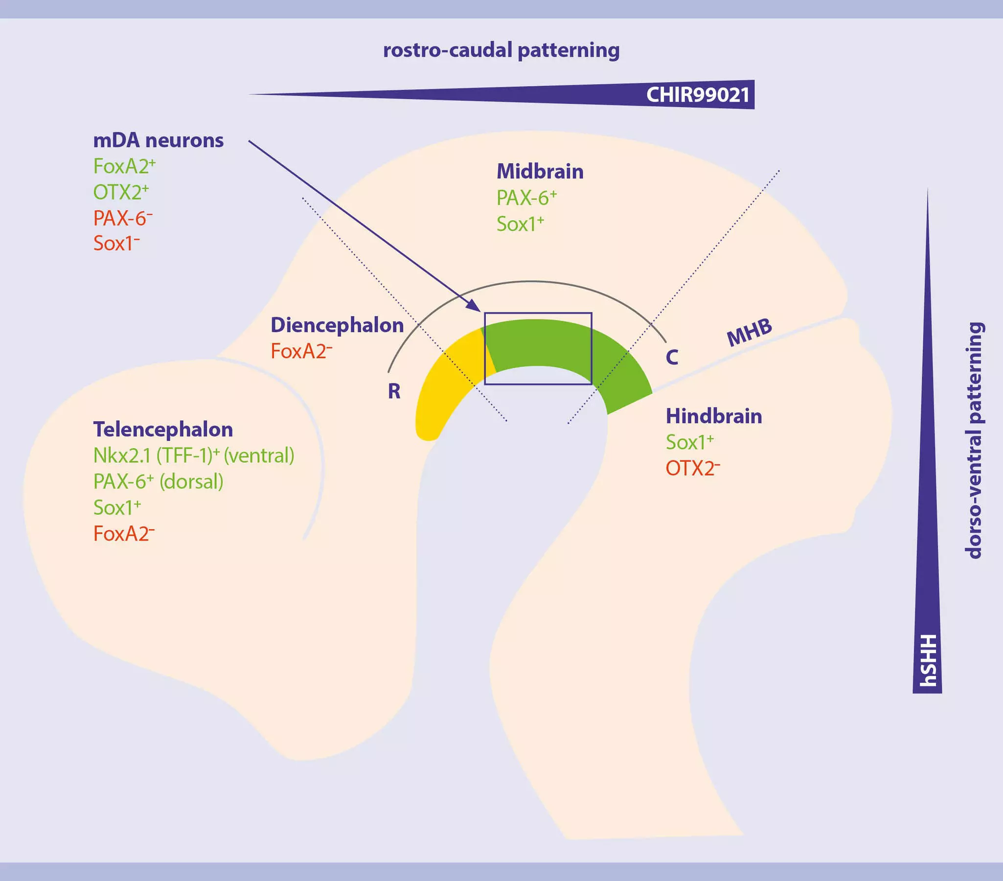
Differentiation of mDA neurons from PSCs

Basal media and supplements for neural differentiation

High-quality, well-defined formulations are essential for promoting robust neural differentiation, supporting healthy growth and long-term survival in vitro. We have developed serum-free cell culture media specifically designed to support PSC differentiation into neural cells.

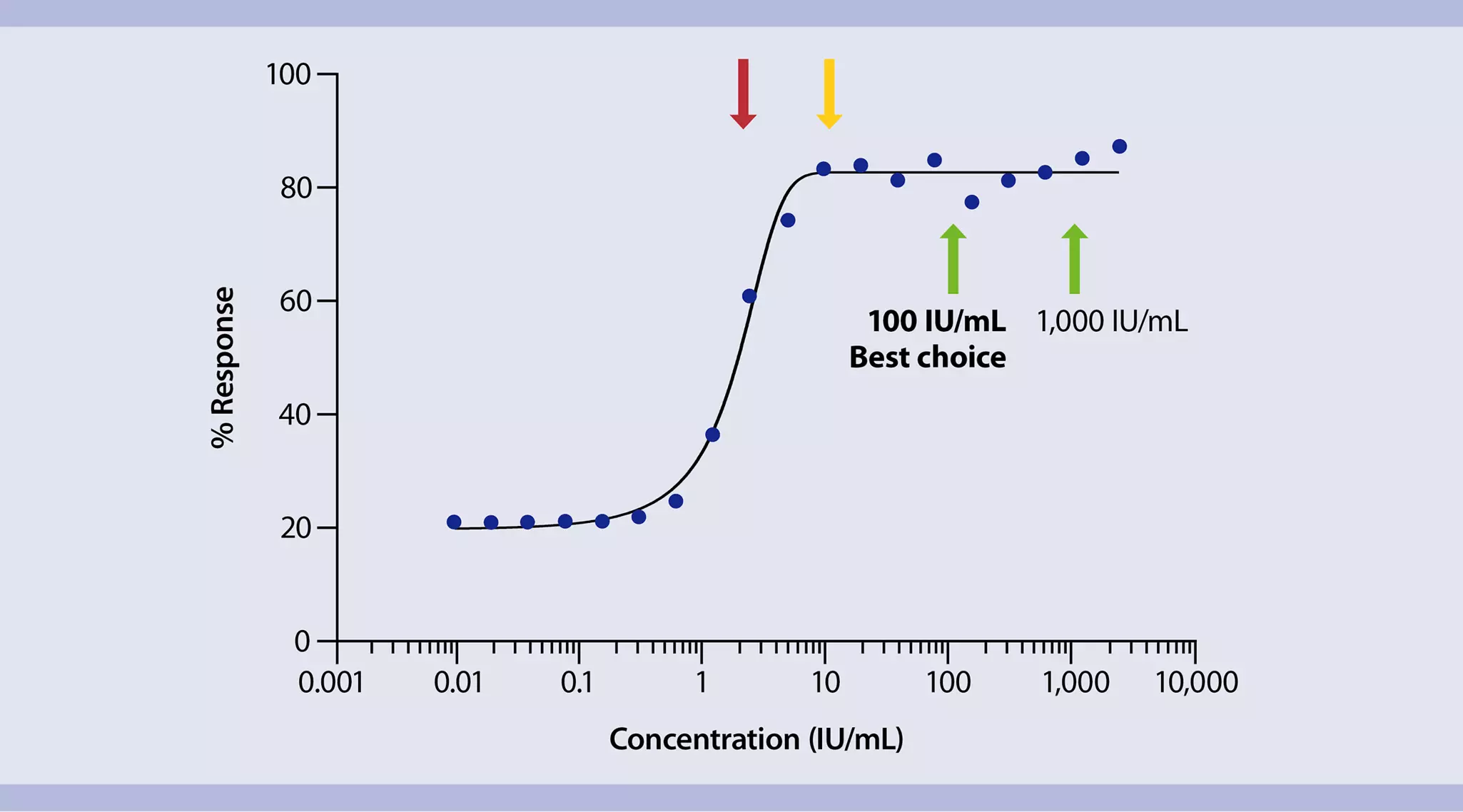
Illustration of line chart showing effective cytokine usage with optimal unit dosing indicates cytokine concentrations that result in maximal cellular response with MACS® Premium-Grade Cytokines
Efficient cytokine usage with specific unit dosing.

Cytokines with lot-specific activity

The generation of PSC-derived neural cells relies heavily on the precise timing and concentration of patterning factors at key stages of differentiation. Among these, cytokines and small molecules play a central role in directing cell fate decisions and promoting lineage-specific maturation.

Cytokine biological activity depends on the specific cytokine and the manufacturing process, with common variations of up to 9-fold between different lots. This variability can compromise reproducibility, lead to inaccurate dosing, and introduce inconsistencies across experiments.

To address this, we provide lot-specific activity data for every batch of our MACS® Premium-Grade Cytokines. This allows researchers to calculate and administer cytokines based on actual biological activity rather than nominal concentration. By avoiding cytokine oversaturation or underdosing, you reduce both experimental variability and unnecessary reagent costs. Moreover, utilizing MACS Premium-Grade Cytokines facilitates a smooth transition to clinical research with MACS GMP Cytokines.

The benefits speak for themselves:

  • Save time – Using MACS Premium-Grade Cytokines, you can rely on lot-specific cytokine activity and skip tedious lot-to-lot testing.
  • Increase reproducibility – Knowing the lot-specific activity, you can precisely dose cytokines based on their biological activity, which increases reproducibility and reliability of experimental results.
  • Reduce costs – Once you’ve determined the optimal cytokine amount for your experiment, you can rely on exact unit dosing with each batch. With no need for oversaturation or repeated testing, you can save reagent and reduce costs.

Illustration showing cell culture reagents required for differentiation of stem cells into neural cells
Cytokines and small molecules required for neural differentiation

Small molecules and MACS Cytokines in research and premium grade, commonly used for differentiation of PSCs into neural cells

Target cell typeSmall moleculesCytokines
Neuronal stem cellsCHIR99021 , DAPT,
Dorsomorphin, Forskolin, LDN193189 , SB431542
FGF-2, Noggin

Neural crestSB431542, LDN-193189, Y27632EGF, FGF-2
Cortical neuronsRetinoic Acid, LDN-193189, SB431542, PD0325901, DAPTFGF-2
Motor neuronsY27632, SB431542, CHIR99021, LDN-193189, DAPT, Retinoic AcidSHH, BDNF, GDNF, CNTF

Dopaminergic progenitorsThiazovivin, CHIR99021, SB431542, LDN-193189, Purmorphamine, DAPTSHH, GDNF, BDNF, FGF-2, FGF-8b

GABAergic neuronsCHIR99021, LDN-193189, SB431542, Retinoic AcidBDNF, GDNF, FGF-2, FGF-8b,IGF-1, SHH
AstrocytesCHIR99021, SB431542, LDN-193189CNTF, EGF, FGF-2
OligodendrocytesCHIR99021, SB431542, LDN-193189CNTF, EGF, FGF-2, PDGF-AA,SHH, IGF-1, NT-3
Microglia BMP-4, FGF-2, VEGF, SCF, IL3 , TPO, IL6, M-CSF, IL34, TGF-β1
Cerebral organoidsDorsomorphin, SB431542, IWP-2
Noggin, FGF-2 , TGFβ, EGF, BDNF, GDNF, NT-3, DKK1
Retinal organoids Noggin, FGF-2 ,DKK1, IGF-1, SHH
Midbrain organoidsCHIR99021, SB431542, LDN-193189BDNF, GDNF, SHH, TGFβ, FGF-8b
Cerebellar organoidsSB431542, CHIR99021Noggin, FGF-8b , BDNF
Neuromuscular organoidsY27632, CHIR99021FGF-2, IGF-1, HGF

Isolation of PSC-derived neural cells

PSC differentiation into neural lineages can result in cell cultures often containing a heterogeneous mix of progenitor and mature cell types. Reliable and efficient cell isolation enables the enrichment of homogeneous populations, minimizes unwanted cell types, and enhances the fidelity of downstream analyses.

Gentle neural cell isolation

Magnetic neural cell isolation

Our MACS MicroBead Technology offers a fast, gentle, and highly specific method for isolating PSC-derived neural cells at any stage of the differentiation process. Based on magnetic cell sorting, this technology enables both positive selection of target cells and depletion of undesired populations, while preserving cell viability, phenotype, and functionality.

Target Starting material Isolation strategyComments Product
Ectoderm
Neural crestSingle-cell suspensionPositive selection of target cellsBased on CD271 expressionNeural Crest Stem Cell MicroBeads, human
Neuronal progenitorsSingle-cell suspensionPositive selection of target cellsBased on PSA-NCAM expressionAnti-PSA-NCAM MicroBead Kit, human and mouse
Glial progenitorsSingle-cell suspensionPositive selection of target cellsBased on A2B5 expressionAnti-A2B5 MicroBead Kit, human and mouse
Immature oligodendrocyteSingle-cell suspensionPositive selection of target cellsBased on O4 expressionAnti-O4 MicroBeads, human, mouse, rat
AstrocytesSingle-cell suspensionPositive selection of target cellsBased on GLAST (ACSA-1) expressionAnti-GLAST (ACSA-1) MicroBead Kit, human,mouse, rat
Mesoderm
Hematopoietic progenitorsSingle-cell suspensionPositive selection of target cellsBased on CD34 expressionCD34 MicroBead Kit UltraPure, human
MicrogliaSingle-cell suspensionPositive selection of target cellsBased on CD11b expressionCD11b MicroBeads, human and mouse
Cellular components
Extracellular vesiclesCell culture supernatantPositive selection Based on CD9, CD63, CD81 expressionEV Starting Kit Pan, human
MitochondriaCell lysatePositive selection of target organelleBased on TOM22 expressionMitochondria Isolation Kit, human
NucleiSingle-nucleus suspensionsPositive selection of target organelleBased on universal nucleus surface epitopeAnti-Nucleus MicroBeads

Streamline cell sorting with flow cytometry

Flow cytometry enables high-throughput sorting of PSC-derived neural cells with precision. By using fluorescently labeled antibodies, you can isolate specific neural cell types while collecting multiparameter data on cell health, identity, and quality. This ensures homogeneous populations for consistent, high-quality downstream applications.

Our MACSQuant® Tyto® Cell Sorter provides a sterile, contamination-free environment with its fully closed cartridge system – protecting both samples and user. Its patented microchip-based technology and integrated temperature control minimize sorter-induced cellular stress, preserving cell viability and functionality – even for the sensitive PSC-derived neural cells.

PSC-derived neural cell characterization

Following PSC differentiation and isolation, characterization of the neural cells is essential to confirm the identity and purity of neural cell populations. This is of particular importance due to the diverse nature of neural cells, including various subtypes within the desired cell lineage. Techniques such as flow cytometry and immunostaining are central to this process, offering insights into cell phenotype, heterogeneity, and function. Additionally, analyzing cell-released factors such as cytokines and extracellular vesicles (EVs), provides valuable information about cell health, activity, and status.

Precise characterization of the PSC-derived neural cells

Our broad portfolio of MACS Antibodies makes for reliable characterization of PSC-derived neural cells. Many of our antibodies for flow cytometry and immunostaining have been crafted using innovative recombinant technology for unmatched reproducibility and standardization.

For EV profiling, the MACSPlex EV Kit Neuro, human, allows simultaneous detection of up to 37 surface markers on EVs that were collected, for example, from the cell culture supernatant – enabling deeper analysis of cell-derived signaling components.

The MACSPlex Cytokine 12 Kit, human, enables the detection of 12 key human cytokines at the same time, from a single sample. Optimized for use with cell culture supernatants, this flow cytometry–based multiplex assay profiles cytokines such as IL-6, TNF-α, and IL-10, enabling researchers to assess microglia activation, polarization states, and functional responses.

The PSC-mDA Neuron Analysis Kit, anti-human, REAfinity® was designed to provide a fast and precise flow cytometry–based quality control assay to assess cell purity and identity after midbrain dopaminergic (mDA) neuron differentiation from PSCs.

Neurons differentiated from human pluripotent stem cells – cultured, stained, and imaged using a workflow from Miltenyi Biotec
PSC-derived neural cells imaged with the MACSima™ Platform. PSC-derived neural cells were stained with antibodies against tyrosine hydroxylase, β-tubulin 3, MAP2, neurofilaments, and GFAP, and imaged with the MACSima Platform. Scale bar: 40 µm.

Step-by-step protocols