3D graphic showing tumor-infiltrating leukocytes (TILs) and a tumor
Logo

Isolation and analysis of tumor infiltrating lymphocytes (TILs)

The amount and composition of tumor-infiltrating leukocytes (TILs) is highly variable. A reliable analysis of TILs and especially TIL subpopulation, calls for efficient isolation strategies. In this workflow, gentle tumor dissociation preserves cell surface epitopes and pre-enrichment allows for improved downstream analysis. It is applicable for manual single-sample separation up to fully automated high-throughput separation.

Content overview

Content

  • Storage of tumor tissue
  • Tumor dissociation
  • Magnetic isolation of human TILs
  • Magnetic isolation of mouse TILs
  • Serial sorting of TIL subpopulations
  • Flow analysis of TILs
  • Omics analysis of TILs
  • Characterization of the TME by ultra high content imaging
  • Step-by-step protocols

Storage of tumor tissue

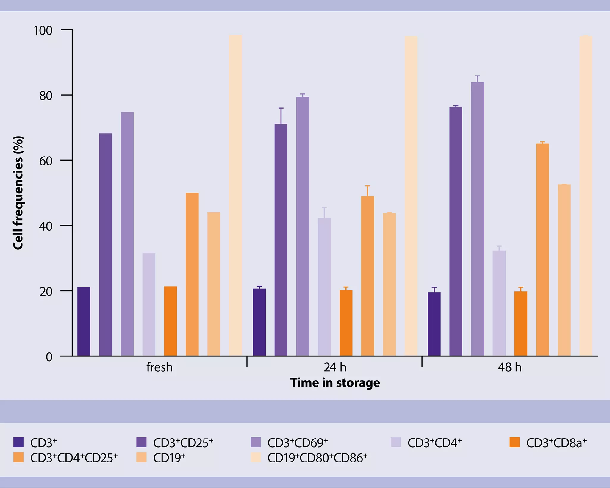
Freshly dissected tumor tissue can be stored for up to 48 h in MACS® Tissue Storage Solution without compromising cell viability or immune cell composition.

The MACS Tissue Storage Solution preserves TIL composition

The MACS Tissue Storage Solution preserves TIL composition

Flow cytometry analysis of TIL populations after storage in MACS Tissue Storage Solution show no changes in TIL composition.

Tumor dissociation

Enzymatic tumor dissociation for efficient recovery of TILs from B16-F10 tumors

Enzymatic tumor dissociation for efficient recovery of TILs

Standardized processing of tumor samples using the Tumor Dissociation Kit together with the gentleMACS® Dissociator enhances reproducibility and was essential for recovery of key populations of TILs.

Cell types identified after tumor dissociation

Analysis of tumor heterogeneity

Single-cell gene expression analysis of mouse tumors enables the clear identification of the different cellular populations present in the tissue.

IFN- γ release of TILs prepared with the gentleMACS Dissociator compared to overnight digestion

Preserved anti-tumor functionality of TILs

Secretion of IFN-γ is one of the hallmark functions of antigen-specific T cells encountering the respective tumor antigen. Combined mechanical and enzymatic processing of TILs with the gentleMACS Dissociator is gentle to cells and results in profound secretion of IFN-γ upon tumor cell co-culture.

Magnetic isolation of human TILs

The amount and composition of TILs is highly variable, complicating the analysis of individual subpopulations. When working with large cohort sizes, even immunophenotyping of TILs by flow cytometry is time consuming and data processing highly work intensive. Therefore, pre-enrichment of TILs is desirable to increase the sensitivity of analysis and save time and effort during downstream analysis such as sequencing and flow cytometry. To this end, Miltenyi Biotec offers optimized MicroBeads for tumor tissue samples.

Automated isolation of human CD45+ TILs from xenograft tumors

Automated isolation of human CD45+ TILs from xenograft tumors with MACS Technology

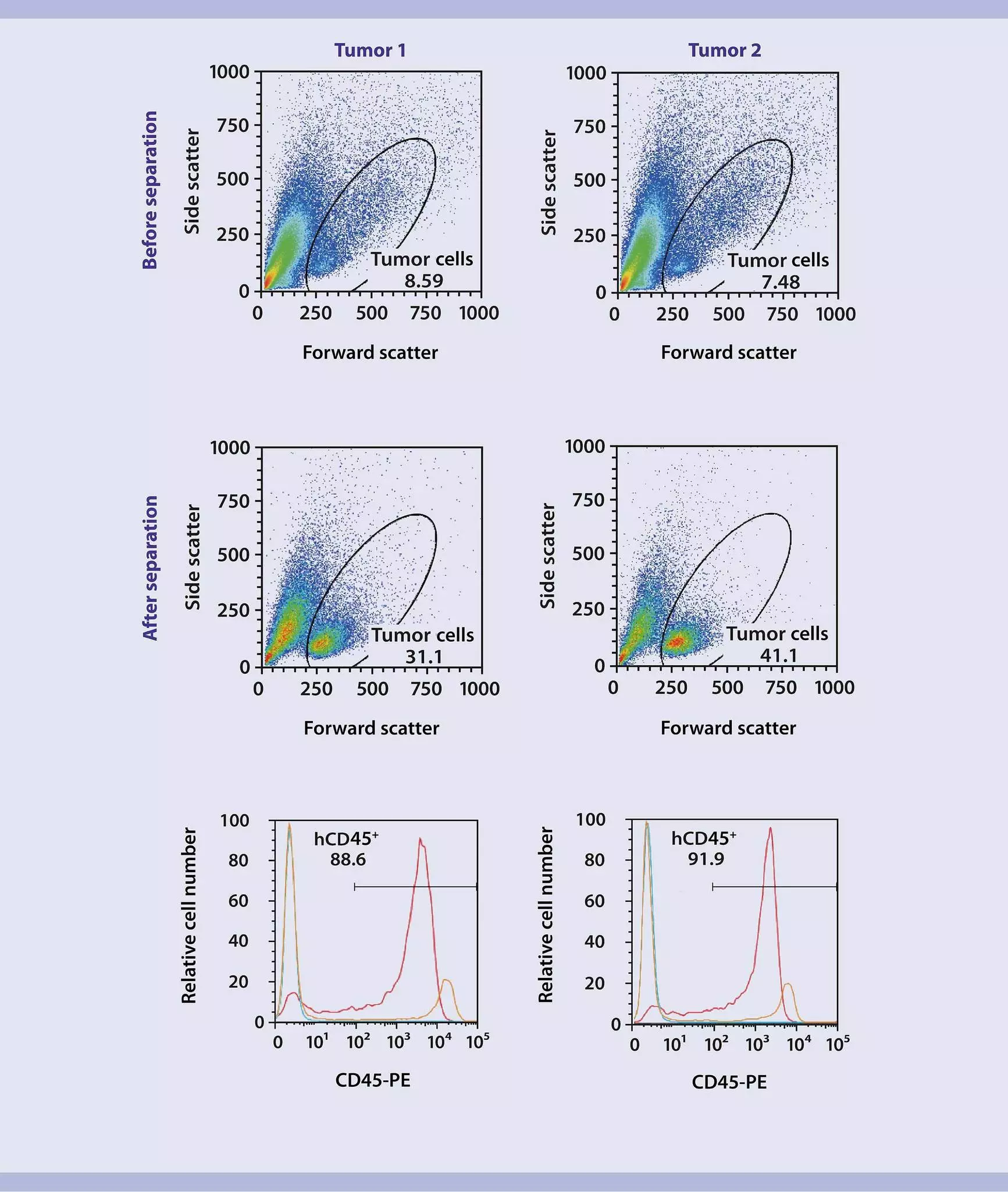
Automated isolation of CD45+ TILs using CD45 (TIL) MicroBeads and the autoMACS® Separator results in an increase of CD45+ TIL frequencies from 29.6% and 29.3% (Tumor 1 and 2) to 88.6% and 91.9%, respectively.

Isolation of MicroBead- and label-free CD45+ TILs from a dissociated ovarian tumor

REAlease Technology delivers beads- and label-free CD45+ human TILs

Isolation of human CD45+ TILs using the REAlease® CD45 (TIL) MicroBead Kit results in high cell purities of 89%. Directly after isolation, cells show staining of biotin (MicroBead-free CD45+ cells), whereas the label-free CD45+ cells after the REAlease Biotin Complex release are negative for biotin similar to the non-labeled cells before separation, indicating efficient MicroBead and label release when applying REAlease Technology.

Isolation of CD4+/CD8+ TIL subpopulation with REAlease Technology followed by dead cell removal improves cell vitality

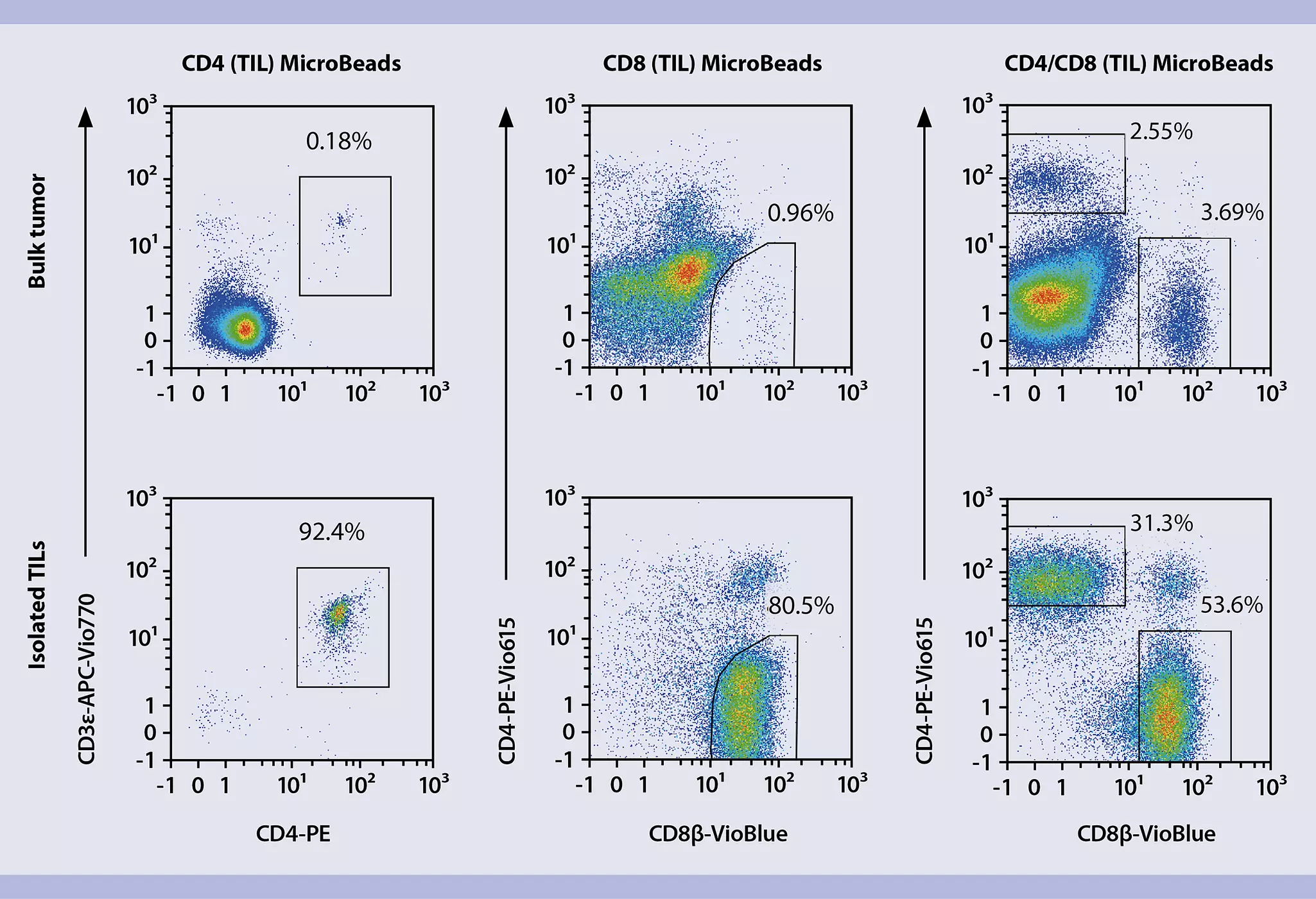
Using the REAlease CD4/CD8 (TIL) MicroBead Kit, human directly on dissociated tumor tissue provides highly pure CD4+/CD8+ human TILs and delivers MicroBead- and label-free cells. Isolated cells are suitable for further processing, e.g., with the MicroBead-based Dead Cell Removal Kit, human, thereby improving the vitality of CD4+/CD8+ TILs from 80% to nearly 90%.

Isolation of CD4+/CD8+ TIL subpopulation with REAlease Technology followed by dead cell removal improves cell vitality

Magnetic isolation of mouse TILs

Automated isolation of human CD45+ TILs from xenograft tumors

Isolation of CD4+, CD8+, and pan T cells from syngeneic mouse tumors

Using CD4+, CD8+, and pan T cell specific MACS MicroBeads for magnetic cell isolation (MACS Technology) directly from dissociated tumor tissue delivers high purities of isolated TILs above 80%.

This pre-enrichment of TILs greatly reduces time and costs of downstream analysis while the quality of data analysis significantly increased.

Flow cytometric analysis showing isolated CD45+ cells
Isolation and analysis of CD45+ cells

Isolation of CD45+ T cells from syngeneic mouse tumor

Isolation of TILs directly from dissociated tumor tissue can be improved by using the CD45 (TIL) MicroBeads for magnetic cell sorting.

Pre-enrichment of TILs significantly improves subset analysis and does not affect the composition of infiltrating immune cell populations.

Serial sorting of TIL subpopulations

One major goal in immunotherapies is to understand how the composition of tumor-infiltrating myeloid and lymphoid cells contributes to patient stratification. However, TIL numbers can be very low and small subpopulations might be lost in background noise, particularly in single-cell analyses. Therefore, serial sorting of TIL subpopulations is highly advantageous in order to investigate the immune cell composition.

Sequential sorting of tumor-infiltrating NK and B cells

Gentle serial sorting of B and NK cells from syngeneic mouse tumors after a CD45+ TIL pre-enrichment

CD45+ TILs can be sorted into single TIL subpopulations using the MACSQuant® Tyto® Cell Sorter. Thanks to releasable antibodies and to the MACSQuant Tyto Cartridge, a serial sorting of tumor-infiltrating NK and B cells is possible in two steps. Flow cytometry analysis using the MACSQuant Analyzer demonstrates a selective enrichment of target cell populations with purity of >94%, without affecting cell viability.

Flow analysis of TILs

Using hybridoma-derived antibodies to identify TILs leads to a gross overestimation of the frequency of immune cell subpopulations present in the tumor. This experimental artifact is most likely caused by unspecific binding of hybridoma-derived antibodies to FcγRs on immune cells, as it is significantly reduced when using an FcR blocking reagent. In contrast, assessment of TIL frequency using REAfinity™ Recombinant Antibodies is unchanged in the presence or absence of FcR blocking, providing a more exact analysis of respective cell populations, even in the absence of FcR blocking. REAfinity Antibodies are highly specific recombinant antibodies that provide superior lot-to-lot consistency and purity compared to mouse or rat hybridoma-derived monoclonal antibodies.

Background-free flow analysis of TILs

Optimized multicolor flow cytometry panels

Phenotypic characterization of TILs by flow cytometry can be performed using optimized panels of fluorochromes-conjugated REAfinity Recombinant Antibodies. Our Miltenyi Biotec tested panels (MBTPs) for flow cytometry applications are developed and validated by Miltenyi Biotec’s R&D experts, as well as by customers who use our antibodies.

Relevant panels for TILs:

AntigenCloneFluorochrome
CD8βREA793VioBlue®
Viobility™ 405/520
Lag-3REA776Vio® Bright 515
TIM-3REA602PE
CD4REA604PE-Vio 615
PD-1REA802APC
CD44REA664APC-Vio 770

AntigenCloneFluorochrome
CD8βREA793VioBlue®
Viobility™ 405/520
CD103REA789Vio® Bright 515
CXCR3REA724PE
CD4REA604PE-Vio 615
CD39REA870APC
CD44REA664APC-Vio 770

Miltenyi Biotec Tested Panels (MBTPs) for T cells

Pre-tested antibody panels that allow for easy and accurate flow cytometry analysis.

Omics analysis of TILs

TIL numbers can be very low and small subpopulations might escape analysis when they are lost in background noise. This can be especially challenging when performing single-cell analysis where a debris-free single-cell suspension with a viability above 80% is required. We offer innovative tools to eliminate debris and enrich viable cells that will consequently improve your single-cell sequencing results.

Magnetic isolation of TILs increases the sensitivity of single-cell receptor sequencing

Magnetic isolation of TILs increases the sensitivity of single-cell receptor sequencing

Single-cell TCRs (A) and BCRs (B) were sequenced prior to and after the isolation of T or B cells from a tumor sample. The graphs show the top 50 TCR (A) and top 25 BCR (B) CD3 clonotypes ranked by their abundancy in the isolated T or B cell fraction (dark purple). The light purple bars show the number of cells containing the same CDR3 clonotype of the respective receptor in the bulk sample.

Characterization of the tumor microenvironment by ultra high content imaging

Understanding the complex interactions between TILs, stroma fibroblasts, resident immune cells, endothelial cells, and extracellular matrix (ECM) proteins in the tumor microenvironment (TME) still remain a challenge in cancer research. Miltenyi Biotec’s MICS (MACSimaTM Imaging Cyclic Staining) technology impressively overcomes these limits as it allows the fluorescence microscopic analysis of hundreds of markers on a single sample, without any harm, in a fully automated manner.

Spatial distribution and in-depth phenotyping of a colon carcinoma sample
MACSima Imaging System

Video
Powerful and automated: Ultra high content imaging with the MACSima™ Imaging Platform

Learn how the MACSima Imaging System can stain hundreds of markers on a single sample, and stain multiple samples at a time, all in an entirely automated fashion. See more than ever before: Unlock new insights and advance your research further, faster!

Step-by-step protocols for TIL research

Get acquainted with our protocols for the isolation, enrichment, and analysis of TILs.