The Direct Lineage Cell Depletion Kit and the indirect Lineage Cell Depletion Kit have been developed for the efficient depletion of mature hematopoietic cells, including T cells, B cells, monocytes/macrophages, granulocytes, erythrocytes and their committed precursors. Both kits are optimized for untouched isolation of lineage-negative cells from mouse bone marrow samples. Using the direct isolation method, depletion can be automated and completed in 20 minutes.


Related products for Lineage Cell Depletion Kit, mouse

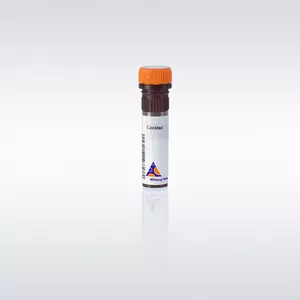
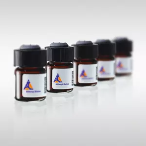
The Direct Lineage Cell Depletion Kit and the indirect Lineage Cell Depletion Kit have been developed for the efficient depletion of mature hematopoietic cells, including T cells, B cells, monocytes/macrophages, granulocytes, erythrocytes and their committed precursors. Both kits are optimized for untouched isolation of lineage-negative cells from mouse bone marrow samples. Using the direct isolation method, depletion can be automated and completed in 20 minutes.
For depletion, cells are magnetically labeled with an antibody cocktail against so-called lineage markers (CD5, CD45R (B220), CD11b, Gr-1 (Ly-6G/C), 7-4, and Ter-119). In the case of the Lineage Cell Depletion Kit, mouse, the labeling is performed in a two-step procedure. Cells are first labeled with a cocktail of biotinylated antibodies followed by labeling with Anti-Biotin MicroBeads.
In the case of the Direct Lineage Cell Depletion Kit the antibodies are directly coupled to MicroBeads allowing a time-saving one-step labeling and isolation of lineage-negative cells in only 20 minutes.
The Lineage Cell Depletion Kit has been used for the enrichment of both, hematopoietic stem cells (HSCs)4 and marrow stromal cells (MSCs) from bone marrow5. The labeling procedure leaves lineage-negative cells untouched, allowing further magnetic separation of lineage-negative cells according to expression of markers such as CD1171,2 or Sca-13.
LS Columns, autoMACS® Columns, or Multi-24 Column Blocks.
Isolation of lineage marker-negative cells from mouse bone marrow using the Direct Lineage Cell Depletion Kit, a MidiMACS™ Separator and an LS Column. Cells were fluorescently labeled with the Lineage Cell Detection Cocktail-Biotin and Anti-Biotin-APC and analyzed by flow cytometry using the MACSQuant® Analyzer. In order to evaluate the LSK fraction, cells were additionally labeled with CD117-PE and Anti-SCA-1-FITC. Cell debris and dead cells were excluded from the analysis based on scatter signals and propidium iodide fluorescence.
- Orlic, D. (2002) Stem cell repair in ischemic heart disease: an experimental model. Int. J. Hematol. 76 (suppl.1): 144-145
- Forte, G. et al. (2006) Hepatocyte growth factor effects on mesenchymal stem cells: proliferation, migration, and differentiation. Stem Cells 24: 23-33
- Lagasse, E. et al. (2000) Purified hematopoietic stem cells can differentiate into hepatocytes in vivo. Nat Med 6: 1229-1234
- Tang, H. et al. (2006) Endothelial stroma programs hematopoietic stem cells to differentiate into regulatory dendritic cells through IL-10. Blood 108: 1189-1197
- Orlic, D. et al. (2001) Mobilized bone marrow cells repair the infarcted heart, improving function and survival. Proc. Natl. Acad. Sci. U.S.A. 98: 10344-10349