Pre-enrichment of mouse hematopoietic stem and progenitor cells
This application protocol describes the pre-enrichment of lineage marker-negative, bone marrow-derived cells and their phenotypic analysis using flow cytometry.
Protocol
Pre-enrichment of mouse hematopoietic stem and progenitor cells
1.5 h
Isolation of mononuclear cells
1.5 h
Depletion of lineage-positive cells
3 d
Expansion of lineage-negative cells or LSK cells
45 min
Flow cytometric analysis or further enrichment of lineage-negative cells
Protocol

This protocol includes detailed instructions for preparing necessary reagents and materials and then carrying out each step.
PEB buffer: Prepare a solution containing phosphate-buffered saline (PBS), pH 7.2, 0.5 % bovine serum albumin (BSA), and 2 mM EDTA by diluting MACS® BSA Stock Solution 1:20 with autoMACS® Rinsing Solution. Keep buffer cold (2−8 °C). Degas buffer before use.
Isolate mononuclear cells from bone marrow extracted from mouse femoral bones.
▲Note: All steps should be performed on ice-cold PBE buffer (see "Things to prepare in advance").
- Extract mouse limbs and place in cold PBE buffer.
- Use sterile scissors and forceps to isolate femoral bones. Place them in a 6 cm cell culture dish with 2–3 mL ice-cold PBE buffer.
- Isolate bone marrow cells either by grinding isolated femoral bones in a mortar or by flushing the cells out of the bone with a syringe.
- Pass cells through a 30 µm MACS® SmartStrainer into a 50 mL centrifuge tube to remove cell clumps that can clog the column.
▲Note: Wet the filter with PBE buffer before adding the cells. - Adjust the final volume of cell suspension to 15–30 mL, depending on estimated cell number.
- Take 10 µL of cell suspension to determine the cell number using a Neubauer chamber or another cell counting device.
- Store a 200 µL aliquot of the cell suspension on ice for later flow cytometric analysis.
- Centrifuge the 50 mL centrifuge tube containing the isolated bone marrow cells at 400×g for 10 minutes.
Deplete cells expressing lineage markers from total bone marrow mononuclear cells using either the Direct Lineage Cell Depletion Kit, mouse or the Lineage Cell Depletion Kit, mouse. The first generates a high cell yield. The latter enables a more stringent depletion. Follow the protocol of the corresponding data sheet.
▲ Note: Lineage depleted cells can be further enriched, e.g., to obtain LSK cells, using fluorescence-activated cell sorting.
▲ Note: It is recommended to filter the magnetically labeled cells before separation to guarantee a single-cell suspension either manually or via the automated protocol using the autoMACS® Pro Cell Separator.
Download data sheet
Direct Lineage Cell Depletion Kit, mouse
 View details
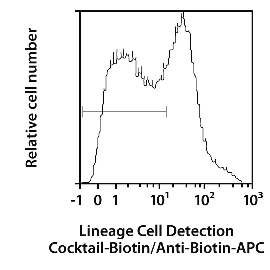
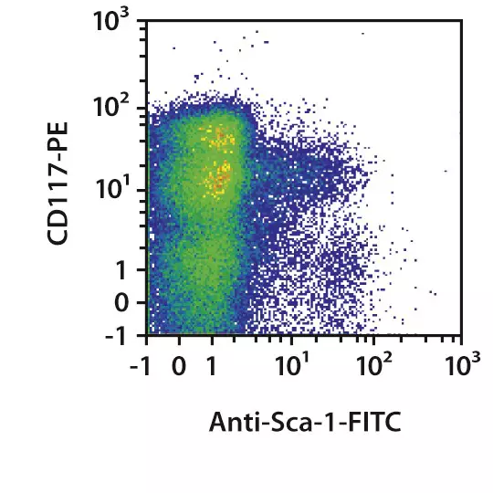
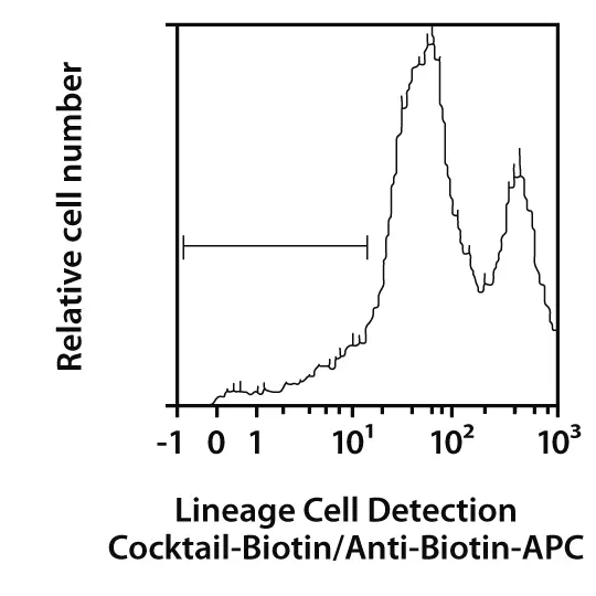
Figure 1: Isolation of untouched lineage-negative cells from a mouse bone marrow cell suspension. After isolation with the DIrect Lineage Cell Depletion Kit, mouse, lineage-negative cells were fluorescently stained with the Hematopoietic Lineage Labeling Cocktail, anti-mouse, Biotin and Biotin Antibody-APC and analyzed by flow cytometry using the MACSQuant® Analyzer. To evaluate the LSK (Lin–Sca-1+c-kit+) fraction, cells were further stained with CD117-PE (c-kit) and Sca-1 Antibody-FITC. Cell debris and dead cells were excluded from the analysis based on scatter signals and propidium iodide fluorescence.
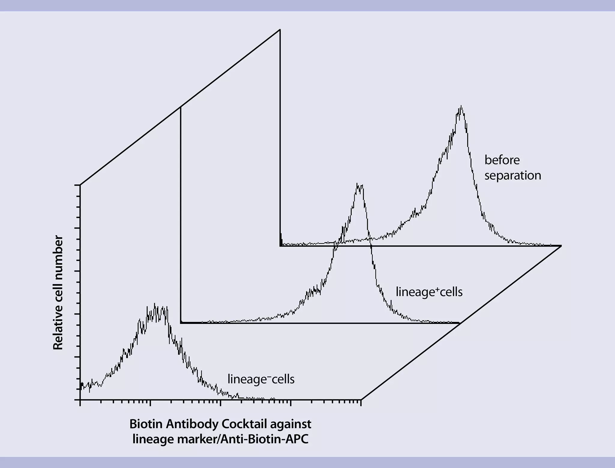
 View details
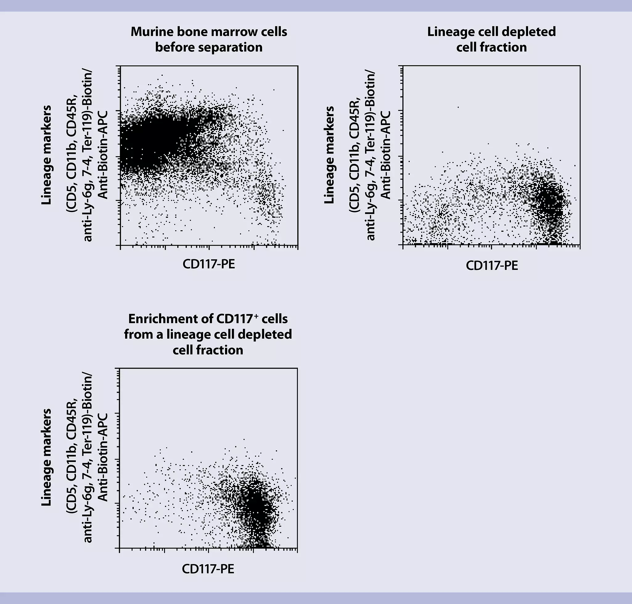
Lineage-negative cells can be expanded in appropriate mouse HSC expansion medium supplemented with cytokines such as SCF, TPO, Flt3-Ligand, IL-3, and IL-6 according to standard cell culture protocols.
Preparation of staining cocktail
Lineage-negative cells can be further analyzed or sorted for HSC-specific surface markers by flow cytometry using MACS® Antibodies or recombinantly engineered REAfinity™ antibodies.
If cell separation was performed with the Direct Lineage Cell Depletion Kit, mouse, use Labeling Check Reagent, APC for the flow cytometric analysis. For separations performed with the Lineage Cell Depletion Kit, mouse, use Streptavidin, APC.
Prepare the staining cocktail as described in the following table:
| Reagent | Original cell fraction | Lineage marker-positive cell fraction | Lineage marker-negative(depleted) cell fraction |
| Sca-1 Antibody-FITC | 10 µL | 10 µL | 10 µL |
| CD117-PE | 10 µL | 10 µL | 10 µL |
| Labeling Check Reagent, APC or Streptavidin, APC | 10 µL | 10 µL | 10 µL |
| FcR Blocking Reagent | 20 µL | 20 µL | 20 µL |
| PEB buffer | 50 µL | 50 µL | 50 µL |
Use the MACS Flow Cytometry – Multicolor Panel Builder to quickly assemble the antibodies for analysis.
Staining procedure
- Centrifuge aliquots of the different cell fractions (original, positive, and negative fractions) at 300×g for 10 minutes.
- Discard supernatant.
- Resuspend each cell fraction in 100 µL of staining cocktail. Carefully pipet the suspension up and down.
- Incubate the resuspended cells at 4 °C for 10 minutes.
- Add 100 µL PBE buffer to each cell fraction and mix thoroughly by pipetting up and down.
- Centrifuge cells at 300×g for 10 minutes.
- Resuspend cell fractions in 200 µL PBE buffer for flow cytometric analysis.
The following is a list of relevant resources that might be of interest: