Neuroscience
Immuno-oncology
Organoid research
Other fields of research
Get expert consultation
Content
- Neuroscience
- Immuno-oncology
- Organoid research
- Other fields of research
- Get expert consultation
See how scientists like you achieve research excellence with the UltraMicroscope Platform.
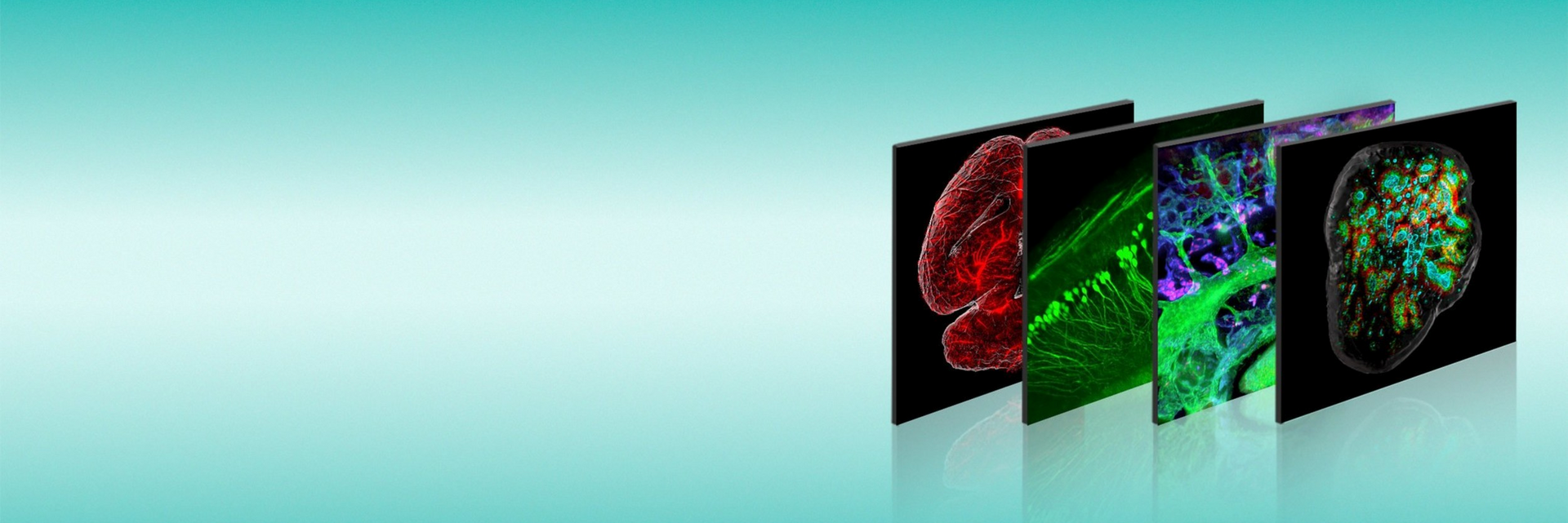

Understanding the inner workings of biological systems requires more than molecular detail – it calls for a true 3D perspective. That’s where the UltraMicroscope Platform comes in, enabling large-scale 3D imaging with cellular resolution. With optimized antibodies, reagents, and tissue-clearing solutions, the platform supports reliable sample preparation and helps you generate high-quality, reproducible 3D imaging results. From entire organs to individual cells, it delivers stunning clarity and the spatial context needed to truly understand intricate biology. Until today, the UltraMicroscope has been featured in over a thousand publications. Here, we highlight a few of the most recent examples.
Discover how the UltraMicroscope Platform is transforming research in neuroscience, immuno-oncology, and beyond – unveiling sophisticated biological processes and unlocking insights once out of reach.
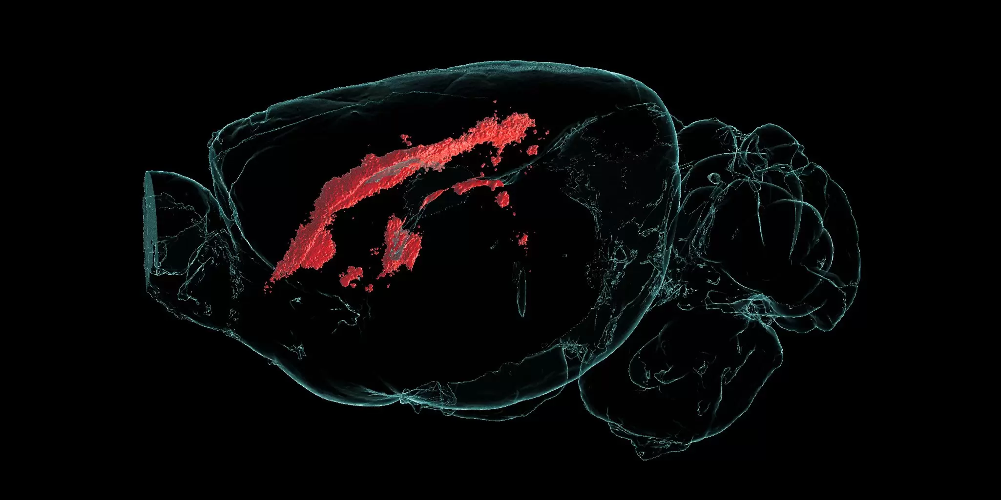
In this study, researchers introduce an integrated imaging workflow for the deep spatial characterization of a somatic mouse glioblastoma model. By combining optical tissue clearing, 3D light sheet microscopy, and targeted 2D multiplex imaging, the workflow enables precise identification and molecular profiling of tumor regions within intact brains. This multimodal approach provides a powerful tool for studying glioblastoma biology and the tumor microenvironment in unprecedented spatial detail.
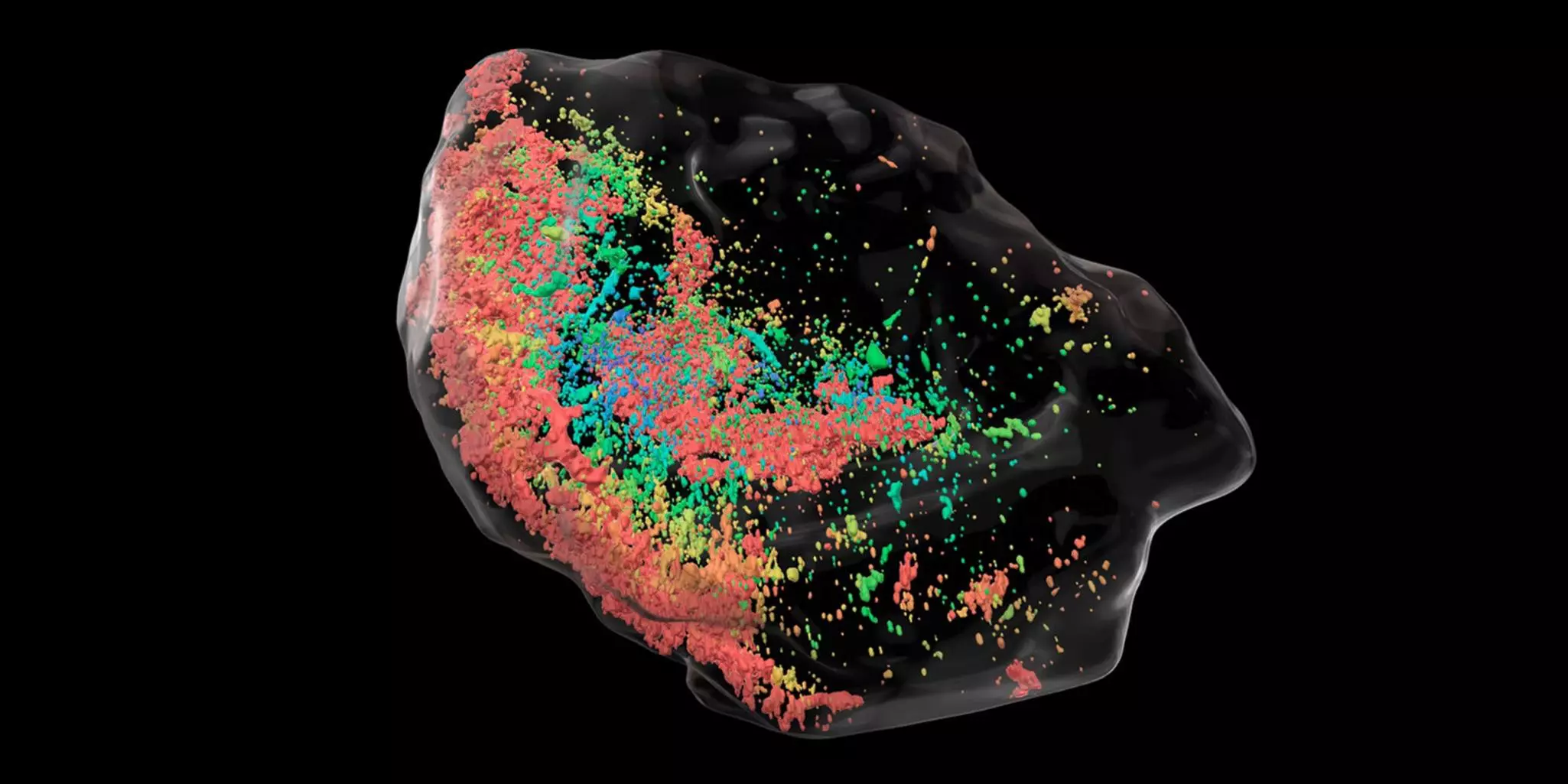
In this study, researchers combined single-cell RNA sequencing, spatial transcriptomics, AI-assisted 3D imaging, and functional studies to reveal a developmental program that drives breast cancer metastasis. They identified metastatic trabecular morphogenesis (MTM), a process that enables metastatic tumors to form complex 3D structures. By uncovering key molecular regulators and therapeutic targets, the findings open new possibilities to specifically inhibit metastatic outgrowth and support the development of treatment strategies for advanced breast cancer.
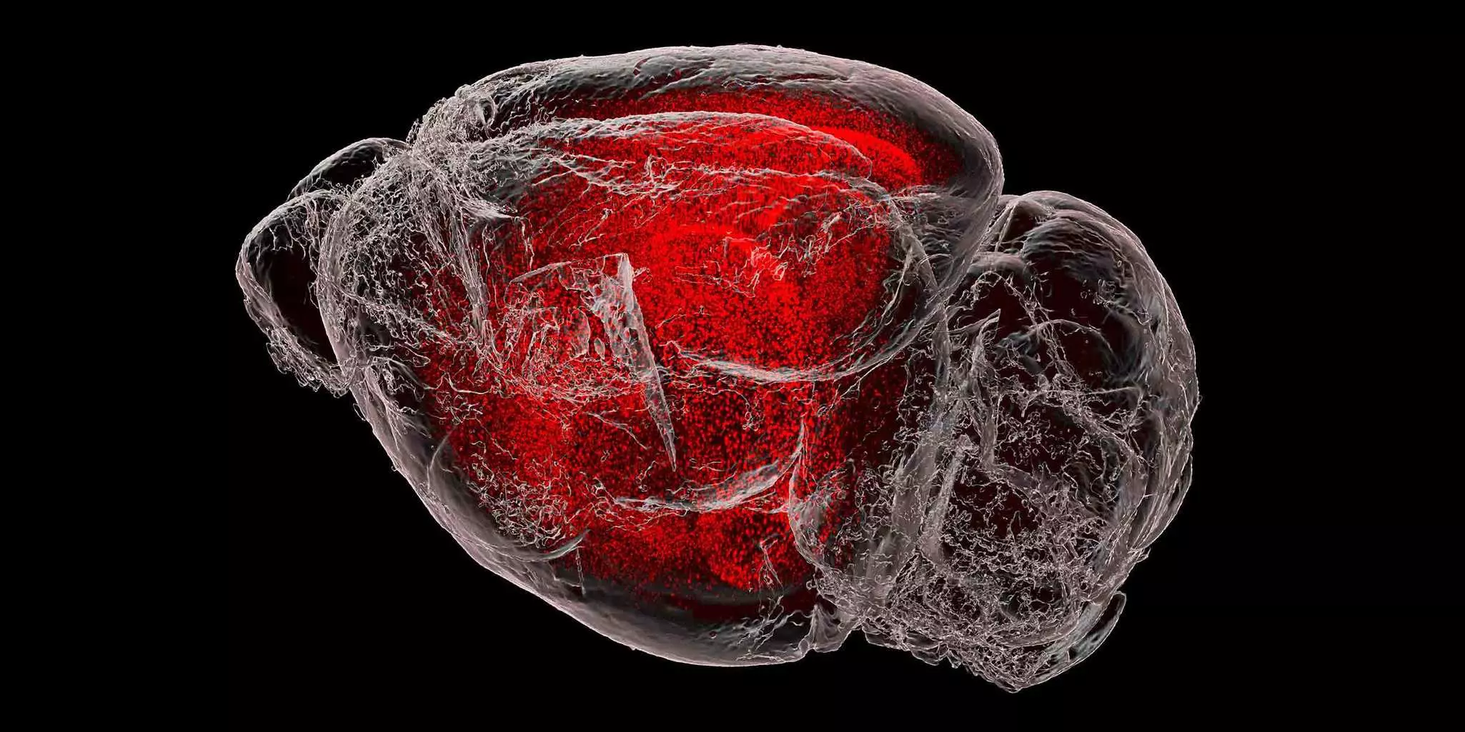
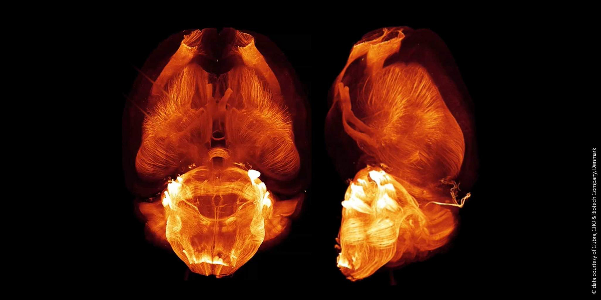
To map mRNA distribution across the entire mouse brain at cellular resolution, Kanatani and his team developed TRISCO, a tissue-clearing method compatible with light sheet microscopy. This approach enabled 3D in situ hybridization in intact brain tissue, revealing gene expression patterns in remarkable detail. Applying the method, the researchers tracked immediate early gene activity after treatment with the anti-obesity drug semaglutide, detecting increased signals in regions linked to obesity. The combination of TRISCO and light sheet microscopy offers a powerful tool for whole-brain transcriptomic analysis at single-cell resolution.
Whole brain image courtesy of Yue Li and Shigeaki Kanatani, Karolinska Institute.
Neuroscience
Immuno-oncology
Organoid research
Other fields of research
Get expert consultation
The video showcases eight whole mouse brains acquired with the UltraMicroscope Blaze and using a fully loaded MACS UltraMount 8 Sample Holder. The samples were stained and cleared with the MACS Deep Clearing Kit. Various conjugated antibodies in different colors (Vio dyes G570, R667, and 780) were employed to stain antigen targets: Calretinin, NeuN, CD31, Neurofilament, Tyrosine Hydroxylase, MBP, and Parvalbumin Antibodies.
Validated 3D-IF antibodies for neuroscience discovery
Finding antibodies that are compatible with whole-brain clearing can be challenging. Explore our selection of fluorochrome-conjugated antibodies validated for cleared tissues, helping you streamline sample preparation and generate reliable 3D imaging results.
| Antibody | Target | Clone | Reactivity |
| β-Tubulin 3 | Developing neurons /neurons | REA1152 | Human/mouse |
| NeuN | Neurons | REA1131 | Human/mouse/rat |
| Neurofilament | Neuronal axons | REA1127 | Human/mouse |
| Tyrosine Hydroxylase | Dopaminergic neurons | REA1159 | Human/mouse/rat |
| Iba1 | Microglia | REA1448 | Human/mouse/rat |
| GFAP | Astrocytes | REA335 | Human/mouse/rat |
| GLAST (ACSA-1) | Astrocytes | ACSA-1 | Human/mouse/rat |
| MBP | Oligodendrocyte- myelin sheet | REA1154 | Human/mouse |
| Calretinin | GABAergic interneurons | REA1157 | Mouse/rat |
| GFP | Expressed GFP | GG4-6F6 | Mouse |
The MACS Deep Clearing Kit and dedicated protocol provide a fast, efficient approach for mouse brain preparation and clearing.
Organic solvent–based tissue clearing at its best.
The MACS Deep Clearing Kit offers an optimized solution for immunolabeling and clearing whole mouse brains.
A human ovarian cancer sample was labeled with Miltenyi Biotec recombinant CD326 (EpCAM) Antibody, Vio R667, REA764 (red) and propidium iodide (cyan). The sample was processed and optically cleared using the MACS Clearing Kit and imaged with an UltraMicroscope Blaze Light Sheet Instrument. Data was processed as a 3D-rendered maximum intensity projection (autofluorescence in grey).
This video shows a solid tumor sample derived from a pancreatic xenograft model that was used to test the efficacy of CAR T cell therapies. For visualization and analysis, the following channels were used: tumor surface (autofluorescence), tumor vasculature (rhodamine lectin), and CAR T cells (CD3-Vio667). The sample was cleared with the MACS Clearing Kit and imaged with the UltraMicroscope Blaze in MACS Imaging Solution. For more information, please refer to Pfeifer et al. (2022): A multimodal imaging workflow for monitoring CAR T cell therapy against solid tumor from whole-body to single-cell level." Theranostics 12.11: 4834-4850.
Validated 3D-IF antibodies for immune-tumor profiling
To truly understand immune-tumor interactions, you need to preserve spatial context while enabling deep antibody penetration into complex tissues. Together, tissue clearing and antibody selection are essential for generating meaningful 3D datasets.
Our workflow brings together the MACS Deep Clearing Kit and fluorophore-conjugated antibodies commonly used for immune and tumor profiling in cleared tissues.
| Antibody name | Specific target | Clone name | Reactivity |
| CD274 (PD-L1) Antibody | Immune checkpoint | REA1197 | Human |
| CD279 (PD1) Antibody | Immune checkpoint | REA1165 | Human |
| CD3 Antibody | T cells | REA613 | Human |
| CD8 Antibody | Cytotoxic T cells | REA734 | Human |
| CD4 Antibody | Helper T cells | REA623 | Human |
| CD45 Antibody | Immune cells | REA747 | Human |
| CD68 Antibody | Macrophages | REA835 | Mouse |
| Ki-67 Antibody | Cell proliferation | REA183 | Human / Mouse |
| Pan-Cytokeratin Antibody | Tumor cells | REA1141 | Human |
| CD326 (EpCAM) Antibody | Tumor cells | REA764 | Human |
The MACS Deep Clearing Kit and dedicated protocol provide a fast, efficient approach for primary human tumor preparation and clearing.
3D image of a cerebral brain organoid derived from pluripotent stem cells (PSCs), captured using the UltraMicroscope Blaze Light Sheet System. N-Cadherin (cyan) highlights the intricate connections between cells, Sox2 (red) pinpoints neural stem cells, and Ki67 (green) identifies actively dividing cells. The surface was generated using a β-Tubulin 3 staining.
Validated 3D-IF antibodies for organoid imaging
Visualizing cellular organization within organoids requires robust antibody staining across the entire sample volume. Explore our selection of primary conjugated 3D-IF antibodies validated for cleared tissues.
| Antibody name | Specific target | Clone name | Reactivity |
| Ki-67 Antibody | Cell proliferation | REA183 | Human/mouse |
| β-Tubulin 3 | Developing neurons /neurons | REA1152 | Human/mouse |
| OTX2 | Development studies | REA1178 | Human |
| PAX-6 | Neural progenitor cells | REA507 | Human |
| Sox2 | Neural stem cell | REA320 | Human/mouse |
| GLAST (ACSA-1) | Astrocytes | ACSA-1 | Human/mouse/rat |
The MACS Deep Clearing Kit and dedicated protocol provide a fast, efficient approach for organoid sample preparation and clearing.
Our support team is happy to provide you with additional information, technical specifications, instrument demonstrations, or any other type of assistance you may need. Simply leave your contact details and we will get in touch with you.