Human CD4+ T helper cells


1 Introduction 2 CD4+ T helper cells in peripheral blood 2.1 Miltenyi Biotec application protocols for CD4+ T helper cells from peripheral blood 2.2 Sample preparation of peripheral blood 2.3 Magnetic separation of CD4+ T cells from peripheral blood and blood products 2.4 Characterization of CD4+ T helper cells by flow cytometry 2.5 Cell culture of CD4+ T helper cells
3 CD4+ T helper cells from other tissues
1 Introduction
T helper cells (TH cells) play a pivotal role in adaptive immunity. Following antigen recognition, naive TH cells are activated, undergo further differentiation, and secrete cytokines to promote ('help') and steer the immune response. All TH cells are positive for CD4, a coreceptor that complements the T cell receptor (TCR) to communicate with an antigen-presenting cell (APC).
2 CD4+ T helper cells in peripheral blood
In human peripheral blood, 15–30% of all CD45+ leukocytes are T cells, with CD4+ T cells accounting for approximately two thirds of the total T cell population, and CD8+ T cells making up the remaining one third. In isolated PBMCs, T cells amount to 45–70% of the total cells and are therefore by far the most abundant cell type. Up to 60% of total PBMCs are CD4+ T cells, and up to 30% are CD8+ T cells.
Cell subsets, frequencies, and marker expression
At a glance: CD4⁺ TH cell subsets
| Subset | Secreted cytokines | Function | |||
| TH1 | IFN-γ, IL-2, TNF-α | • Promote cellular immune response • Activate macrophages • Promote proliferation of cytotoxic T cells | |||
| TH2 | IL-4, IL-5, IL-6, IL-13 | • Promote the humoral immune response • Activate B cells for expansion and antibody production | |||
| TH17 | IL-17A, IL-17F, IL-21, IL-22, IL-26 | • Maintain mucosal barriers • Support pathogen clearance at mucosal surfaces | |||
| TH9 | IL-9 in high amounts, IL-10 | • Important in various inflammatory scenarios: asthma, ulcerative colitis, helminth infections | |||
| TH22 | IL-13, IL-22, TNF-α | • Recruited mainly to the skin, contributing to host defense against microbial pathogens • Influence epithelial and stromal cells • Promote tissue repair and remodeling | |||
| Tfh | IL-6, IL-10, IL-12, IL-21 | • Essential for germinal center formation, affinity maturation, and development of most high-affinity antibodies and memory B cells | |||
Depending on the cytokine environment, naive CD4+ TH cells might differentiate into several subsets, including TH1, TH2, TH17, TH9, TH22 cells, and T follicular helper cells (Tfh). Naive CD4+ T cells can also differentiate into induced regulatory T cells (iTreg; PMID: 26688349). See chapter Regulatory T cells.
Most T cell subtypes can undergo memory differentiation steps after activation by their respective antigen. Apart from differentiating into effector T cells, some naive T cells (TNAIVE) may differentiate into various memory T cell subsets, such as stem cell-like memory T cells (TSCM), central memory T cells (TCM), effector memory T cells (TEM) and effector memory RA+ T cells (TEMRA). Each differentiated subset is defined by distinct surface markers. Antigen-inexperienced T cells express naive marker CD45RA, as well as homing receptors CD62L and CCR7, but lack CD45RO and CD95 expression. With ongoing differentiation towards memory phenotypes, CD45RA, CD62L, and CCR7 are down-regulated, while memory marker CD45RO and activation marker CD95 are gradually up-regulated. With progressive differentiation towards the memory phenotype, antigen-dependency, tissue tropism, effector function, and senescence increase (PMID: 24258910, 26999211).
Shared features of all memory T cell subtypes are that they are long-lived and can quickly expand to large numbers of effector T cells upon re-exposure to their cognate antigen, thereby mounting a faster and more potent immune response than the first immune response to a given pathogen. The different subtypes exert different functions and exhibit different properties, such as tissue tropism or capacity for self-renewal, reflecting the specific immune-related circumstances that led to their differentiation into a given memory subtype.

 View details
Typically, the frequency of naive T cells specific for a given antigen is very low, ranging between 0.01 and 0.001% of the total T cell count, depending on the respective specificity. When a naive T cell encounters its cognate antigen and is consequently activated, clonal expansion begins, boosting the frequency of those antigen-specific T cells by several orders of magnitude. This way, they can efficiently fulfill their role as effectors in the immune response (PMID: 22517866, 17707129).
Most clonally expanded antigen-specific T cells die after the termination of the immune response, but a small percentage survive as memory T cells. Memory T cells have a long lifespan and can quickly expand to large numbers of effector T cells upon re-exposure to their cognate antigen. At birth, the T cell repertoire is almost exclusively composed of naive T cells. With progressing age and antigen-experience, memory T cells may become the most abundant T cell population, constituting up to 35% of all circulating T cells (PMID: 24336101).
Notably, laboratory mice carry almost exclusively naive T cells due to their specific keeping conditions and relatively young average age. This, of course, changes dramatically in certain disease-related experimental settings. In humans, the frequency of naive and memory T cells greatly depends on age, living conditions, and individual history of immune responses.
 Related PDFs:
2.1 Miltenyi Biotec application protocols for CD4+ T helper cells from peripheral blood
Miltenyi Biotec has created dedicated application protocols for the isolation of different CD4+ T cell subsets.
2.2 Sample preparation of peripheral blood
Miltenyi Biotec offers various kits for the direct isolation of CD4+ T cells from peripheral blood and blood products. No specific sample preparation is needed when using those kits. CD4+ T cells can also be isolated from PBMCs, which can be generated either by density gradient centrifugation or using the MACSprep™ PBMC Isolation Kit, human. For details, see the MACS Handbook chapter Human blood.
 MACS Handbook:
2.3 Magnetic separation of CD4+ T cells from peripheral blood and blood products
Miltenyi Biotec has developed numerous products for the straightforward magnetic separation of CD4+ T cells and corresponding subsets. T cells can be isolated either straight from whole blood, buffy coat, leukoreduction system chambers (LRSC), or Leukopak® without density gradient centrifugation and erythrocyte lysis, or from PBMCs.
For details on MACS® Cell Separation Technology, see the MACS Handbook chapter Magnetic cell separation.
MACS Handbook:
2.3.1 Isolation of CD4+ T cells from whole blood, buffy coat, LRSC, and Leukopak®
| Starting material | Isolation strategy | Comments | Automation | Product |
| Pan CD4+ T cells | ||||
| Whole Blood | Positive selection of target cells | Suitable for smaller sample volumes (total capacity: 40 mL whole blood). | Yes* | StraightFrom® Whole Blood CD4 MicroBeads, human |
| Buffy coat | Positive selection of target cells | Allows direct processing of an entire buffy coat (up to 80 mL) without density centrifugation and includes the needed columns. | Yes** | StraightFrom Buffy Coat CD4 MicroBead Kit, human |
| LRSC | Positive selection of target cells | Allows direct processing of an entire LRSC (up to 40 mL) without density centrifugation and includes the needed columns. | Yes** | StraightFrom LRSC CD4 MicroBead Kit, human |
| Lenkopak® | Positive selection of target cells | Allows direct processing of a ½ LeukoPak® without density centrifugation and includes the needed columns. | Yes** | StraightFrom® Leukopak® CD4 MicroBead Kit, human |
| Whole blood | Depletion of non-target cells | Isolation of CD4+ T cells directly from whole blood in less than 30 minutes (total capacity: 3x30 mL whole blood). | No | MACSxpress® Whole Blood CD4 T Cell Isolation Kit, human |
| Buffy coat | Depletion of non-target cells | Isolation of CD4+ T cells directly from an entire buffy coat in less than 30 minutes. | No | MACSxpress Buffy Coat CD4 T Cell Isolation Kit, human |
| LRSC | Depletion of non-target cells | Isolation of CD4+ T cells directly from LRSC (up to 40 mL) in less than 30 minutes. | No | MACSxpress LRSC CD4 T Cell Isolation Kit, human |
| Naive/memory subsets | ||||
| Whole blood | Depletion of non-target cells | Isolation of CD4+CD45RO+ cells directly from whole blood in less than 30 minutes (total capacity: 3x30 mL whole blood). | No | MACSxpress Whole Blood CD4 Memory T Cell Isolation Kit, human |
| *Automation options range from fully automated benchtop solutions such as the autoMACS® NEO Separator to high-throughput platforms such as the MultiMACS™ Cell24 Separator Plus or MultiMACS X. **Semi- or fully automated high-throughput cell separation with the MultiMACS™ Cell24 Separator Plus or MultiMACS X. | ||||
The StraightFrom® MicroBead Kits were developed for the rapid positive selection of target cells directly from whole blood, buffy coat, LRSC, or Leukopak®. None of the kits require any sample preparation, such as density centrifugation, erythrocyte lysis, or cell count. The appropriate kit is chosen based on the starting material.
With the StraightFrom Buffy Coat CD4 MicroBead Kit, human, CD4+ cells are separated from an entire buffy coat in less than 30 minutes. The kit can be used in combination with a QuadroMACS™ Separator for manual separation, but is best combined with the MultiMACS™ Cell24 Separator Plus.
StraightFrom Buffy Coat CD4 MicroBead Kit, human

 View details
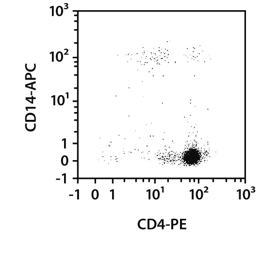
Isolation of CD4+ T cells directly from buffy coat. Separation of a buffy coat sample using the StraightFrom Buffy Coat CD4 MicroBead Kit and the MultiMACS™ Cell24 Separator Plus with the Single-Column Adapter and Whole Blood Columns. Cells were fluorescently stained with CD4-PE, CD14-APC, as well as CD45-VioBlue® and analyzed by flow cytometry using the MACSQuant® Analyzer. Cells were triggered via CD45-VioBlue. Cell debris and dead cells were excluded from the analysis based on scatter signals and propidium iodide fluorescence.
Alternatively, the MACSxpress® Cell Isolation Kits allow the column-free isolation of target cells from freshly drawn anticoagulated whole blood, buffy coat, or LRSC by depletion of non-target cells. MACSxpress Whole Blood Kits are ideal for the processing of larger sample volumes (total capacity 3×30 mL) and are available for the isolation of CD4+ cells and CD4+CD45RO+ memory T cells. Specialized MACSxpress Kits enable the isolation of CD4+ T cells from an entire buffy coat or LRSC.
The MACSxpress Whole Blood CD4 T Cell Isolation Kit was developed for the fast and easy isolation of highly pure CD4+ T cells directly from whole blood. Non-target cells are removed by immunomagnetic depletion using MACSxpress Beads. Simultaneously, erythrocytes are sedimented, yielding target cells of high purity.
2.3.2 Isolation of CD4+ T cells from PBMCs
| Starting material | Isolation strategy | Comments | Automation | Product | |||
| PBMCs | Positive selection of target cells | CD4 is predominantly expressed on TH cells. Suitable for the depletion or enrichment of CD4+ cells form a PBMC sample. | Yes* | CD4 MicroBeads, human | |||
| PBMCs | Positive selection of target cells and subsequent label removal | The kit allows the isolation of label-free CD4+ cells, because the complete labeling complex can be released from the cell surface after separation. | No | REAlease CD4 MicroBead Kit, human | |||
| PBMCs | Depletion of non-target cells | Isolation of CD4+ TH cells via depletion of CD8+ T cells, monocytes, neutrophils, eosinophils, B cells, dendritic cells, NK cells, granulocytes, TCRγ/δ+ T cells, and erythroid cells | Yes* | CD4+ T Cell Isolation Kit, human | |||
| *Automation options range from fully automated benchtop solutions such as the autoMACS® NEO Separator to high-throughput platforms such as the MultiMACS™ Cell24 Separator Plus or MultiMACS X. | |||||||
Instead of working directly with whole blood or blood products, samples can be processed over a density gradient centrifugation to pre-enrich peripheral blood mononuclear cells (PBMCs) as starting material for subsequent TH cell isolation.
CD4 MicroBeads, human enable positive selection or depletion of CD4+ cells by direct magnetic labeling.
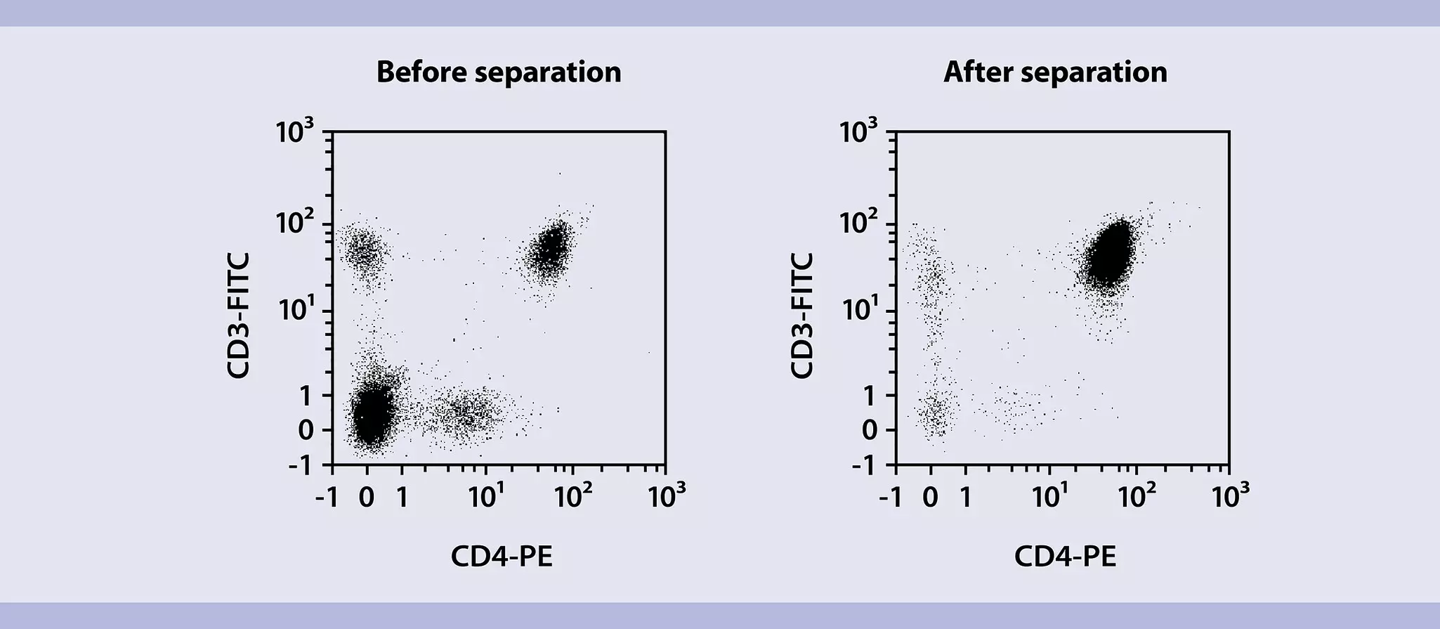
The CD4+ T Cell Isolation Kit, human enables fast isolation of untouched cytotoxic CD4+ T cells from PBMCs in only 18 minutes. This updated kit offers even better performance and a significantly shorter protocol, replacing its well-known predecessor.
CD4+ T Cell Isolation Kit, human
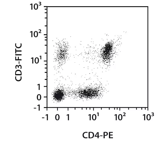
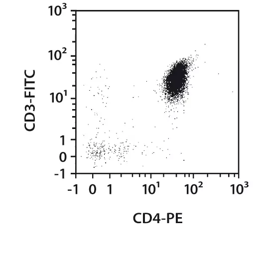
Isolation of untouched CD4+ T cells from PBMCs. Samples were processed using the CD4+ T Cell Isolation Kit, LS Column, and a MidiMACS™ Separator. Cells were labeled with CD4-PE and CD3-FITC and analyzed by flow cytometry using the MACSQuant Analyzer.
The REAlease® CD4 MicroBead Kit, human offers a rapid solution for the positive selection of CD4+ T cells from PBMCs with the option to fully remove the MicroBead and antibody labeling. Cells are thus completely label-free and ready for any downstream application, including a second round of magnetic labeling and separation for further subset isolation.
REAlease® CD4 MicroBead Kit, human
a) Cell purity
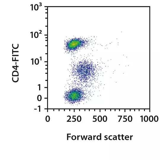
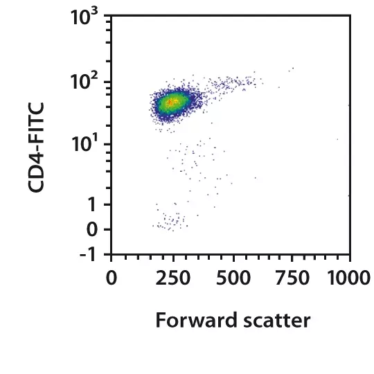
Label-free, highly pure CD4+ T cells.
CD4+ T cells were isolated from human PBMCs using the REAlease CD4 MicroBead Kit, MS Columns, and a MiniMACS Separator. Cells were fluorescently stained with CD4-FITC and analyzed by flow cytometry using the MACSQuant Analyzer X. Cell debris and dead cells were excluded from the analysis based on scatter signals and propidium iodide fluorescence.
b) Label-free cells: REAlease Biotin Complex release
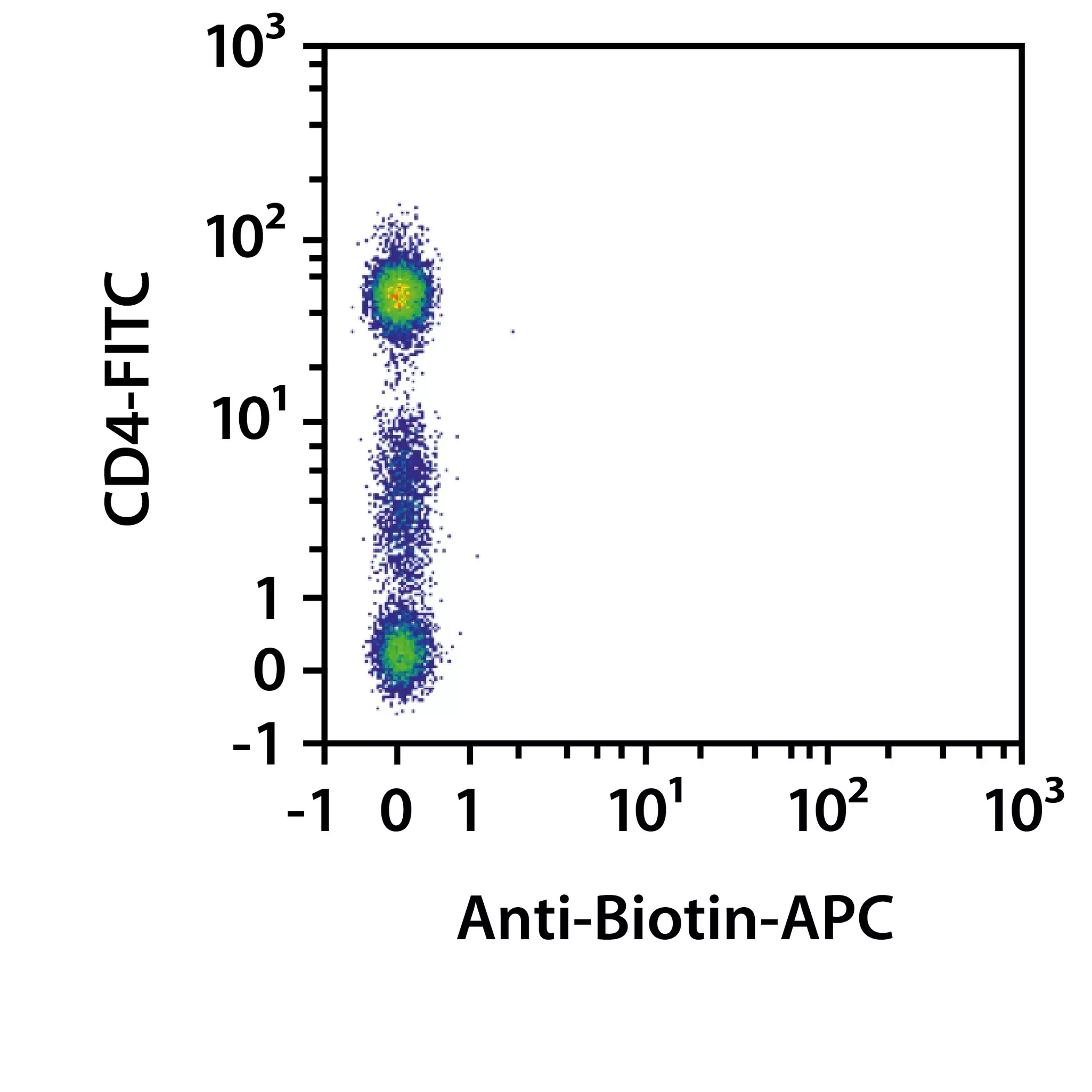
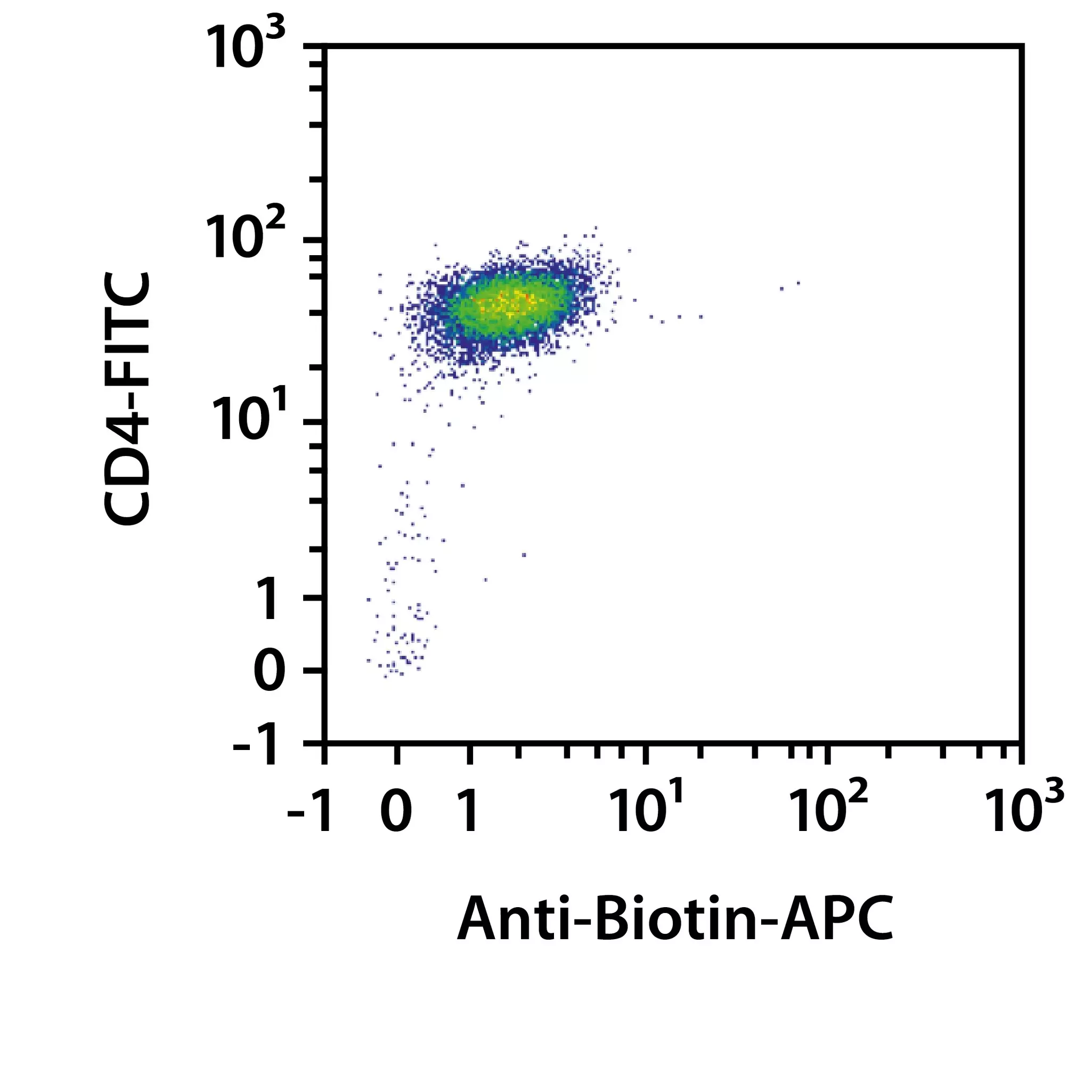
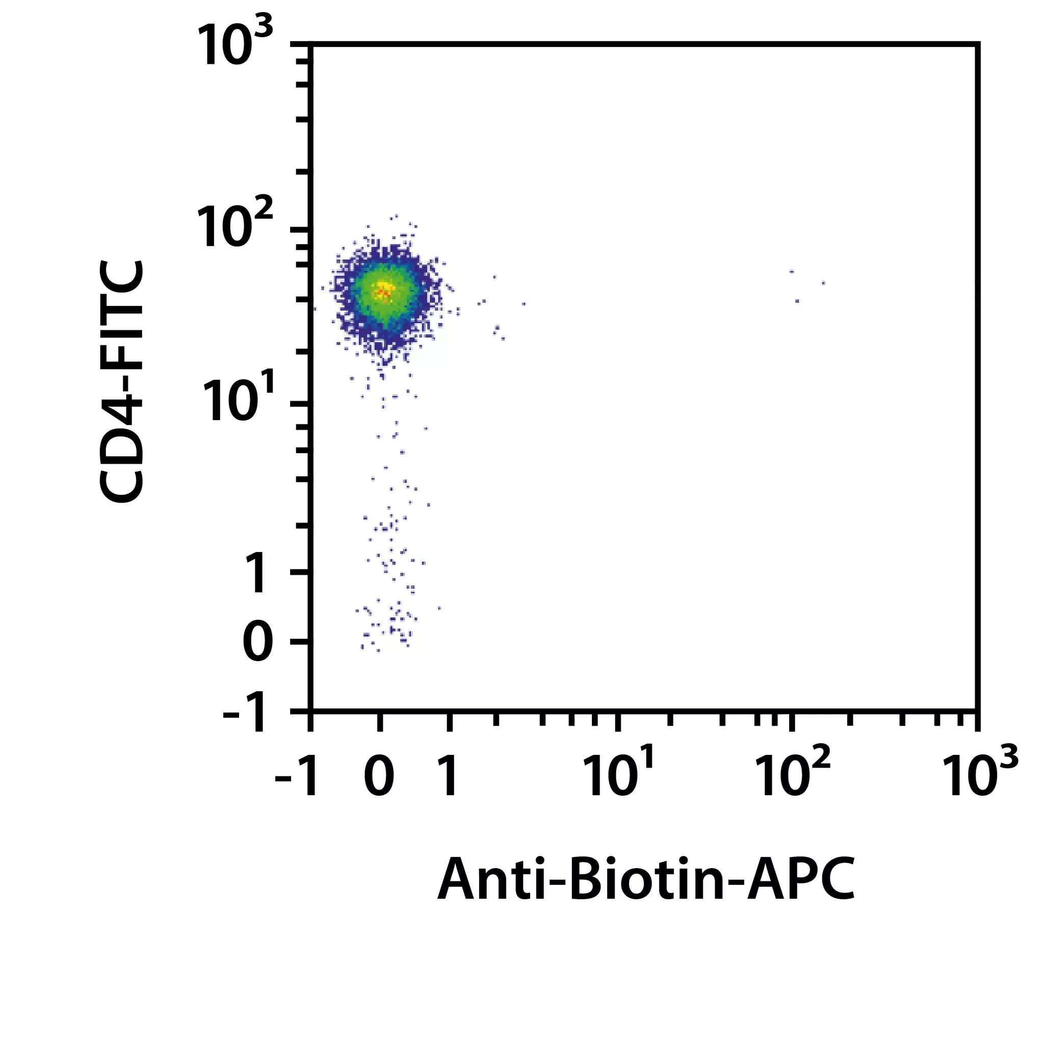
Efficient removal of all labels was shown by using Anti-Biotin-APC to analyze the cells by flow cytometry for the presence of REAlease Biotin Complex. Directly after isolation, the cells showed biotin staining (MicroBead-free CD4+ cells), whereas the label-free CD4+ cells after release of REAlease Biotin Complex were negative for biotin similar to the non-labeled cells before separation. Release efficiency was higher than 99% for the REAlease Anti-Biotin MicroBeads (CD4).
2.3.3 Isolation of naive/memory TH cell subsets from PBMCs
At a glance: Kits and reagents for the separation of naive/memory CD4⁺ TH cell subsets from PBMCs
| Starting material | Isolation strategy | Comments | Automation | Product | ||
| PBMCs | Depletion of non-target cells | Isolation of all naive CD4+ T cells by depletion of CD45RO+ and non-T cells. | Yes* | Naive CD4⁺ T cell Isolation Kit II, human | ||
| PBMCs | Depletion of non-target cells | Isolation of all memory CD4+ cells by depletion of CD45RA+ and non-T helper cells. | Yes* | Memory CD4⁺ T cell Isolation Kit, human | ||
| PBMCs | Depletion of non-target cells | Isolation of all CD4+ effector memory T cells by depletion of non-T helper, CD45RA+, and CCR7+ cells. | Yes* | CD4⁺ Effector Memory T cell Isolation Kit, human | ||
| PBMCs | Sequential separation | Depletion of non-CD4+ cells and naive CD4+ T cells, followed by separation of TCM based on CD197. | Yes* | CD4⁺ Central Memory T cell Isolation Kit, human | ||
| PBMCs | Positive selection of target cells | CD62L (L-Selectin) is a naive/early memory T cell marker. | Yes* | CD62L MicroBeads, human (after pre-selection with CD4⁺ T cell Isolation Kit, human) | ||
| PBMCs | Positive selection of target cells | CD45RA is expressed on naive T cells, but not on memory T cells | Yes* | CD45RA MicroBeads, human (after pre-selection with CD4⁺ T cell Isolation Kit, human) | ||
| PBMCs | Positive selection of target cells | CD45RO is expressed on memory T cells, but not on naive T cells | Yes* | CD45RO MicroBeads, human (after pre-selection with CD4⁺ T cell Isolation Kit, human) | ||
| *Automation options range from fully automated benchtop solutions such as the autoMACS® NEO Separator to high-throughput platforms such as the MultiMACS™ Cell24 Separator Plus or MultiMACS X. | ||||||
The Naive CD4+ T Cell Isolation Kit II, human was developed for the isolation of untouched naive CD4+ T cells from PBMCs. Memory CD4+ T cells and non-CD4+ T cells are labeled with a cocktail of biotinylated CD45RO, CD8, CD14, CD15, CD16, CD19, CD25, CD34, CD36, CD56, CD123, Anti-TCRγ/δ, Anti-HLA-DR, and CD235a (Glycophorin A) antibodies, and then magnetically labeled with Anti-Biotin MicroBeads for subsequent depletion.
Naive CD4+ T Cell Isolation Kit II, human
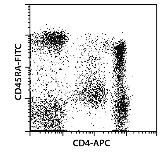
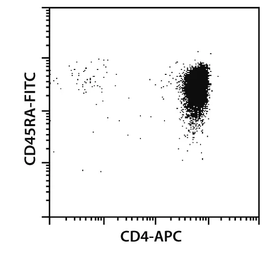
Isolation of untouched naive CD4+ T helper cells from PBMCs. Samples were processed using the Naive CD4+ T Cell Isolation Kit II, an LS Column, and a MidiMACS Separator. Cells were stained with CD4 and CD45RA antibodies.
The Memory CD4+ T Cell Isolation Kit, human is used to isolate untouched memory T helper cells from PBMCs by depletion of naive T cells, CD8+ T cells, B cells, NK cells, TCRγ/δ⁺ T cells, monocytes, DCs, granulocytes, platelets, and erythroid cells. PBMCs are incubated with a cocktail of biotinylated CD45RA, CD8, CD14, CD16, CD19, CD56, CD36, CD123, Anti-TCRγ/δ, and CD235a (Glycophorin A) antibodies. Non-target cells are then magnetically labeled with Anti-Biotin MicroBeads for subsequent depletion.
Memory CD4+ T Cell Isolation Kit, human


Isolation of untouched memory CD4+ T helper cells from PBMCs. Samples were processed using the Memory CD4+ T Cell Isolation Kit, an LS Column, and a MidiMACS Separator. Cells were stained with CD4 and CD45RO antibodies.
The CD62L MicroBeads, CD45RA MicroBeads, or CD45RO MicroBeads are used to positively select the respective cells according to the given marker.
CD62L MicroBeads, human
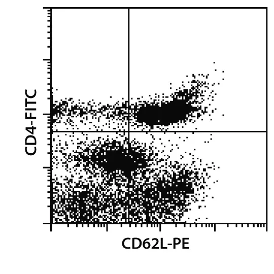

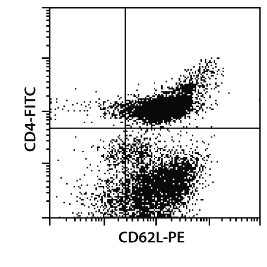
Cell separation with CD62L MicroBeads, human. CD62L+ cells were separated from PBMCs using CD62L MicroBeads. For positive selection, CD62L+ cells were isolated using an MS Column and a MiniMACS™ Separator. For depletion of CD62L+ cells, the sample was separated over an LD Column in a MidiMACS Separator.
2.3.4 Isolation of other CD4+ T cell subsets from PBMCs
At a glance: Kits and reagents for the separation of various CD4⁺ T cell subsets from PBMCs
| T cell subset | Isolation strategy | Comments | Automation | Product | ||||
| TH2 | Direct isolation of target cells | CD294 (CRTH2) is a marker for TH2 cells. | Yes* | CD294 (CRTH2) MicroBead Kit, human | ||||
| Skin-homing T cells | Direct isolation of target cells | Cutaneous lymphocyte-associated antigen (CLA) is a marker for skin-homing T cells | Yes* | Anti-CLA MicroBead Kit, human | ||||
| Skin-homing CD4+ T cells | Sequential separation | First isolate CD4+ T cells and then select cells expressing the skin-homing receptor CLA | Yes* | Combine: CD4+ T Cell Isolation Kit, human Anti-CLA-PE, human (clone HECA-452) Anti-PE MicroBeads | ||||
| Recent thymic emigrant T cells | Sequential separation | First non-CD4+ cells and memory CD4+ T cells are depleted. Then RTEs are separated via CD31. | Yes* | CD4+ Recent Thymic Emigrant Isolation Kit, human | ||||
| *Automation options range from fully automated benchtop solutions such as the autoMACS® NEO Separator to high-throughput platforms such as the MultiMACS™ Cell24 Separator Plus or MultiMACS X. | ||||||||
2.4 Characterization of CD4+ T helper cells by flow cytometry
CD4+ T cell subsets and their differentiation status can be determined by flow cytometry based on their expression of cell surface markers, transcription factors, and the cytokines they produce. Miltenyi Biotec offers a vast portfolio of conventional and recombinant REAfinity™ Antibodies for comprehensive analysis.

 View details
Flow cytometry panels
The following surface markers, cytokines, and transcription factors can be used to identify CD4+ T cell subsets by flow cytometry.
At a glance: Markers for the analysis of CD4⁺ TH cell subsets using MACS antibodies.
| T cell development – naive vs. memory (TSCM/TCM/TTM/TEM/TEMRA) | CD4+ TH subsets (TH1/TH2/TH9/TH17/TH22/Tfh) | Activated TH cells | Exhausted TH cells | ||||
| CD3 | CD3 | CD3 | CD3 | ||||
| CD4 | CD4 | CD4 | CD4 | ||||
| CD8 | CD161 (NK1.1) | CD8 | CD8 | ||||
| CD27 | CD183 (CXCR3) | CD25 (IL2RA) | CD96 (TACTILE) | ||||
| CD28 | CD184 (CXCR4) | CD27 | CD152 (CTLA-4) | ||||
| CD45RA | CD185 (CXCR5) | CD28 | CD160 (NK1) | ||||
| CD45RO | CD194 (CCD4) | CD69 | CD223 (LAG-3) | ||||
| CD57 | CD196 (CCR6) | CD95 (FasR) | CD244 (2B4) | ||||
| CD62L (L-Selectin) | CD197 (CCR7) | CD134 (OX40) | CD278 (ICOS) | ||||
| CD95 (FasR) | CCR10 | CD137 (4-1BB) | CD279 (PD1) | ||||
| CD127 | IL-2 | CD154 (CD40L) | CD366 (TIM-3) | ||||
| CD197 (CCR7) | IL-4 | Ki-67 | TIGIT | ||||
| IL-5 | KLRG1 | VISTA | |||||
| IL-9 | EOMES | ||||||
| IL-10 | CD272 (BTLA) | ||||||
| IL-13 | |||||||
| IL-17 | |||||||
| IL-21 | |||||||
| IL-22 | |||||||
| IL-26 | |||||||
| IFN-γ | |||||||
| T-bet | |||||||
| RORγT | |||||||
| GATA2 | |||||||
Analysis of surface markers and cytokines
Miltenyi Biotec offers a range of solutions for the analysis of T cell–associated surface markers and cytokines:
- MACS Antibodies are the ideal solution for the staining of T cell surface markers or the intracellular staining of cytokines.
- MACS Cytokine Secretion Assays allow detection and enrichment of viable cytokine-secreting cells. The human kits are available for a large number of cytokines, including TNF-α, GM-CSF, IFN-α, IFN-γ, IL-2, IL-4, IL-5, IL-10, IL-12, IL-13, IL-17, and IL-22, and can be used for further characterization of TH cell subsets
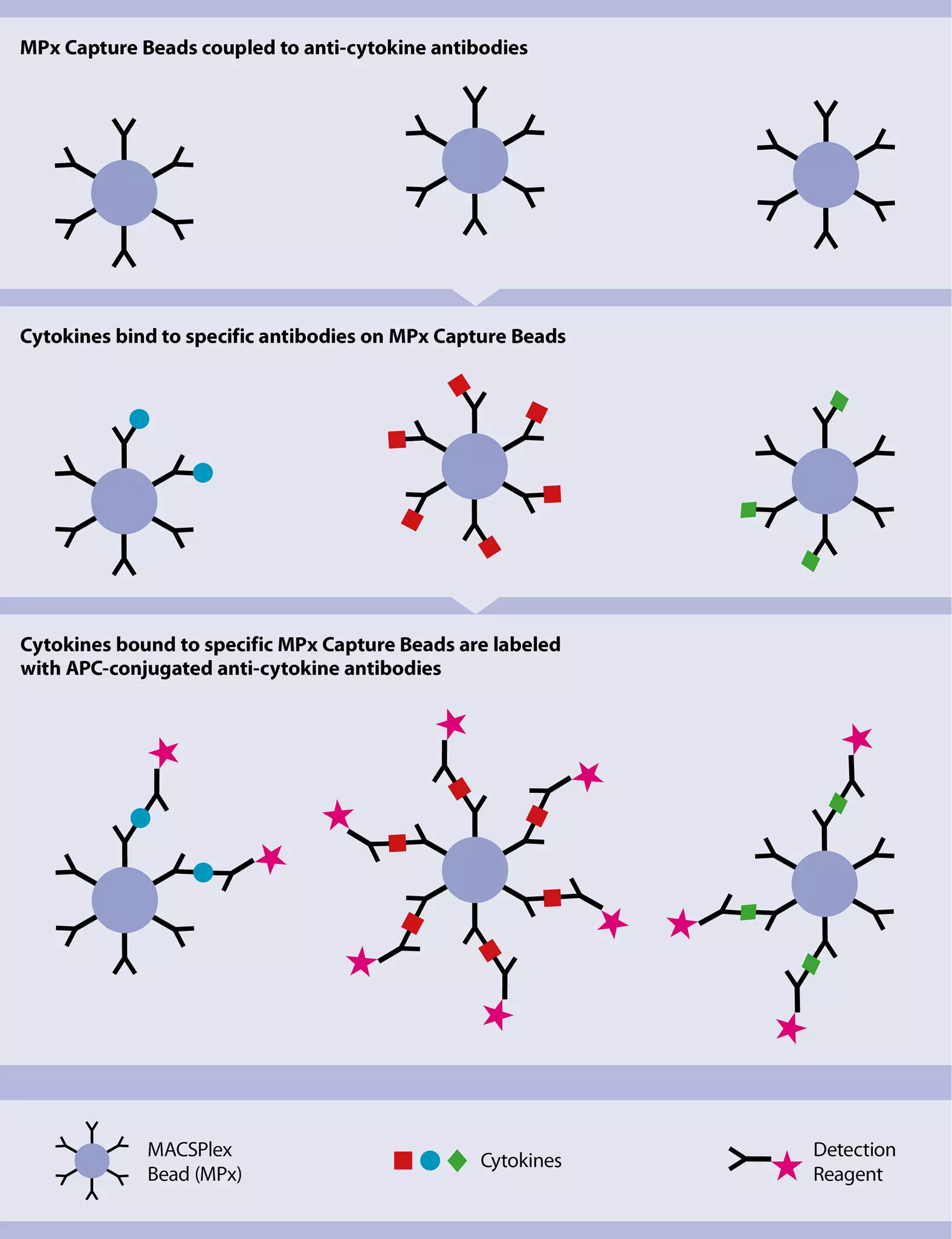
- The human MACSPlex 12 Cytokine Kits are used for multiplex analysis of secreted cytokines in serum and cell culture supernatants using a standard flow cytometer. Cytokines that can be analyzed in a single sample include: GM-CSF, IFN-α, IFN-γ, lL-2, IL-4, IL-5, IL-6, IL-9, IL-10, IL-12p70, IL-17A, and TNF-α.
- The Rapid Cytokine Inspectors allow for rapid analysis of activated T cells by combining surface marker analysis (e.g., CD4, CD8, CD154) with intracellular cytokine staining (e.g., IFN-γ, TNF-α, IL-2, and IL-17).

 Related videos:
 Related products:
2.5 Cell culture of CD4+ T helper cells
The Miltenyi Biotec cell culture and stimulation portfolio offers a specialized and versatile range of culture media and reagents for the stimulation, activation/expansion, and differentiation of T cells.
Cultivation, activation, and expansion of T helper cells
At a glance: Kits and reagents for the cultivation, activation, and expansion of T cells
grade
TexMACS™ Medium, research grade is a serum-free cell culture medium developed specifically for T cells. It has been used in a variety of applications and, in combination with MACS Cytokines, is an ideal starting point for reliable cultivation conditions. The medium is also available in MACS GMP Grade, and with or without phenol red.
For detailed information about Miltenyi Biotec media optimized for T cells, see the chapter Cell culture media.

T cell activation is essential for a variety of downstream application. Miltenyi Biotec offers polyclonal stimulation reagents that have been carefully designed to ensure optimal stimulation conditions.
T Cell TransAct™ is a ready-to-use reagent that is applied volumetrically, eliminating the need for bead-to-cell ratio calculations. Excess reagent is simply removed via culture wash. T Cell TransAct is available in both research and GMP grades, for a seamless transfer of workflows into clinical settings.

The T Cell Activation/Expansion Kit, human is based on large cell-sized particles loaded with biotinylated antibodies of choice to activate and expand primary cells. The large cell-sized particles mimic antigen-presenting cells and lead to efficient T cell activation, when loaded with CD2, CD3, and CD28 antibodies and applied in a specific bead-to-cell ratio. The following decision tree serves as a guide to choose the appropriate T cell activation product for a given project.

CytoStim™ is an antibody-based reagent that rapidly stimulates T cells. It can be used as a non-toxic alternative to SEB, for example, for the positive control of antigen-specific T cell stimulation assays or intracellular cytokine staining experiments to detect cytokine or activation marker expression.

PepTivator® Peptide Pools enable the antigen-specific stimulation of both CD4+ and CD8+ T cells with an extensive panel of tumor-, virus-, fungi-, and microbiota-specific antigens. Consisting of 15-mer peptides with 11-amino-acid overlaps, PepTivator Peptide Pools cover the complete sequence of the respective antigen. Available in research and premium grades as well as MACS GMP Grade. The most popular PepTivator Peptide Pools are also available in a 96-well cell culture plate format for high-throughput cell activation.
MACS Cytokines are available in three different grades – research and premium grades as well as MACS GMP Grade – to provide best flexibility in any assay setup. Notably, premium-grade MACS Cytokines exhibit well-defined biological activities, normalized to international reference standards (IU/mg) that allow exact unit dosing for reproducible results without laborious pretesting
Finally, the CD3 and CD28 pure – functional grade antibodies, human are suitable for in vitro T cell activation and expansion. The CD3 (OKT3) and CD28 (15E8) antibodies recognize the respective human receptors. Upon receptor binding, a stimulatory signal is transferred that, in combination with additional cytokines (e.g., IL-2 or IL-7/IL-15), leads to the activation and expansion of T cells.
Differentiation of CD4+ T helper cells
At a glance: Kits and reagents for the differentiation of T helper cells
| Use | Comments | Product | ||
| Differentiation | Consistent, high-quality recombinant cytokines for successful cell culture. Available in premium and research grades as well as MACS GMP Grade. | MACS Cytokines | ||
| Differentiation | Functional-grade antibodies effectively mimic or inhibit ligand-receptor interactions. | Functional-grade antibodies, human | ||
By combining polarizing cytokines and blocking or activating antibodies (‘functional-grade’), naive CD4+ cells can be differentiated into a variety of different TH cell subtypes. With the optimized TexMACS Medium, premium-grade cytokines, and a vast array of functional-grade antibodies, Miltenyi Biotec offers multiple tools for the efficient polarization of naive CD4+ T cells.
MACS Handbook:
3 CD4+ T helper cells from other tissues
T cells can also be obtained from tissues other than peripheral blood. Miltenyi Biotec offers a variety of solutions for the preparation of human tissues, as well as the subsequent separation, cultivation, and analysis of the respective T cell populations.
3.1 Miltenyi Biotec application protocols for T cells from other tissues
Miltenyi Biotec has created dedicated application protocols to enrich T cells from various human tissues:
3.2 Sample preparation of different tissues
Tissues must be dissociated into a single-cell suspension for many downstream applications, including isolation of cell subpopulations, cell culture, or flow cytometry analysis. The combination of mechanical dissociation and enzymatic treatment based on the gentleMACS™ Dissociator with Heaters and specific Tissue Dissociation Kits enables reproducible T cell isolation. T cells can be obtained with excellent yield, high viability rate, and preserved cell epitopes even from hard-to-process tissues, including tumor, skin, and brain tissue, amongst others.