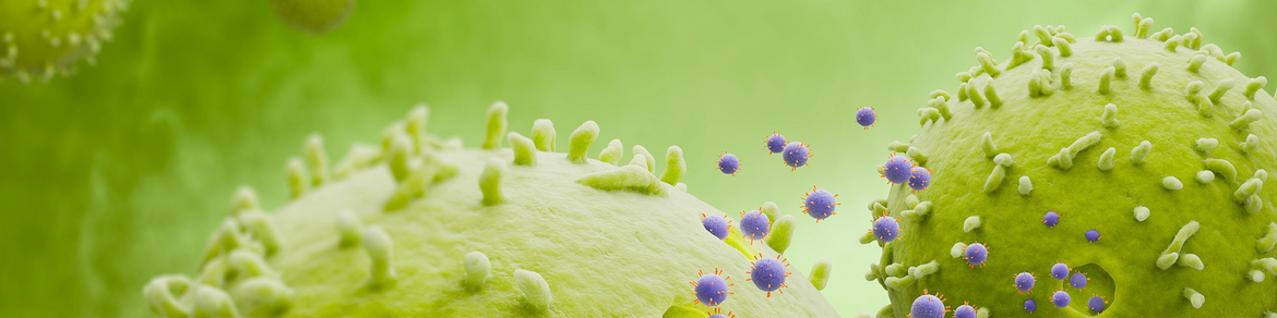
Simple isolation and analysis of extracellular vesicles (EVs)
Already investigating EVs or ready to explore these intercellular messengers that hold great promise for diagnostics and therapeutics? Either way, we have the right tools for you. Isolate highly pure EVs, such as exosomes, directly from any source and simply screen surface markers on EVs at once by flow cytometry. Learn more about our research solutions for EVs, such as exosomes.
Magnetic isolation of highly pure EVs

Accelerate EV isolation with our easy-to-use EV Isolation Kits for human and mouse samples, a true alternative for conventional ultracentrifugation.
- Selection of EVs according to surface markers i.e. CD9, CD63, CD81, or all three markers combined
- Low starting sample volume (0.5 to 2 mL)
- Isolation from multiple sample types e.g. cell culture supernatant, urine, plasma, serum, cerebrospinal fluid (CSF), ascites etc.
- Complete workflow in <2 h
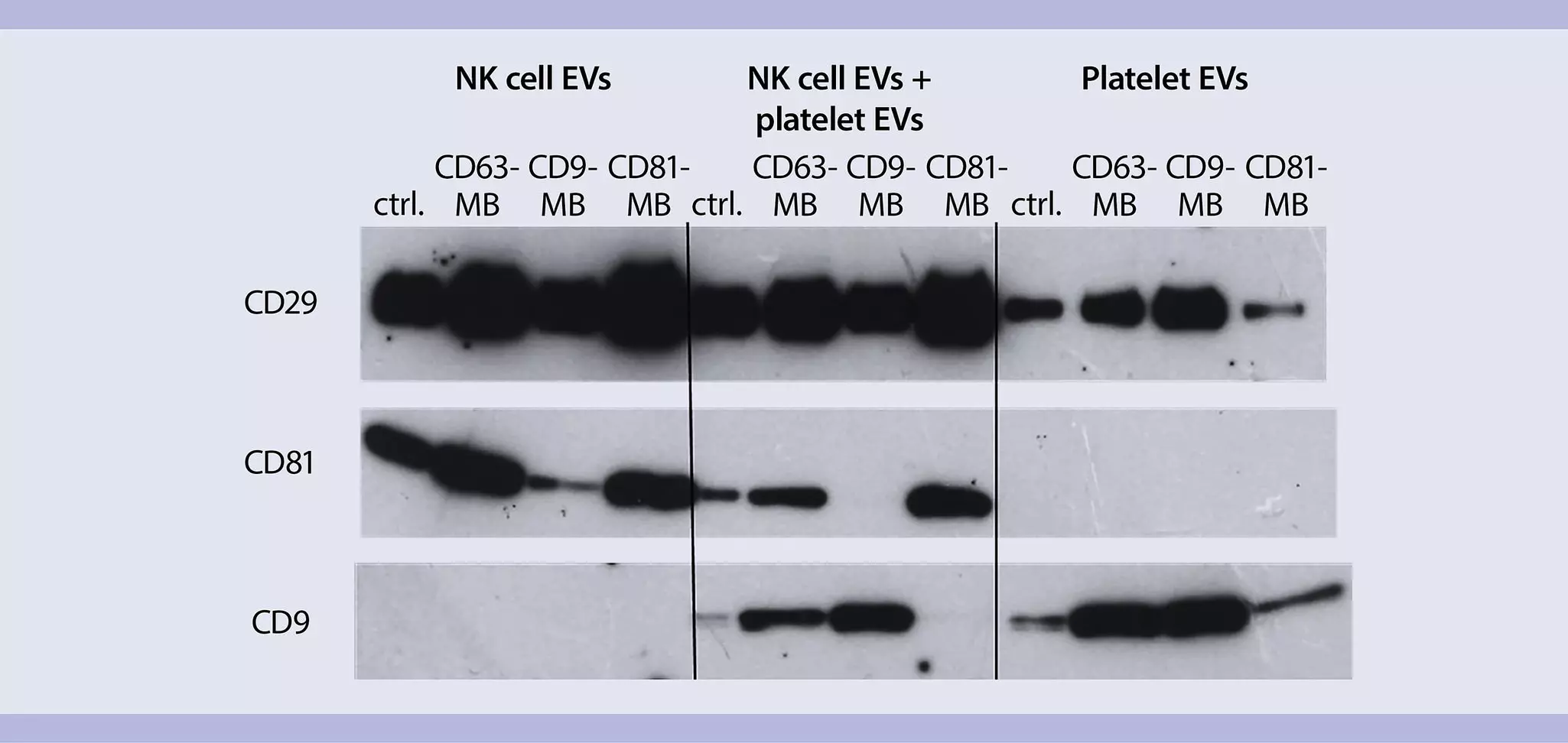
Separation of EV subpopulations
MACS® Technology enables the physical separation and enrichment of different EV subpopulations such as exosomes, based on their surface markers. EVs isolated with our kits are suitable for downstream molecular and genomic analysis.
Comprehensive flow cytometric analysis of EV surface markers

Advance your EV analysis with our MACSPlex EV Kits for human and mouse samples. Simply screen and profile multiple surface markers on EVs by flow cytometry in one go.
- Unique multiplex bead platform for protein profiling of EVs by flow cytometry
- Sensitive and semi-quantitative analysis of 37 markers in parallel (+ 2 isotype controls)
- Fast (<2 h) and easy protocol
- Works on any flow cytometer able to discriminate FITC, PE, and APC fluorescence
- Direct detection possible; no pre-enrichment required
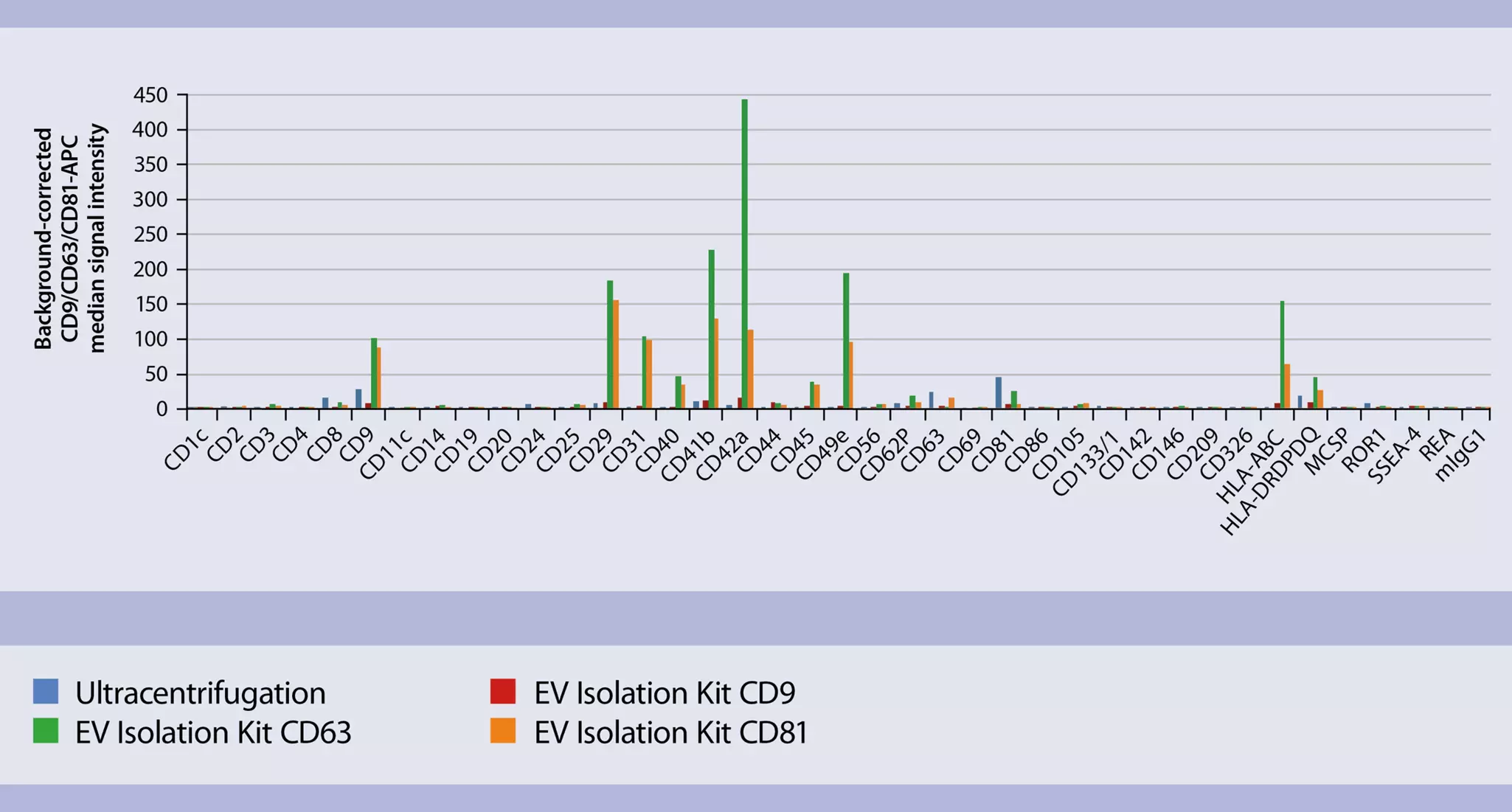
Our kits make a difference
Plasma EVs isolated with our EV Isolation Kits* and analyzed with the MACSPlex EV Kit, human reveal strong signals in comparison to EVs isolated by ultracentrifugation.
Video
Extracellular vesicle analysis by flow cytometry using the MACSPlex EV Kits
This video provides step-by-step instructions on how to use the MACSPlex EV Kit, human. It explains how to prepare the extracellular vesicle (EV) sample, couple the EVs to the capture beads, and stain the EV sample with the detection reagents. The video also provides practical tips for improved sample handling.