The MACSPlex EV Kit IO (for immuno-oncology) allows the detection and characterization of extracellular vesicles (EVs), such as exosomes, using 37 different surface epitopes and two isotype controls. The MACSPlex EV Kit IO comprises a cocktail of various fluorescently labeled bead populations, each coated with a specific antibody binding the respective surface epitopes. The 39 bead populations can be distinguished by different fluorescence intensities via flow cytometry.


Related products for MACSPlex EV Kit IO, human
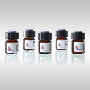

The MACSPlex EV Kit IO (for immuno-oncology) allows the detection and characterization of extracellular vesicles (EVs), such as exosomes, using 37 different surface epitopes and two isotype controls. The MACSPlex EV Kit IO comprises a cocktail of various fluorescently labeled bead populations, each coated with a specific antibody binding the respective surface epitopes. The 39 bead populations can be distinguished by different fluorescence intensities via flow cytometry.
Extracellular vesicles (EVs) are incubated with the antibody-coated MACSPlex EV Capture Beads. Subsequently or in parallel, EVs bound to the MACSPlex EV Capture Beads are labeled with the MACSPlex EV Detection Reagents. The MACSPlex EV Detection Reagents can also be combined to create a cocktail comprising of MACSPlex EV Detection Reagent for CD9, CD63, and CD81. Consequently, sandwich complexes are formed between the MACSPlex EV Capture Bead, extracellular vesicle, and the detection reagent (see Data and images, figure 1). These complexes can be analyzed based on the fluorescence characteristics of both the MACSPlex EV Capture Bead and the detection reagent. Positive signals indicate the presence of the respective surface epitope within the EV population (see Data and images, figure 2). It is also possible to compare different EV samples using the MACSPlex EV Kits allowing semi-quantitative analysis of differential surface epitopes.
Minimal information for studies of extracellular vesicles (MISEV2023)
For more information on the latest state of the art extracellular vesicle (EV) research, including currently available technologies and expert guidelines for separation and characterization of EVs from different sources, please refer to the Minimal information for studies of extracellular vesicles (MISEV) provided by The International Society for Extracellular Vesicles (ISEV)
Our MACSPlex EV Kits are included and recommended within the MISEV guidelines for bead-based flow cytometry characterization of EVs.
The MACSPlex EV Kit IO has been developed for the simultaneous flow cytometric detection of 37 surface epitopes that are known to be present on different extracellular vesicles plus two isotype control beads.
To access a Certificate of Analysis (CoA) follow the link below and enter the specific lot of your product.
Should you wish to request additional documentation, such as a Safety Data Sheet (SDS) in other languages than English or Swedish, an SDS-replacement document, or a Certificate of Origin (CoO), please direct your inquiry to your local technical support team.
- Collino, F. et al. (2020) Extracellular vesicles derived from induced pluripotent stem cells promote renoprotection in acute kidney injury model. Cells 9 (2): 453
- Martin-Jaular, L. et al. (2021) Unbiased proteomic profiling of host cell extracellular vesicle composition and dynamics upon HIV‐1 infection. EMBO J. 40 (8): e105492
- Calleri, A. et al. (2021) Protective effects of human liver stem cell-derived extracellular vesicles in a mouse model of hepatic ischemia-reperfusion injury. Stem Cell Rev Rep. 17 (2): 459
- Pleet, M. et al. (2023) Viral Immune signatures from cerebrospinal fluid extracellular vesicles and particles in HAM and other chronic neurological diseases Front Immunol 14: 1235791
- Bruno, S. et al. (2020) HLSC-derived extracellular vesicles attenuate liver fibrosis and inflammation in a murine model of non-alcoholic ateatohepatitis. Mol. Ther. 28 (2): 479-489
- Welsh, J. A. et al. (2022) MPAPASS software enables stitched multiplex, multidimensional EV repertoire analysis and a standard framework for reporting bead-based assays. Cell Rep Methods. 2 (1): 100136
- Welsh, J. A. et al. (2024) Minimal information for studies of extracellular vesicles (MISEV2023): From basic to advanced approaches. J Extracell Vesicles : doi: 10.1002/jev2.12404
- Görgens, A. et al. (2019) Optimisation of imaging flow cytometry for the analysis of single extracellular vesicles by using fluorescence-tagged vesicles as biological reference material. J. Extracell. Vesicles 8 (1): 1587567
- Gupta, D. et al. (2020) Quantification of extracellular vesicles in vitro and in vivo using sensitive bioluminescence imaging. J. Extracell. Vesicles 9 (1): 1800222
- Wiklander, O. P. B. et al. (2018) Systematic methodological evaluation of a multiplex bead-based flow cytometry assay for detection of extracellular vesicle surface signatures. Front Immunol 9: 1326
- Vacchi, E. et al. (2020) Immune profiling of plasma-derived extracellular vesicles identifies Parkinson disease. Neurol Neuroimmunol Neuroinflamm. 7 (6): e866
- Andaloussi, S. et al. (2017) Reproducible and scalable purification of extracellular vesicles using combined bind-elute and size exclusion chromatography. Sci Rep 7 (1): 11561
- Salvi, S. et al. (2021) Detection and investigation of extracellular vesicles in serum and urine supernatant of prostate cancer patients. Diagnostics 11 (3): 466
- George, M. S. et al. (2023) Extracellular vesicles in COVID-19 convalescence can regulate T cell metabolism and function iScience 26 (8): 107280
- Koliha, N. et al. (2016) Melanoma affects the composition of blood cell-derived extracellular vesicles. Front Immunol 7: 282
- Martire, S. et al. (2021) A first phenotypic and functional characterization of placental extracellular vesicles from wmen with multiple sclerosis. Int J Mol Sci 22 (6): 2875
- Nguyen, V. V. T. et al. (2024) Inter‐laboratory multiplex bead‐based surface protein profiling of MSC‐derived EV preparations identifies MSC‐EV surface marker signatures J Extracell Vesicles 13 (6): doi: 10.1002/jev2.12463
- Forte, D. et al. (2021) Distinct profile of CD34+ cells and plasma-derived extracellular vesicles from triple-negative patients with Myelofibrosis reveals potential markers of aggressive disease. J Exp Clin Cancer Res. 40: 49
- Görgens, A. et al. (2021) A robust and semi-quantitative method for analyzing exosomes by flow cytometry. Trillium Extracellular Vesicles 3 (1): 41-45
- Kaspi, H. et al. (2021) MSC-NTF (NurOwn®) exosomes: a novel therapeutic modality in the mouse LPS-induced ARDS model. Stem Cell Res Ther. 12: 72
- Koliha, N. et al. (2016) A novel multiplex bead-based platform highlights the diversity of extracellular vesicles. J Extracell Vesicles 5: 29975
- Shin, K-O. et al. (2020) Exosomes from human adipose tissue-derived mesenchymal stem cells promote epidermal barrier repair by inducing de novo synthesis of ceramides in atopic dermatitis. Cells 9 (3): 680
- Brahmer, A. et al. (2023) Assessment of technical and clinical utility of a bead-based flow cytometry platform for multiparametric phenotyping of CNS-derived extracellular vesicles. Cell Commun Signal 21 (1): 276
- Brahmer, A. et al. (2019) Platelets, endothelial cells and leukocytes contribute to the exercise-triggered release of extracellular vesicles into the circulation. J. Extracell. Vesicles 8 (1): 1615820
- Pomatto, M. et al. (2021) Differential therapeutic effect of extracellular vesicles derived by bone marrow and adipose mesenchymal stem cells on wound healing of diabetic ulcers and correlation to their cargoes. Int J Mol Sci 22 (8): 3851