Granulocytes


1 Introduction
Granulocytes belong to the myeloid cell family and represent the most abundant cell type in peripheral blood. They are characterized by the presence of cytoplasmic granules and a poly-lobed nucleus, which gives them the name polymorphonuclear leukocytes (PMN, PML, or PMNL). Special staining techniques developed by Paul Ehrlich in the late 1800s allowed the identification of granulocytes in blood and opened the possibility to study these cells in different pathological conditions.
Granulocytes are derived from stem cells in bone marrow. They have a short life span in the periphery, which can be prolonged at sites of infection. Located at strategic points in the body with preformed cytoplasmic granules filled with various effector molecules, they are among the first responders at the onset of innate immune responses. Granulocytes play a well-known role in inflammation and allergy, but are also capable of antigen presentation and immune response modulation (PMID: 23007469). The toxic mediators produced by these cells have been undeniably linked to various pathologies, and therapeutic approaches targeting granulocytes are currently under development to treat allergic diseases and asthma (PMID: 28784414,28283697, 26819959, 23485549, 28099862, 27558343).
 MACS Handbook:
2 Granulocytes in peripheral blood
Granulocytes circulate through the body via peripheral blood and account for approximately 25–75% of CD45+ leukocytes. They can be divided into three subtypes – neutrophils, eosinophils and basophils – named according to their characteristic staining with hematoxylin and eosin. Basophils stain dark blue, eosinophils red, and neutrophils stain pink.
 Related PDFs:
2.1 Cell subsets, frequencies, and marker expression
At a glance: Granulocyte subtypes in peripheral blood
| Cell subtype | Frequency | Markers | Function |
| Neutrophils | 25–75% of total circulating human white blood cells | CD15+, CD16+, FcεRIα+, CD66b+, CD11b+, CD49d– | Destroy pathogens by phagocytosis and release of anti-microbials Recruit other immune cells |
| Eosinophils | 1–10% of peripheral leukocytes | CD15+, CD66b+, CD11b+, Siglec-8+, CD193+, CD16– | Release mediator molecules upon activation Involved in antigen presentation Polarize T cells |
| Basophils | 0.2–1% of white blood cells | CD15–, FcεRIα+, CD123+, CD11b+, CD203c+, CD117– | Release mediators that promote blood flow to site of infection Promote TH2 differentiation Release cytokines |
Neutrophils represent the most abundant type of granulocytes, accounting for 25–75% of all circulating human white blood cells (PMID: 23435331, 22491176). Their mechanisms of action encompass degranulation of antimicrobial proteins, release of chemotactic factors (PMID: 22392929, 20140197, 25512469, 21555529), and formation of neutrophil extracellular traps or NETs (PMID: 23435331, 22491176, 15001782) in response to pathogens. Typical markers are CD15, CD66b, CD16, and the absence of CD49d.
Representing 1–10% of peripheral leukocytes, eosinophils in resting state reside mainly in the periphery, especially in lamina propria (PMID: 11877470, 25049430). They readily release their granule content including eosinophil cationic protein (ECP), eosinophil peroxidase (EPX), and major basic protein (MBP) as well as chemokines, and cytokines in response to pathogens (PMID: 25003763). They play a role in antigen presentation and T cell polarization (PMID: 25003763, 8450230, 20065995, 16551246). A specific marker of eosinophils in human blood is Siglec-8. Other commonly used markers are CD15, CD193, and the absence of CD16.
Basophils are a small population, accounting for only 0.2–1% of white blood cells (PMID: 28506528). They are rich in pre-formed granules containing histamine, leukotrienes (LTs), and platelet-activating factor (PAF). Under specific conditions, they play non-redundant roles as effector cells and promote TH2 differentiation. They can release large amounts of IL-4 and may take part in dendritic cell modulation. A specific marker for blood basophils is CD203c. Other typical markers are FcεRIα, CD123, and the absence of CD117 (PMID: 27135604, 23558889).
2.2 Sample preparation of peripheral blood
Eosinophils and neutrophils can be isolated directly from whole blood, without prior sample preparation. For some applications and for the isolation of basophils, blood, buffy coat, or buffy cone are first processed to generate PBMCs. For additional information about human peripheral blood, see the MACS Handbook chapter Human cell sources – Blood (human).
Miltenyi Biotec has developed a special protocol to generate PBMCs from whole blood for subsequent isolation of granulocytes. This protocol provides step-by-step instructions for performing a density gradient using freshly drawn human blood treated with an anticoagulant (preferably EDTA or citrate, ACD-A, CPD) or fresh defibrinated blood or buffy coat (not older than 8 hours) treated with an anticoagulant. The resulting white blood cell layer directly above the red blood cells is collected and diluted with buffer to wash the cells. Any remaining red blood cells are then lysed and the white blood cells are washed twice. Finally, the cell pellet is resuspended in an appropriate buffer, and cells are counted before proceeding with magnetic cell separation. This protocol can be found as an appendix in the data sheet of the CD16 MicroBeads, human, which you can download from the related resources panel to the right.
MACS Handbook:
Related PDFs:
2.3 Magnetic separation of granulocytes from peripheral blood
Miltenyi Biotec has developed numerous products for the straightforward magnetic separation of granulocyte subsets. Both positive selection and depletion strategies can be pursued for cell isolation directly from blood. Several positive selection strategies are possible to isolate granulocytes from PBMCs.
For details on MACS® Cell Separation Technology, see the MACS Handbook chapter Magnetic cell separation.
MACS Handbook:
2.3.1 Isolation of neutrophils and eosinophils from whole blood
At a glance: Kits and reagents for the separation of neutrophils and eosinophils from whole blood
| Cell subtype | Starting material | Isolation strategy | Comments | Automation | Product |
| Eosinophils and neutrophils | Whole blood | Positive selection of target cells | Reagent recognizes 3-FAL, also designated Lewis X. Antigen is not expressed on basophils and lymphocytes. | Yes* | StraightFrom Whole Blood CD15 MicroBeads, human |
| Eosinophils and neutrophils | Whole blood | Positive selection of target cells | Antigen expressed on eosinophils and neutrophils, but not basophils and lymphocytes. | Yes* | StraightFrom Whole Blood CD66b MicroBeads, human |
| Eosinophils | Whole blood | Depletion of non-target cells | Isolates untouched eosinophils from up to 30 mL anticoagulated whole blood. | No | MACSxpress Eosinophil Isolation Kit, human |
| Neutrophils | Whole blood | Depletion of non-target cells | Isolates untouched neutrophils from up to 30 mL anticoagulated whole blood. | No | MACSxpress Neutrophil Isolation Kit, human |
| *Automation options range from fully automated benchtop solutions such as the autoMACS® NEO Separator to high-throughput platforms such as the MultiMACS™ Cell24 Separator Plus or MultiMACS X. | |||||
StraightFrom® MicroBeads can be used to isolate granulocytes directly from whole blood. Pre-enrichment of white blood cells by density gradient centrifugation is not required. The purified cells are well-suited for further flow cytometry analysis, molecular biology studies, and functional studies.
MACSxpress® Technology is a cell separation platform especially designed for processing large volumes of anticoagulated whole blood. Untouched human leukocytes can be isolated rapidly from up to 30 mL of anticoagulated whole blood by removing immunomagnetically labeled non-target cells. The MACSxpress Eosinophil Isolation Kit, human and MACSxpress Neutrophil Isolation Kit, human have been tested by independent labs and found to be the optimal solutions compared to density gradient and products from other manufacturers (PMID: 28647456, 27567327).
MACSxpress Neutrophil Isolation Kit, human
 View details
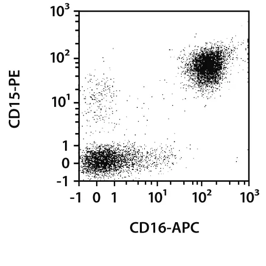
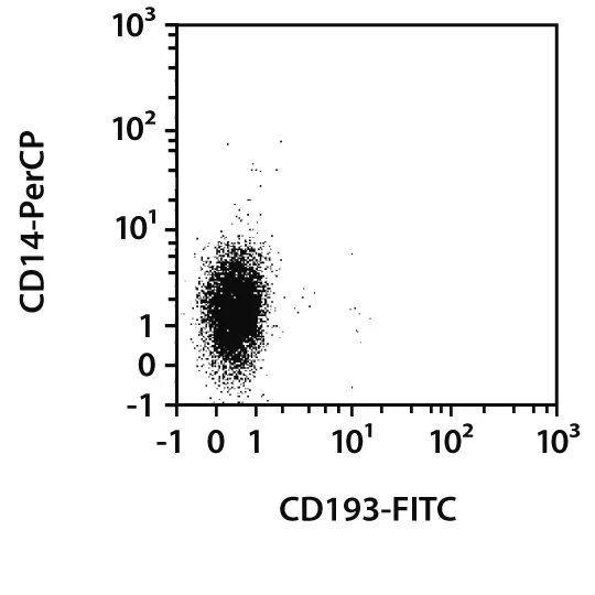
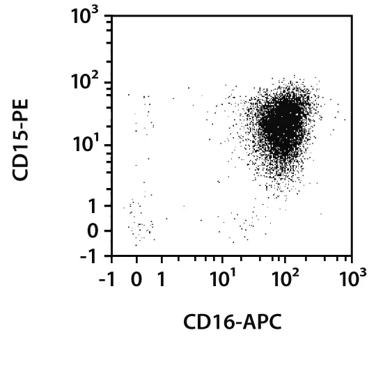
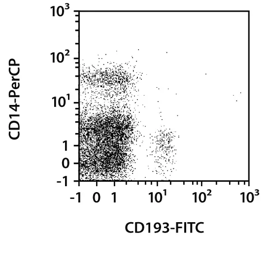
Efficient enrichment of neutrophils from whole blood. Untouched neutrophils were isolated from 8 mL of human EDTA-anticoagulated whole blood using the MACSxpress® Neutrophil Isolation Kit, human, a MACSmix™ Tube Rotator, and a MACSxpress Separator. The isolated cells were fluorescently stained with CD14-PerCP, CD15-PE, CD16-APC, CD45-VioBlue, and CD193-FITC and analyzed by flow cytometry using the MACSQuant® Analyzer. Cell debris, non-leukocytes, and dead cells were excluded from the analysis based on CD45 expression, scatter signals, and propidium iodide fluorescence.
It should be noted that neutrophils are extremely susceptible to non-specific activation. However, isolation of neutrophils with the MACSxpress Neutrophil Isolation Kit, human does not activate the cells, as shown by analysis of surface expression markers, adhesion, migration, phagocytic capacities, and production of reactive oxygen species (ROS). A scientific poster describing results from this analysis can be downloaded from the related resources panel to the right.
2.3.2 Isolation of basophils and eosinophils from PBMCs
| Cell subtype | Starting material | Isolation strategy | Comments | Automation | Product |
| Basophils | PBMCs | Depletion of non-target cells | Uses indirect magnetic labeling to remove non-target cells and achieve high-purity of the basophil population | Yes* | Basophil Isolation Kit II, human |
| Basophils | PBMCs | Positive selection of target cells | Concurrent isolation of basophils and plasmacytoid dendritic cells | Yes* | CD123 MicroBeads, human |
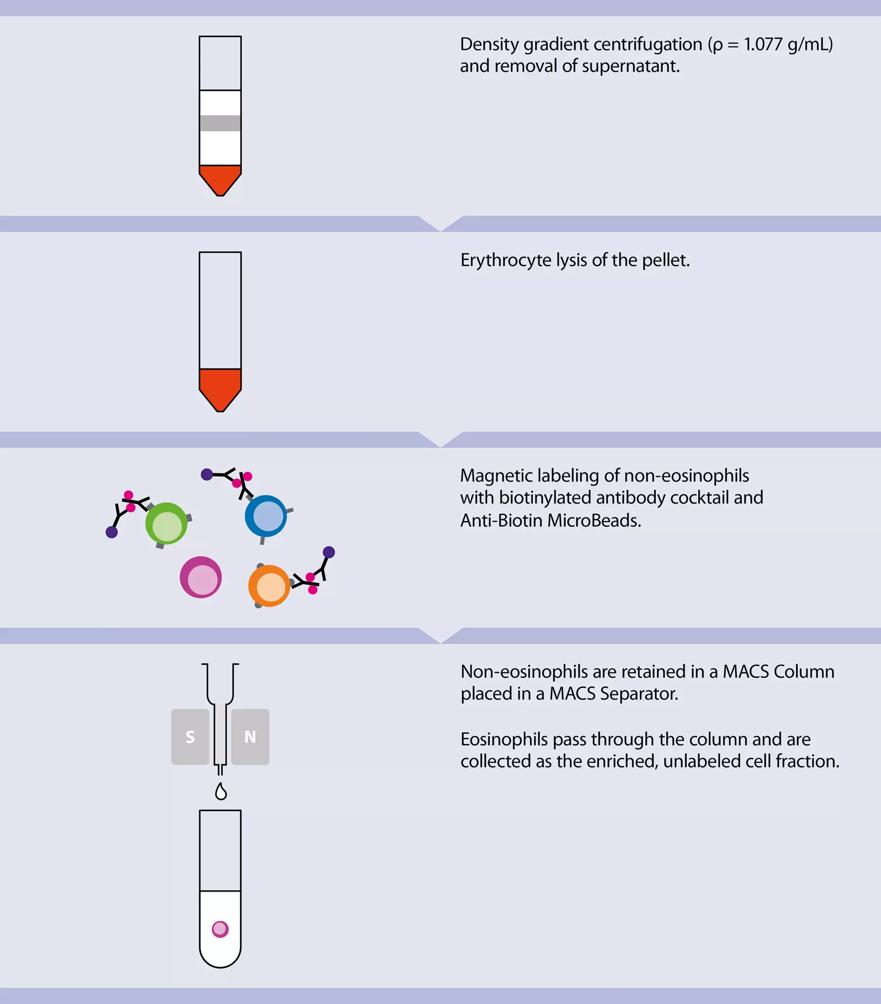
| Eosinophils | PBMCs | Depletion of non-target cells | Uses indirect magnetic labeling system to isolate untouched eosinophils | Yes* | Eosinophil Isolation Kit, human |
| *Automation options range from fully automated benchtop solutions such as the autoMACS® NEO Separator to high-throughput platforms such as the MultiMACS™ Cell24 Separator Plus or MultiMACS X. | |||||
Both positive selection and depletion strategies are possible with different Miltenyi Biotec products. The Basophil Isolation Kit II, human and Eosinophil Isolation Kit, human use a cocktail of biotin-conjugated antibodies to indirectly label non-target cells. The highly pure eosinophils or basophils that result from the depletion of the magnetically labeled cells are suitable for functional studies on signal transduction, activation mechanisms, or cytokine production.
 View details
Eosinophil Isolation Kit, human
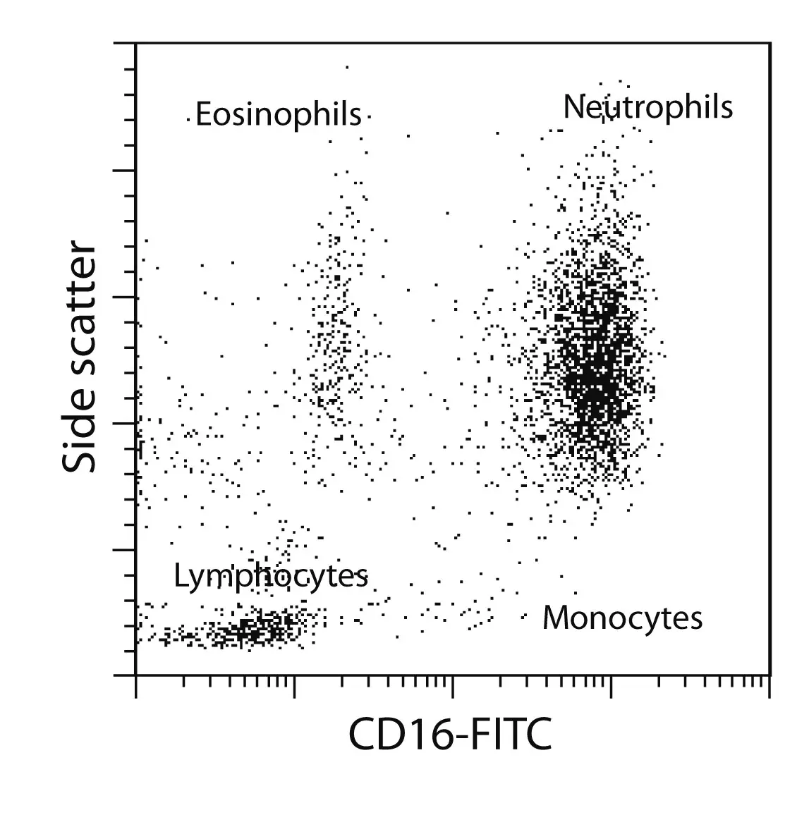
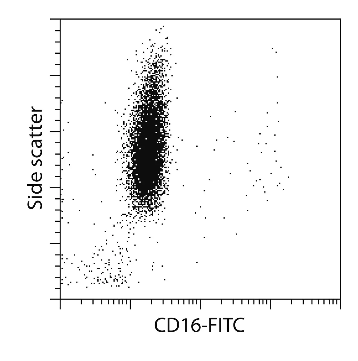
High purity of isolated eosinophil population. PBMCs generated from human peripheral blood served as starting material to isolate untouched eosinophils using the Eosinophil Isolation Kit, human, an LS Column, and a MidiMACS™ Separator. Cells were fluorescently stained with CD16-FITC. Eosinophils do not express CD16.
Related PDFs:
 Related videos:
2.4 Characterization of granulocytes by flow cytometry
Granulocytes can be analyzed by flow cytometry using intracellular and surface staining. Markers commonly used to analyze neutrophils, basophils, and eosinophils are listed in Cell subsets, frequencies, and marker expression.
Like many of the myeloid cell types, granulocytes are quite sticky. Thus, we recommend using REAfinity™ Antibodies whenever possible, to achieve optimal staining for flow cytometry. These recombinantly engineered antibodies are highly specific and require no FcR blocking as the Fc region is specifically mutated to abolish FcγR binding. Therefore, REAfinity Antibodies are especially well-suited to stain myeloid cells. For more information about Miltenyi Biotec antibodies, see Cell analysis reagents – antibodies.
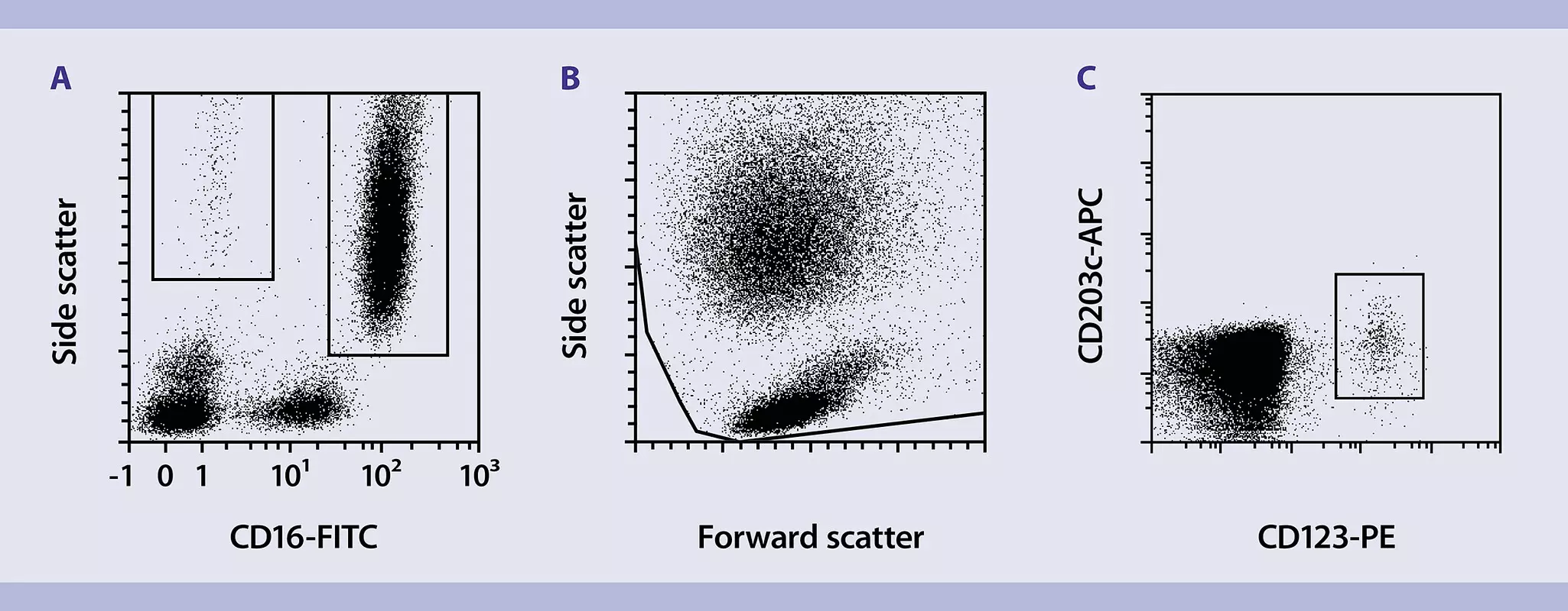
A common problem encountered by researchers studying myeloid cells is the sporadic appearance of markers used to identify and potentially isolate distinct cell subtypes. Granulocytes are no exception in this respect. In general, identification by flow cytometry can be done based on side scatter (SSC) and forward scatter (FSC) characteristics for the most abundant neutrophils and eosinophils, because they are highly granular and thus segregate well from lymphocytes and monocytes. This is not true for basophils. Identification based on the expression of a single specific antigen is restricted to eosinophils (Siglec-8+) and basophils (CD203c+) in blood, whereas neutrophils are commonly identified via marker combinations (CD15+CD16+).
 Related products: