Accelerating spatial biology with community-shared organ mapping antibody panels for the MACSima® Platform
- Tissue- or application-specific spatial biology antibody panels
- Expert-curated and optimized for MACSima Imaging Cyclic Staining (MICS)
Our MICS Panels are expert-curated to address specific scientific questions, providing a strong foundation for multiplex spatial analysis using the MACSima Platform. Discover our published panels and explore their biological context, structure, and marker composition.
From a scientific question to a proven MICS Panel
Our MICS Panels are developed by our R&D Teams, independently or in collaboration with academic partners, to address defined scientific questions. Expert-curated for specific tissues and disease models, these panels are optimized for spatial analysis on the MACSima Platform and supported by thorough and transparent documentation, providing a reliable foundation for reproducible spatial biology research.
By making them available to the research community, we aim to accelerate discovery and foster scientific advancement. Each panel contributes to a growing ecosystem of reusable spatial biology resources, reflecting best practices for reproducibility, transparency, and long‑term value.

Accelerate discovery
Skip months of panel development and start generating data right away with validated panels.

Deepen understanding
Use the proven panels as a starting point for your spatial biology project, and adjust it to your research.

Foster collaboration
Build on openly shared, proven panels to enable reproducible and comparable research.
MICS Panel: Liver
Our MICS Panel: Liver follows the organ mapping antibody panel (OMAP) framework, developed by the spatial biology community to standardize multiplex organ imaging and support the Human Reference Atlas initiative. Initially designed to evaluate CAR target expression in the liver, it enables comprehensive visualization of liver microanatomy and key tissue-resident immune cell populations.
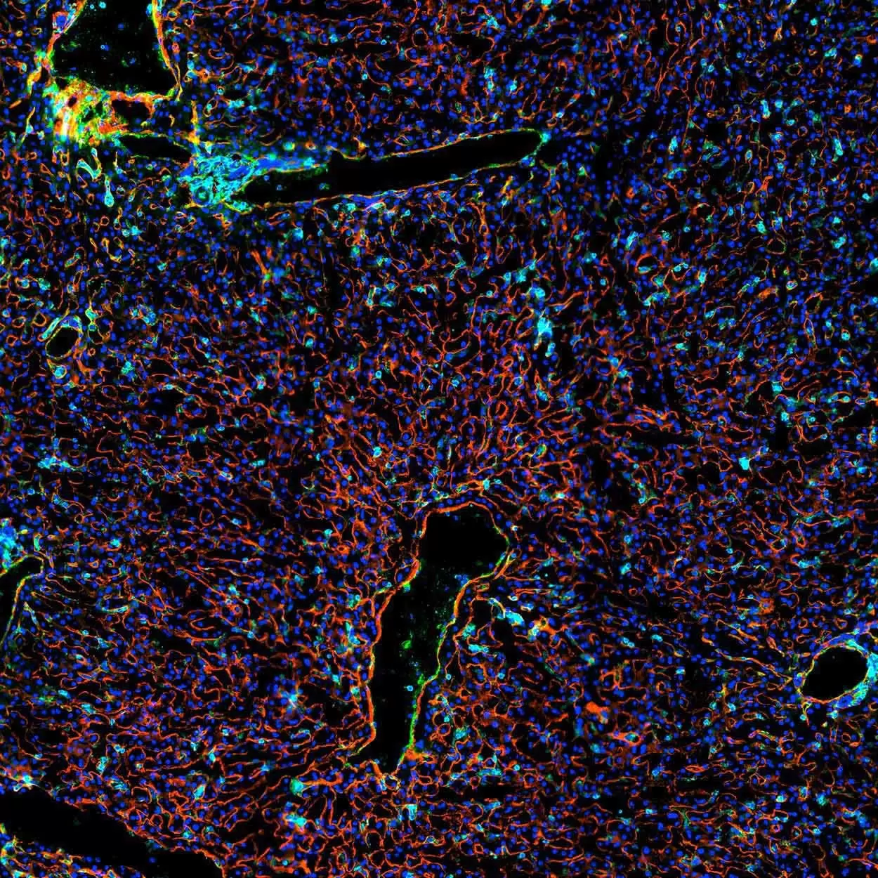
With this panel you can:
- Map spatial alterations in tissue architecture: Visualize liver anatomical structures, including hepatic vasculature and connective tissue in health and disease.
- Characterize the immune microenvironment: Map immune cell types and their spatial interactions to decipher disease mechanisms in fibrosis, cancer, or hepatotoxicity.
- Evaluate drug response and toxicity: Assess drug-induced changes on both cellular and tissue level.
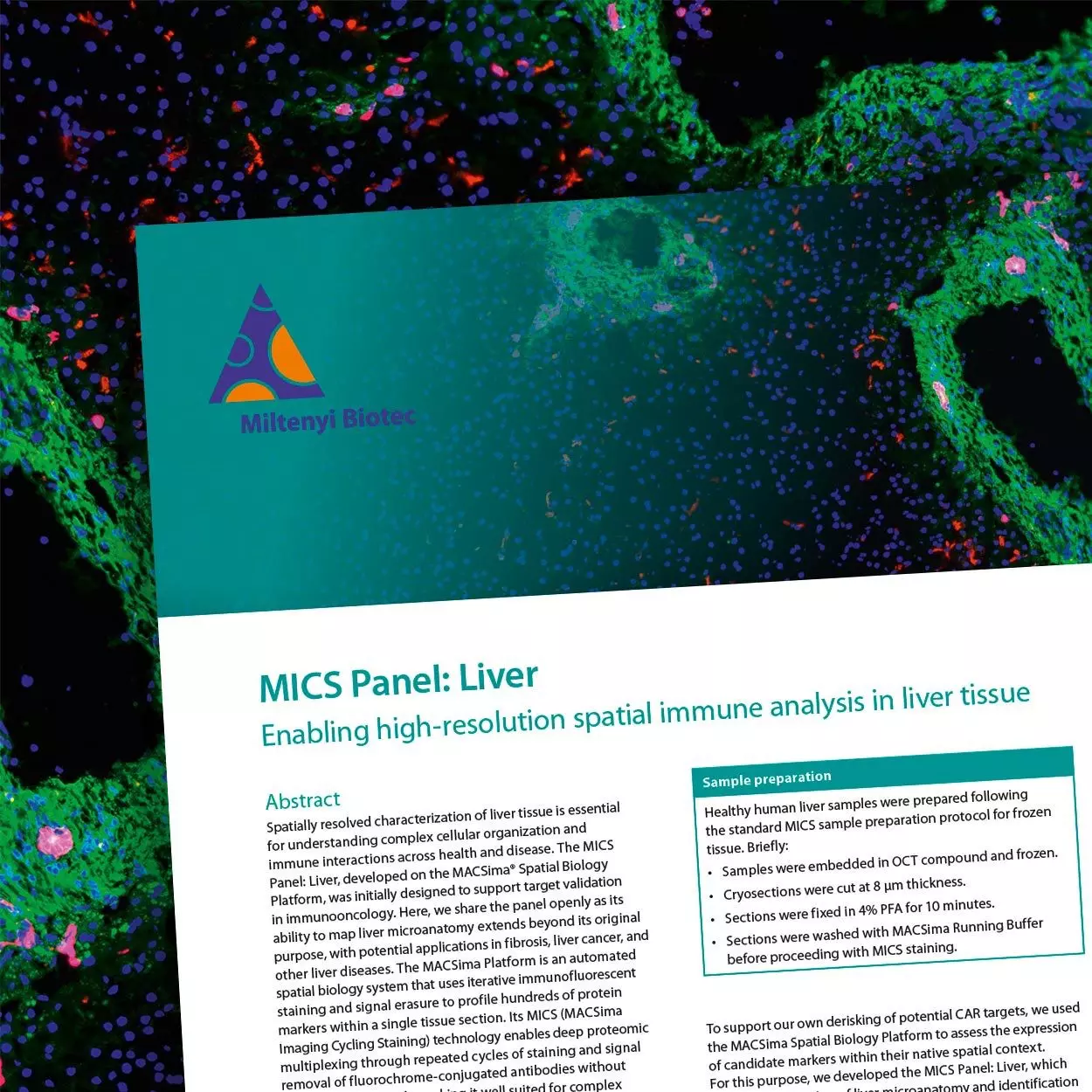
Dive into the MICS Panel: Liver application note to explore the panel composition and scientific rationale, discover additional markers, and get practical insights on how to use it for your research.
Want to know more about the OMAP project?
The OMAP framework was developed to address key challenges in multiplexed tissue imaging and support the construction of a Human Reference Atlas. Read the full study to learn how OMAP panels are designed, validated, and published, and discover how these resources can support your research.
Organ Mapping Antibody Panels: a community resource for standardized multiplexed tissue imaging.
Quardokus, E.M., et al (2023) Nat Methods 20, 1174–1178. | Read paper