Reveal the hidden architecture of the liver
Download the free application note to explore the MICS Panel: Liver
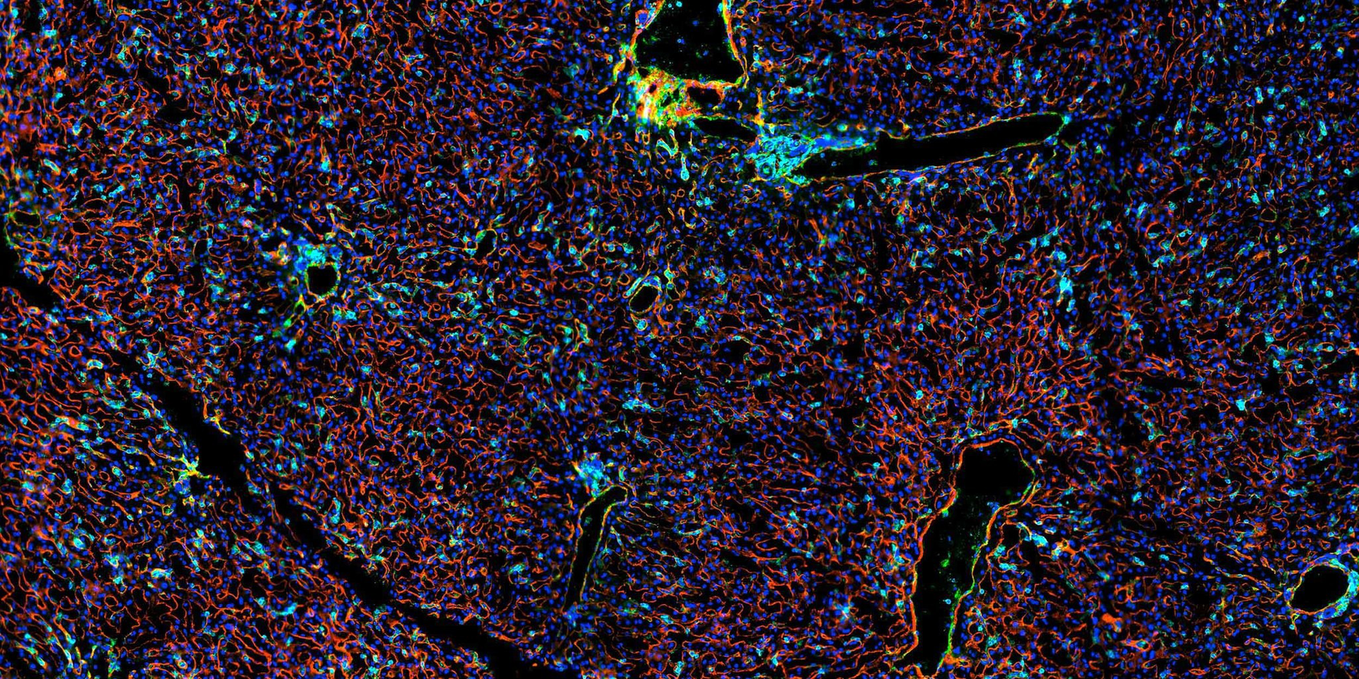

Reveal the hidden architecture of the liver
Map the liver with high-plex spatial antibody profiling
Developed on the MACSima® Spatial Biology Platform, the MICS Panel: Liver makes high-resolution spatial profiling of human liver tissue easy and efficient through high-plex antibody imaging. This innovative panel allows researchers to visualize a wide range of liver and immune cell populations, helping to reveal the complex cellular interactions that drive liver health and disease. With its ability to map liver microanatomy, the MICS Panel: Liver supports studies in areas such as fibrosis, liver cancer, and metabolic dysfunction.
Discover how you can gain deeper insight into tissue architecture, immune microenvironments, and disease mechanisms with this powerful tool for spatial biology.
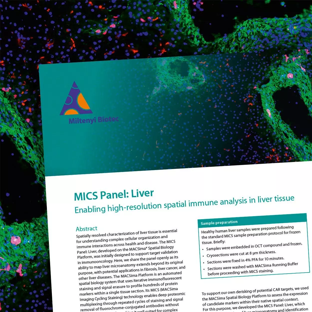
In the application note, you'll explore how to:
- Profile liver tissue using a high-plex antibody panel.
- Visualize immune, endothelial, epithelial, and stromal markers in situ.
- Map vascular and connective tissue structures with high-plex antibody staining.
- Investigate spatial changes in fibrosis, cancer, and liver injury.
- Apply automated high-plex imaging using the MACSima Platform.
Get the free application note
Fill in the form to receive the download link for the application note directly in your inbox and to subscribe to our newsletter.