CD11b (Microglia) MicroBeads have been developed for the positive isolation of primary mouse and human CD11b+ microglia. The purification protocol takes approx. one hour.


Related products for CD11b (Microglia) MicroBeads, human and mouse


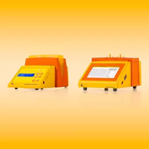


CD11b (Microglia) MicroBeads have been developed for the positive isolation of primary mouse and human CD11b+ microglia. The purification protocol takes approx. one hour.
Microglia, often called brain macrophages, are the resident immune effector cells in the central nervous system (CNS). In addition, activated microglia serve as the major antigen-presenting cells in the CNS. They are morphologically, immunophenotypically, and functionally related to cells of the monocyte/macrophage lineage.
This detailed protocol using CD11b (Microglia) MicroBeads allows the fast isolation of primary microglia within 1 hour. The use of the MACS Neural Tissue Dissociation Kit (P) prior to the purification leads to highest yield and viability.
For the isolation of microglia from myelin-containing tissue samples (human tissue or adult mouse or rat), the use of Myelin Removal Beads II is highly recommended.
CD11b (Microglia) MicroBeads have been used to isolate CD11b+ cells from single-cell suspensions of brain tissue3-6.
MS, LS, LD, autoMACS® Columns or Multi-24 Column Blocks.
Whole-brain tissue from P1 mice was first dissociated to single cells using the Neural Tissue Dissociation Kit (T) and then microglia were isolated using CD11b (Microglia) MicroBeads, an MS Column, and a MiniMACS™ Separator. Cells are fluorescently stained with CD11b-APC and CD45-FITC.
- Lauren, L. et al. (2011) Microglia and memory: modulation by early-life infection. J. Neurosci. 31: 15511-15521
- Justin, A. et al. (2012) The Ifng gene is essential for Vdr gene expression and vitamin D3-mediated reduction of the pathogenic T cell burden in the central nervous system in experimental autoimmune encephalomyelitis, a multiple sclerosis model. J Immunol 189: 3188-3197
- Lee, J. K. et al. (2008) Regulator of G-protein signaling 10 promotes dopaminergic neuron survival via regulation of the microglial inflammatory response. J. Neurosci. 28: 8517-8528
- Su, W. et al. (2014) The p53 transcription factor modulates microglia behavior through microRNA-dependent regulation of c-maf. J Immunol 192: 358-366
- Kleinberger, G. et al. (2014) TREM2 mutations implicated in neurodegeneration impair cell surface transport and phagocytosis. Sci Transl Med 6: 243ra86
- Woodling, N. S. et al. (2014) Suppression of alzheimer-associated inflammation by microglial prostaglandin-E2 EP4 receptor signaling. J. Neurosci. 34: 5882-5894
- Zeiner, P. S. et al. (2014) MIF receptor CD74 is restricted to microglia/macrophages, associated with a M1-polarized immune milieu and prolonged patient survival in gliomas. Brain Pathol. 25: 491-504
- Kiris, E. et al. (2015) Dysregulation of glutamine transporter SNAT1 in rett syndrome microglia: a mechanism for mitochondrial dysfunction and neurotoxicity. J. Neurosci. 35: 2516-2529
- Kremer, M. et al. (2010) Reduced immunoproteasome formation and accumulation of immunoproteasomal precursors in the brains of lymphocytic choriomeningitis virus-Infected mice. J Immunol 185: 5549-5560
- Zhang, X. et al. (2014) Peripheral role of cathepsin S in Tʜ1 cell-dependent transition of nerve injury-induced acute pain to a chronic pain state. J. Neurosci. 34: 3013-3022
- Cho, S.-H. et al. (2015) SIRT1 deficiency in microglia contributes to cognitive decline in aging and neurodegeneration via epigenetic regulation of IL-1β. J. Neurosci. 35: 807-818