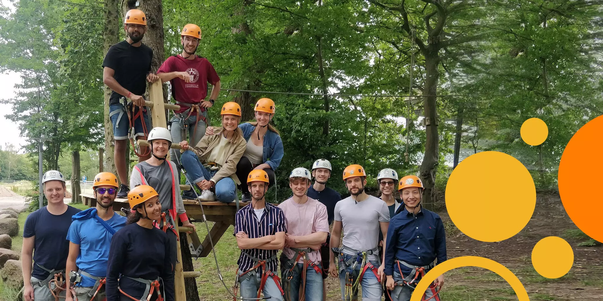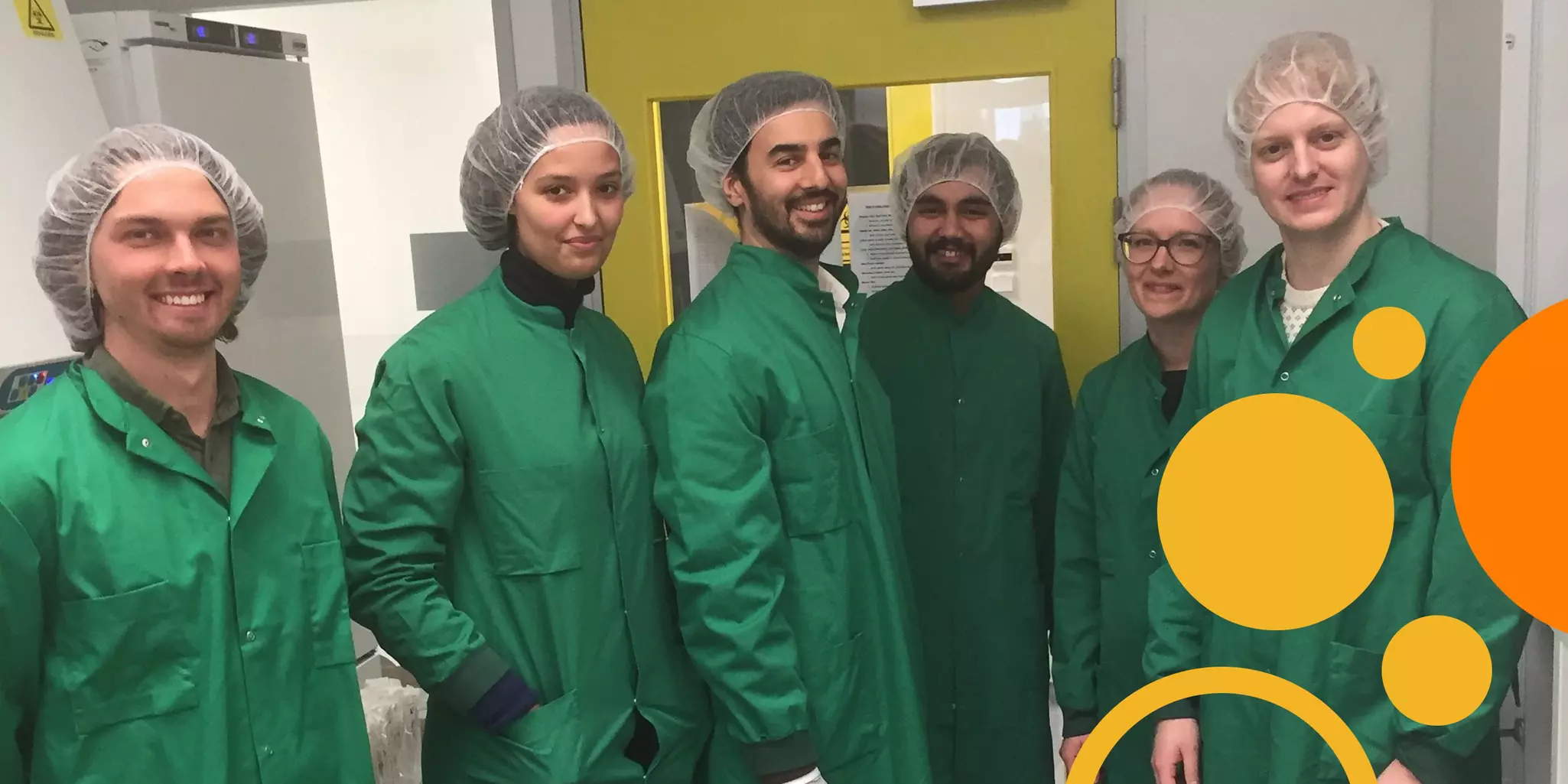
Pause. Take a step back. Learn about people.
Miltenyi Biotec is a community of colleagues fascinated by science and dedicated to empowering cell research. That is our drive. But what makes many of us go the extra mile are the personal contacts we make with researchers around the world. While their work undoubtedly motivates us, their stories as human beings make us smile, reflect, admire, and at times, laugh out loud. Some of them are accomplished athletes. Others lead very different lives outside of the lab. And others yet pursue unexpected hobbies and interests. Each story is unique. Each contact is an opportunity to pause, take a step back, and learn something about people.
The interviews of our Scientific Spotlight are a glimpse into the lives of scientists like you. We hope you enjoy each conversation and find inspiration from their thoughts and experiences exploring the frontiers of the biological sciences.
Interviewees of the Miltenyi Biotec Scientific Spotlight receive no financial or other compensation for their participation.
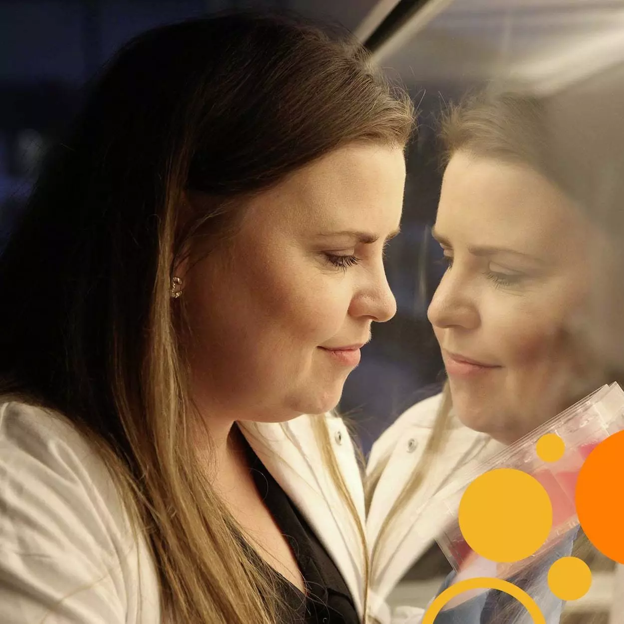
Just do it: building a life, a lab, and a community from the ground up
Interview with Dr. Maria Ormhøj, Assistant Professor in Experimental & Translational Immunology, Department of Health Technology at the Technical University of Denmark (DK)
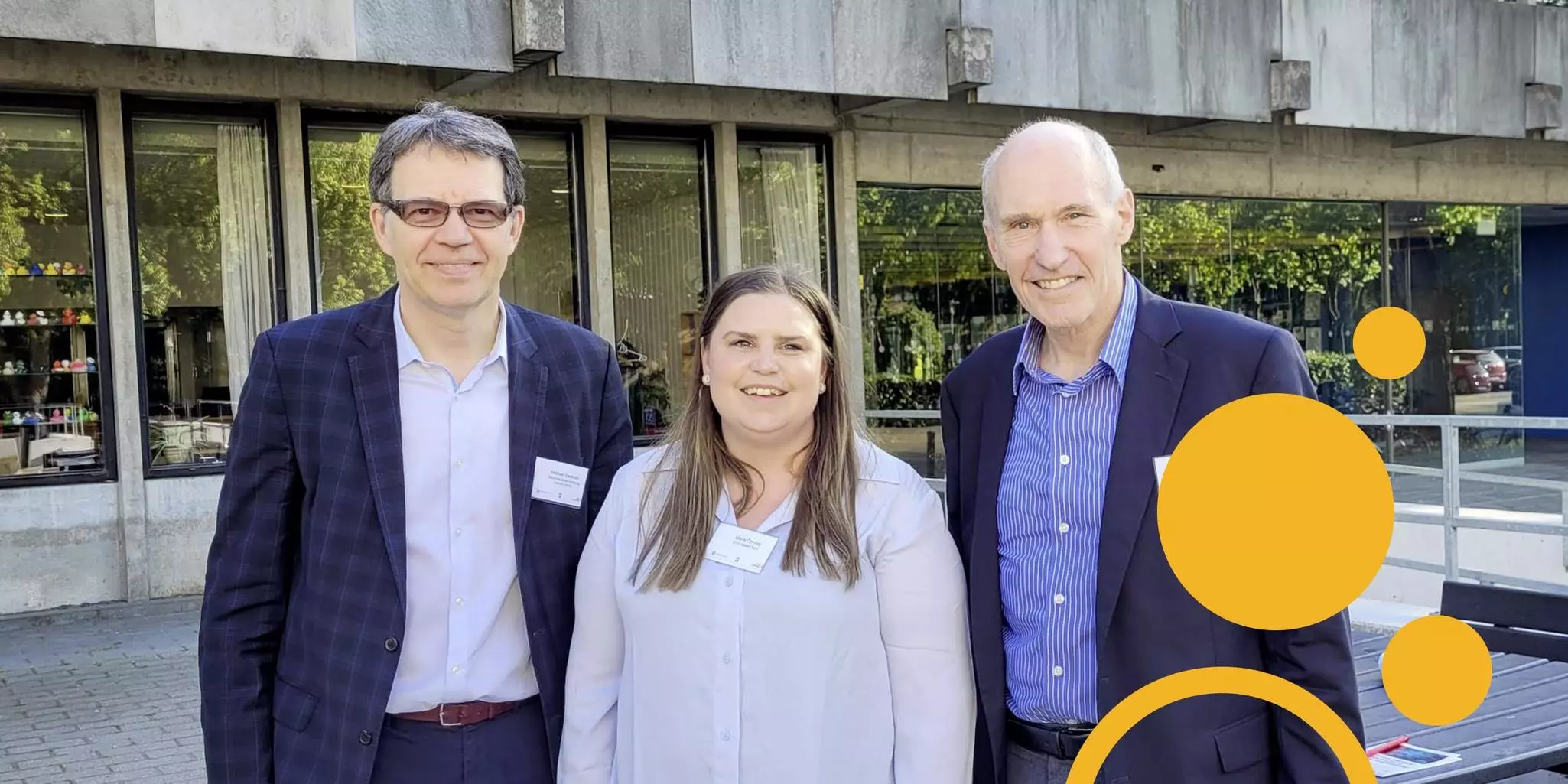
When you think about Denmark, you might think about Vikings, Lego, the Little Mermaid, and sleek furniture. If Dr. Maria Ormhøj has anything to say about it, you’ll soon add engineered cell therapies to the list. Maria is an Assistant Professor in the Department of Health Technology at the Technical University of Denmark (DTU), and she is eager to make the fledging Danish cell therapy community into a global powerhouse. “It’s a really good thing that we are such a small country because we work well together. I think that can bring us to a lot of important clinical discoveries because we can effectively bridge basic science to clinical practice. That is what I really want to do in Denmark: to bring all of that together and build a strong community within the field.” Maria is a mover and shaker among cell therapy researchers. She organizes conferences for networking. She supervises projects to reimagine how cell therapies work. She built the facilities to translate those reimagined concepts into workable therapies. Becoming that impetus took the occasional leap of faith and the tenacity to see it through, but as one mentor described her, Maria is “as persistent as a T cell.”
 View details
Of networks and opportunities
“My philosophy is that no one person can be an expert on everything. So, I always try to think about who has skills that can be brought into a project to make it go from basic science to the production of a treatment. I think that requires a lot of different people, and I try to advocate that when it comes to CAR T cell therapy in Denmark.” Bringing people together is at the heart of much of what Maria does. The national conferences she has helped organize were aimed at establishing collaboration between experienced investigators, young scientists, and clinicians. She is also working with DTU on a PhD Summer Program to train students in cell engineering. Finally, she strives to connect her own students with researchers in academia and industry who can help them reach their goals.
This pursuit of research alliances is a thread through Maria’s own professional development. Looking to challenge herself, she gained experience and colleagues by visiting other labs. As a PhD student, Maria crossed paths with Dr. Marcela Maus, who was setting up her lab at Massachusetts General Hospital and seeking help to launch her research program. So, Maria crossed the Atlantic, husband and young daughter in tow, to spend a year and a half in Boston. “I’m grateful for everything I learned from her and her lab,” says Maria. “It also was a truly amazing environment. You’d have a normal Monday morning meeting and Michel Sadelain or Stanley Riddell would come in. It was fantastic to be a part of that and experience science in such a translational environment. Plus, from a personal point of view, it was very special to bring my family with me and experience a completely new culture with them.” A few years and a second daughter later, Maria’s family would repeat the adventure in Germany while Maria visited the lab of Prof. Dr. Hinrich Abken at the Leibniz Institute for Immunotherapy in Regensburg.
In a quest to understand the immune system, Noah Allen took his research to the sky
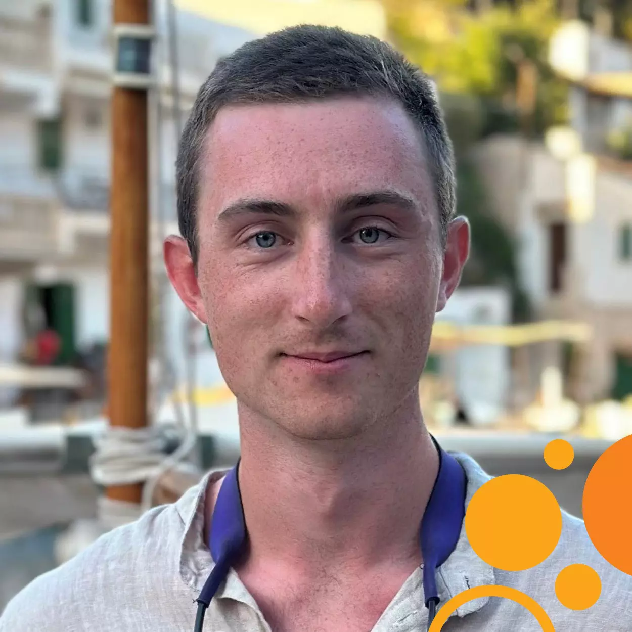
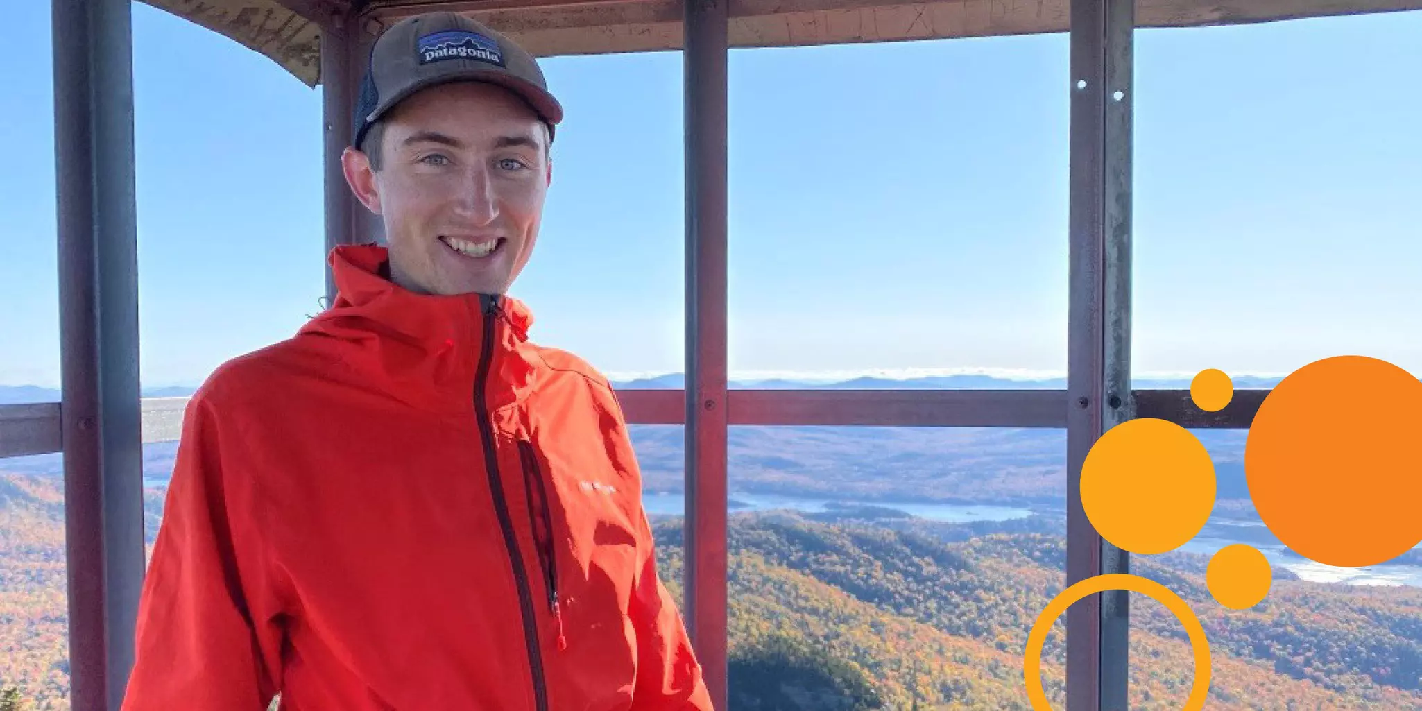
Interview with Noah Allen, Research Assistant and PhD candidate, Rensselaer Polytechnic Institute (US) and winner of the Immunology Innovator Award
Diagnosed with a fused bicuspid aortic valve as a teenager, Noah Allen understood at an early age that medical breakthroughs hold life-changing power. At the time, Noah’s heart defect didn’t warrant immediate action, and since the treatment option was extremely invasive, he decided to take a “wait-and-see approach.” The 26-year-old’s condition ignited his passion for medical advancement and has shaped his ambitions, leading him down a path into the uniquely associated study of galaxies. Now, Noah is dedicated to uncovering the intricacies of the human immune system and how it is influenced by inflammatory environments, ultimately shedding light on this complex network of organs, cells, and proteins.
Out-of-this-world research
As a child growing up on the idyllic 1,400-acre Shelburne Farms property in Vermont, space may have seemed light years away but for Noah Allen it’s been the topic of dinnertime conversations as long as he can remember.
“My dad is always saying ‘OK, OK, but what is gravity?’ ”
It seems quite apropos that Noah would take home the 2023 Miltenyi Immunology Innovator Award in that exact subject, “Does spaceflight induce excessive immune-mediated liver damage?”
The 26-year-old — who was initially interested in pursuing medical school — decided to shift his aspirations while attending Wheaton College in Massachusetts, after developing a passion for biochemistry. Noah ended up graduating with a major in biochemistry and minor in physics.
Fast forward to graduate school at Rensselaer Polytechnic Institute, where his advisor opened the door to NASA and the study of spaceflight research, and Noah quickly discovered that he liked the idea that this work concerns more than celestial objects.
Seeking answers for a healthy life after childhood leukemia
Interview with Dr. Kees-Jan Pronk, Head of Pediatric Oncology, Hematology, Immunology, and Nephrology at Skåne University Hospital in Lund (SE)
In many ways, pediatric cancers are counterintuitive. Children suffer from an insidious disease typical of old age, where developing rather than aging tissues derail and make them very sick. Treatment is harsh and debilitating as rapidly dividing cells are systemically poisoned or irradiated. And yet, kids recover. In high-income countries, over 80% of children with cancer are cured. As Head of Pediatric Oncology, Hematology, Immunology and Nephrology at Skåne University Hospital in Lund, Dr. Kees-JanPronk is at the epicenter of life-changing events for families confronting childhood cancer.
Providing patient care, managing administrative duties, and running a research lab, Kees-Jan witnesses a patient’s journey from different angles. He works with colleagues to administer therapy and manage treatment side effects. Concurrently, he documents the long-term impact of chemotherapy and explores ways to do things better. From that confluence of what is and what could be done, Kees-Jan unpacks the “counterintuitive” of childhood cancers during our interview and reveals how unique aspects of children and their malignancies have defined cancer care over time.
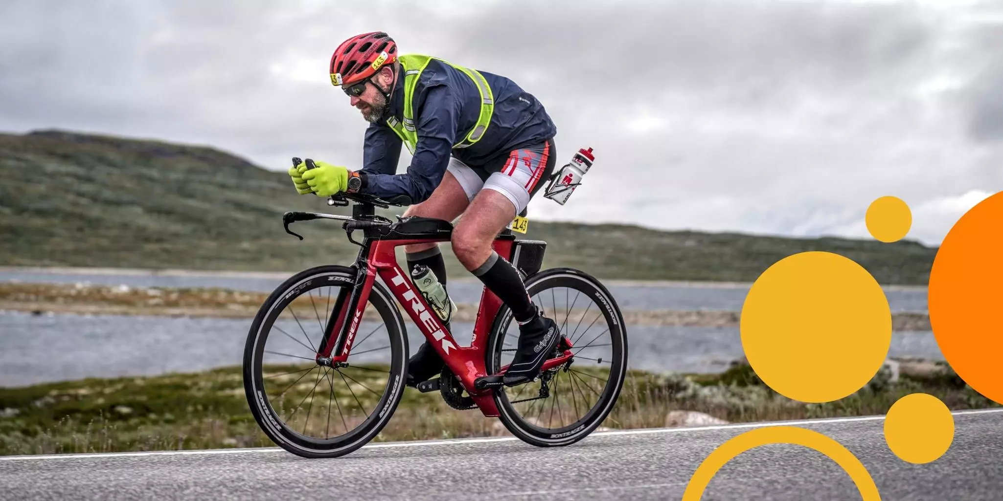
Rare, resilient, and unexplained
Three aspects of cancer in children stand out. First, it is rare. The Cancer Atlas reports a global annual incidence of about 140 new cases per million children under the age of 15. “In Sweden,” Kees-Jan expands, “we have about 350 children diagnosed per year, compared to 55,000–60,000 adults.” Second, children are strikingly resilient to the assault of chemotherapy. Kees-Jan explains that “their bodies are healthier compared to adults, and so, children tolerate higher doses of chemotherapy. Typically, we give chemotherapy and then wait for patients to recover from the effects before we can give the next course. Children just recover much faster. In that sense, we can keep a higher pace and intensify treatment more in pediatric patients.” And finally, although we oftentimes know the genetic insults that drive towards neoplastic transformation, childhood malignancies harbor few known genetic aberrations that can be pursued as targets for small molecule or biologic therapies. New cancer treatments generally serve “the larger number of adult patients that belong to these cytogenetic high-risk groups compared to children.”
The past: chemotherapy debuts
Those three features – rare, resilient, and unexplained – have defined the evolution of pediatric oncology. “Let me start at the beginning,” says Kees-Jan. “The first cancer patients to be treated with chemotherapy were children. In 1947, Sidney Farber in Boston treated children with acute lymphoblastic leukemia [ALL] with aminopterin, a predecessor of methotrexate. While the children did eventually relapse, Farber was able to show that chemotherapy could produce a temporary remission. At the time, this was very controversial. Many people said ‘how can you expose these poor children to these tough medicines? They suffer hugely and die anyways.’ But then, with combinations of chemotherapy, children started to survive. This form of treatment then trickled over to adults.”
The same occurred with acute myeloid leukemia. The initial successful treatment of children subsequently led to the use of chemotherapy also in adults. Arguably, the early successes observed in children are at least partially attributable to their biological resilience after treatment. One can speculate whether the concept of chemotherapies would have persisted as a viable therapy had they been tested first in adults.
The present: a generational gap
Today, however, the focus of cancer drug development has shifted. “A lot of classical chemotherapy regimens and strategies were born in the pediatric world and adopted by adult care,” says Kees-Jan. “Now, though, many of the more modern treatments are developed, tested, or given to adults and then trickle down to children.” In fact, a 2019 analysis of cancer drugs approved by the U.S. Food & Drug Administration revealed a median lag of 6.5 years between their first-in-human and first-in-child clinical trials.
Again, the unique aspects of childhood cancers underlie this 180-degree turn. With comparatively small patient populations and high success rates of first-line chemotherapy treatments, demonstrating the superior efficacy of alternative treatments takes very long. “Consider ALL where we can cure about 92% of patients,” describes Kees-Jan. “If you want to go to 94 or 95%, you need a large number of patients to show statistically that a new drug is better than the old one. That complicates the whole thing.”
Targeted therapies do benefit children. Imatinib, for example, showed efficacy in treating adult chronic myeloid leukemia, but its target, the BCR-ABL translocation, is also present in a subset of pediatric ALL cases. “Children with BCR-ABL translocated ALL went from nearly all going through transplantation and many relapsing, to a situation today where transplantation is not always needed, and survival numbers are much better.” Therefore, authorities have launched programs, such as the Pediatric Investigation Plans (PIP) of the European Medicines Agency and the Research to Accelerate Cures and Equity for Children Act (RACE Act) in the U.S., that incentivize drug developers to include pediatric cancers.
The future: the long tail of chemotherapy
While the survival rates achieved in pediatric oncology are an extraordinary testament to decades of hard work by collaborating researchers and clinicians, it shrouds a critical need at the center of Kees-Jan’s own research. “From the late 1960s to the mid-1980s, there was this drastic increase in survival. In those days, the goal was to cure the patients, but we knew nothing about the late effects of therapy,” Kees-Jan highlights. “With time, those patients became adults, and it became clear that they had paid a rather high price. The numbers today – if you take all who received cancer treatment as a child – are that roughly 1/3 have side effects that mildly impact daily life, and 1/3 have side effects that severely affect their life.”
Kees-Jan is involved in a multi-country study following a cohort of children who were treated for ALL according to the same protocol. “We have this homogeneous cohort of survivors, and we have a lot of knowledge about them because we recorded a lot of variables; we know exactly what type of disease they had and the acute side effects they experienced.” These fortuitous circumstances allow collaborators in Denmark, Sweden, Norway, and Finland to answer longitudinal questions through regular health checkups and surveys.
The team at Lund is responsible for assessing immune reconstitution and kidney health. First, they assess how immune cells recover and protect patients against infection over time. Chemotherapy wipes out at least part of the effects of childhood vaccinations. Thus, patients are re-vaccinated. The team examines how well immunity is reconstructed to achieve the desired protection. Then, to investigate if high levels of methotrexate impair kidney function later in life, the team uses methotrexate clearance profiles recorded during therapy to see if slow-clearing patients are more likely to develop renal problems. If so, clearance rates could be used to identify patients for post-treatment monitoring.
Expanding treatment strategies
Ensuring a good life quality for childhood cancer survivors is a key focus at Lund. However, as Kees-Jan explains, we may need to think differently about future therapies. “Many improvements in chemotherapy have come from increased treatment intensity, made possible by better supportive care. We can treat infections better. We can treat pain much better. We treat bleeding and so on. But we are approaching a limit in classical chemotherapy. What comes next are smarter treatments. Thus, we are looking to de-intensify treatments and implement targeted therapies that achieve equal efficacy but with less toxicity.”
So, in addition to understanding the life-long effects of chemotherapy, Kees-Jan is also exploring a potential target to stop leukemic cells directly. Collaborating with his former advisor, David Bryder, and postdoctoral fellow Isabel Hidalgo, Kees-Jan is exploring the potential role of Bmi1 in leukemogenesis. Bmi1 is an epigenetic regulator of hematopoietic stem cells that is reportedly overexpressed in several hematological malignancies. Using a mouse model system to control the expression of Bmi1 very precisely, the three researchers observed that Bmi1 overexpression led to an altered phenotype not only in stem cells but also in lymphocytes. In some organs of these mice, long-term Bmi1 overexpression produced tissue patches with lymphocytic infiltration resembling pre-lymphoma states. Could Bmi1 be involved in the development of leukemia, and could knocking it down halt the process? They aim to answer these questions in mouse models and pediatric human cells. If the answer is yes, then already-marketed Bmi1 inhibitors may be an alternative to or enhancer of chemotherapy.
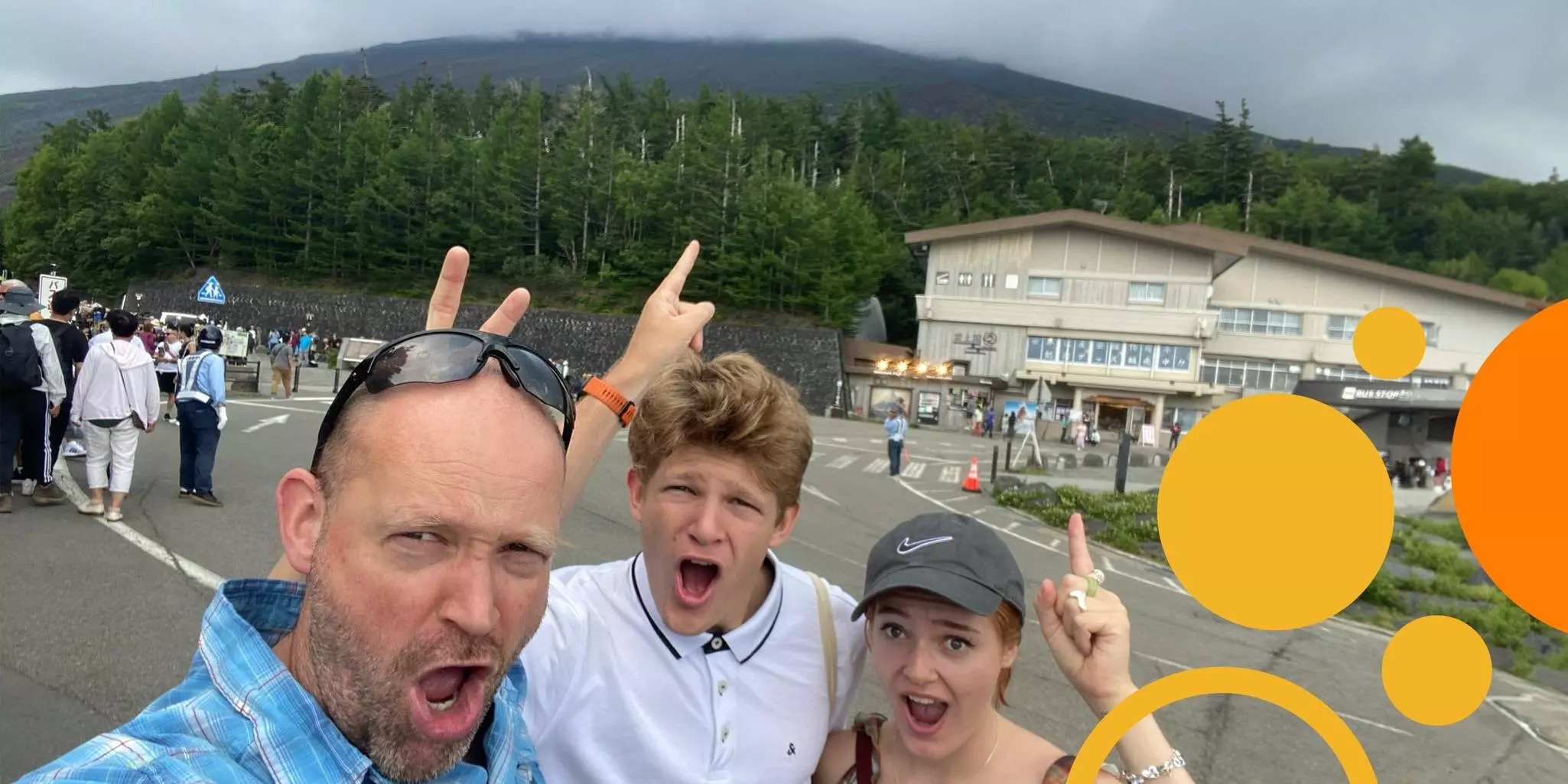
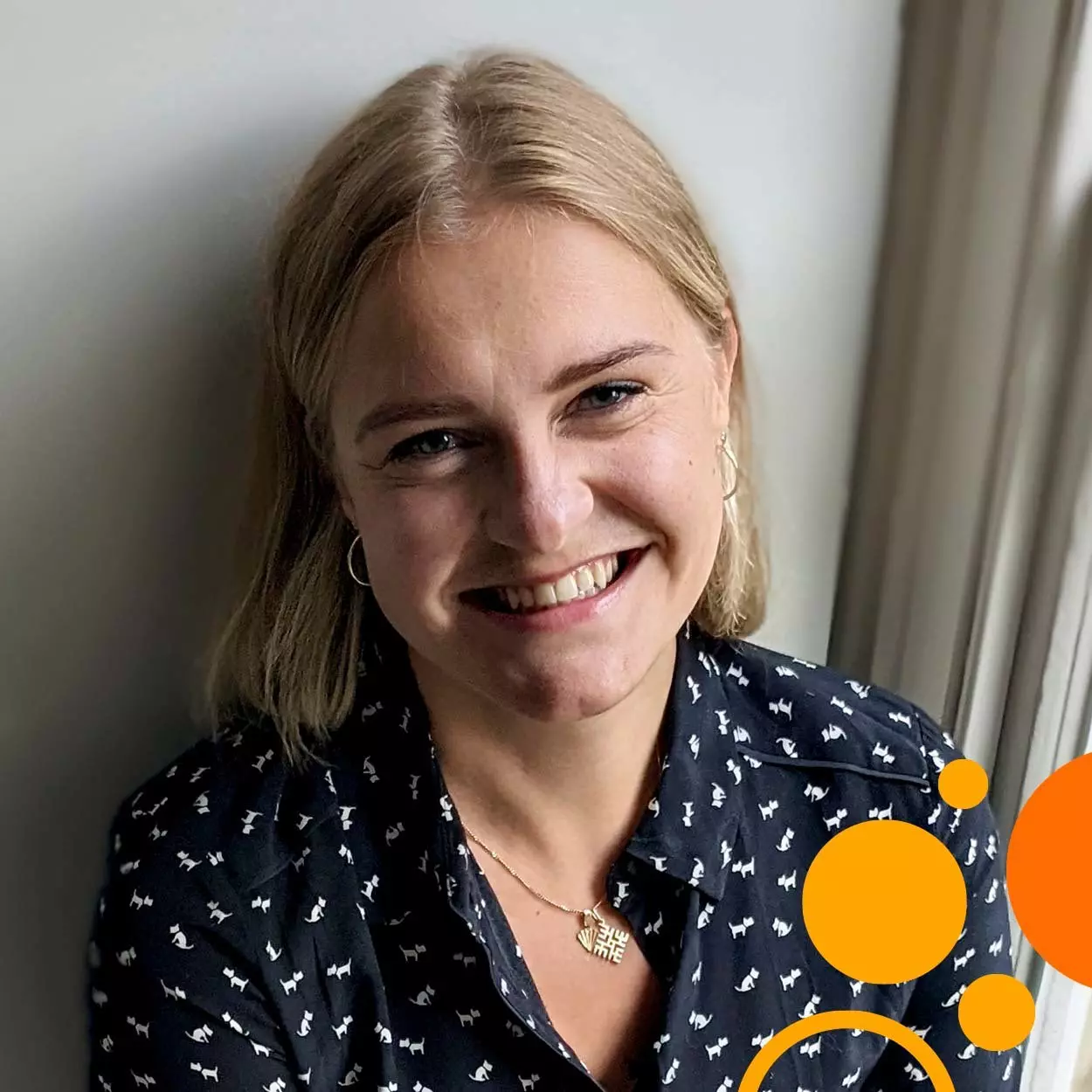
Fierce curiosity and the temerity to pursue it relentlessly
Interview with Dr. Marta Barisa, Senior Research Fellow at the Great Ormond Street Institute of Child Health University College London (UK)
The term ‘innovation’ gets thrown around quite a bit. Influencers splash it across the internet; consulting firms sell its promises; some 70,000 books map out how to achieve it. In that onslaught of advice and business plans, the word has lost meaning. So, it’s refreshing to hear Dr. Marta Barisa talk about her research. She and her colleagues at the UCL Great Ormond Street Institute of Child Health tackle difficult-to-treat cancers by engaging in bold exploration and creative detective work. What Marta calls experimental pediatric oncology is really a foray into the thousand unknowns of why, when, and how cell therapies work. Using every tool at their disposal, Marta and her colleagues leave no idea untried and no assumption uncontested to turn around the paucity of successful cell therapies for solid tumors. In conceding how little we understand, Marta and her colleagues see every attempt to coax immune cells into antitumor action as an opportunity to effect change. And that is true innovation.
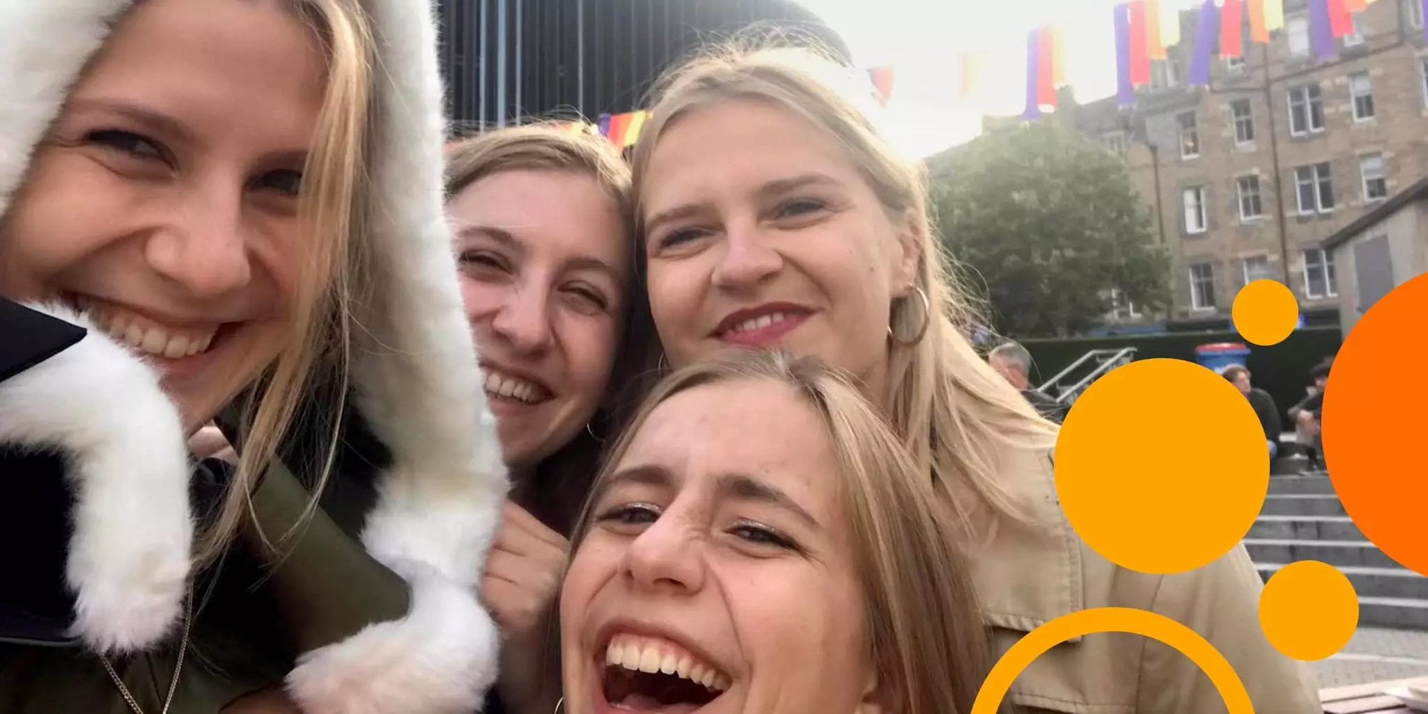
“Shamelessly run with your inner geeky passions”
Marta comes from an international family that lives and moves around the world. Her parents had international careers and raised Marta and her sisters on three different continents. Upon finishing school in Norway, Marta wanted to take on the world. “Norway is beautiful. But to an ambitious, bored 18-year-old, it was suffocating,” she smiles. “I wanted excitement and fun. I decided I would study at St George’s Medical School in London because it was big and exhilarating and scary. I was completely swept up by it all, but I did realize two things. One, that science is super fun. And two, that I’m not cut out for clinical medicine. Death, imperfection, and disappointment are very much par for the course in oncology, and I could not distance myself emotionally.” So, Marta left clinical ambitions behind. A one-year deep-dive into basic research at MIT in Boston eventually led her back to London to study and graduate in a burgeoning new field: cellular immunotherapy.
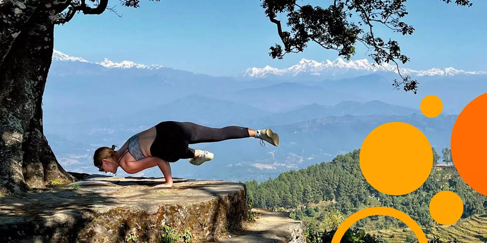
The complexity and scientific audacity of that field has captivated Marta for nearly 10 years. She pursues her research zealously, driven by “the geeky, fun science stuff.” But that’s how she is about everything. To disconnect from work, she devours books on art history, political philosophy, and anthropology. She loves yoga and plays the piano and flute. “For me, that reset comes from immersion into different worlds. I guess I’m not good at doing nothing. I just think that the greatest pleasure and joy in life is to find something that's fun and challenging and then just throw yourself into it completely. I often tell students to have zero shame and just run with their inner geeky passions. Obsessive curiosity will reward you a thousand-fold.”
It's hard to convey Marta’s fervor in sharing thoughts about her work, but perhaps her own words are evidence enough.
You mentioned the opsonin-secreting T cells. How do they work?
Those cells are an example of the multiplex engineering I mentioned. We know that a cell therapy needs a strong signal that engages the effector cell. That is the kill switch. As it turns out, the intracellular signaling of that kill switch is remarkably redundant among immune cells. The bits that differ are on the surface of the cell. So, what if you can use that same kill switch but in a way that engages not only the effector but also other cells? I mean, why keep the signal to yourself when you can share it with friends? That’s what we’ve done with these γ/δ T cells. We’ve engineered them with opsonin-secreting constructs, and the opsonin tags cancer cells for destruction.
The thinking here is to secrete enough opsonin to engage all effectors but also wake up some sleepy myeloid cells, for example. Better yet, we’ve armed our γ/δ T cells also to secrete the inflammatory cytokine IL-15. So, we first engage effectors with opsonin. The effectors kill tumor cells which then release pro-inflammatory mediators. At the same time, our effector is pumping out IL-15 into the tumor microenvironment, tagging tumor cells with opsonin, and killing more tumor cells. It’s punching in every direction inside the solid tumor, and in response, anergic or even protumorigenic myeloid cells, neutrophils, or NK cells redifferentiate and start killing tumor cells. We have shown that our γ/δ T cells can engage myeloid and lymphoid cells residing in the tumor microenvironment. This shared signal concept recruits the patient’s immune system – which is especially relevant for allogeneic immunotherapy – while retaining the functionality of a CAR.
What developments do you anticipate in cell therapies for solid tumors?
At least in the short term – perhaps the next five years – I think we will almost certainly see greater success in autologous α/β T cells and solid tumor immunotherapy. I think we're very close and that the conversation about solid tumor immunotherapy will soon change. In the long-term, my aspiration and main task is to deliver not only solid tumor drugs that work, but affordable drugs that work. While scientifically exciting and attractive, autologous, personalized therapies are not the way to treat pediatric cancers worldwide. If we can make them more accessible and off-the-shelf, if we can mass-manufacture at a reasonable cost, then we must. For that reason, I put my faith in appropriately engineered γ/δ T cells. With some effort, we can make α/β T cells allogeneic, but why not just use a cell type that isn’t alloreactive? So, my medium-term vision is first early wins in efficacy with α/β T cells and then plug those learnings into γ/δ T cells to offer efficacy and affordability. I’d say that the 10-year story is off-the-shelf, properly multicistronic-engineered γ/δ T cells, and that will be a game-changer.
Longer term than that, who knows, really! This field moves so fast that it’s difficult to predict where it will be in 20 years. And that’s what makes this fun.
On resetting rogue immune cells and oneself
Interview with Dr. Nazia Matto, Research Associate and Lead Production Scientist in the Department of Hematology at King’s College London (UK)
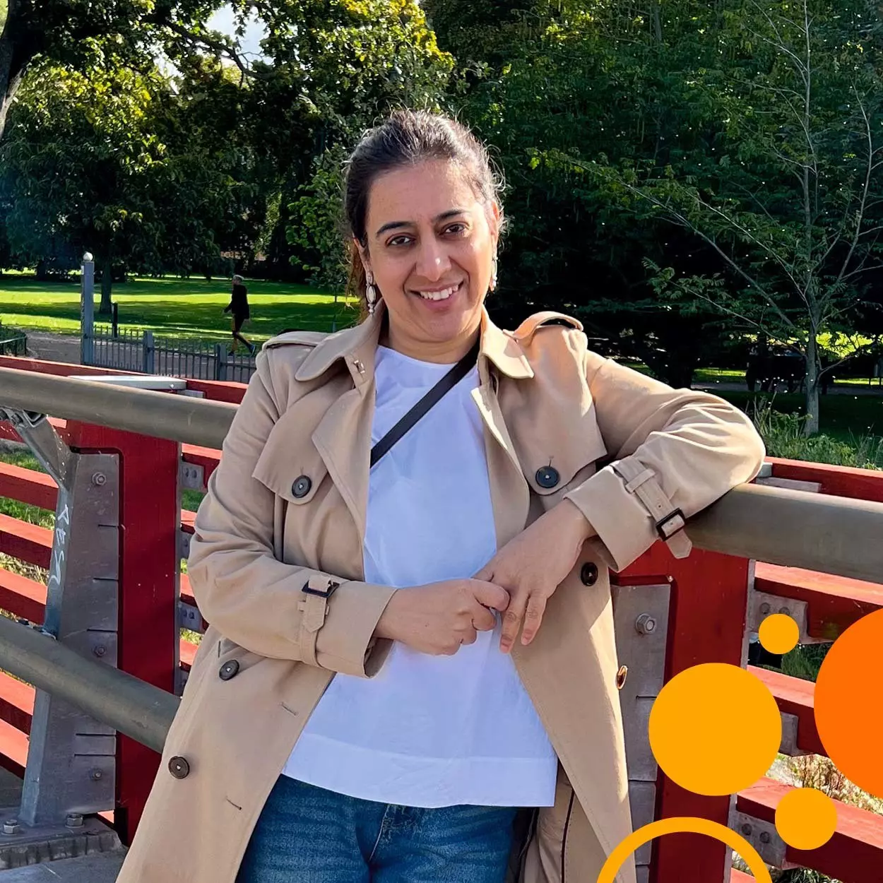
Nazia Matto sits comfortably in front of her computer. She smiles almost imperceptibly as she evaluates how best to describe her current position at King’s College London. It was meant to be a split position. Some of her time, she would harvest and expand regulatory T cells from patients with aplastic anemia as part of a Phase I dose study to treat their condition. The rest of her time, she would conduct the analysis of the samples collected from these patients before and after the treatment infusion. And yet, at this point, she finds herself working full-time on production.
“The challenge that I face is that we are harvesting these cells from anemic patients,” Nazia explains. “So, even though we are using a leukapheresis product as starting material for each patient, their overall deficiency compared to normal subjects means that we are starting expansion from very low numbers of T cells. That raises the possibility that we might not achieve the desired dose.” Nazia doesn’t mind the change in plans. The loads of paperwork she manages or the 12 to 14 hours on her feet when cells are collected from patients also don’t bother her. But she does think a lot about the responsibility of producing what is a last-resort treatment for the patients in the study.

Keeping effector T cells in check
Aplastic anemia is an autoimmune condition that can progress to myelodysplastic syndrome and acute myeloid leukemia. The patients that Nazia treats are unresponsive to immunosuppressives and ineligible for a stem cell transplant. The hope is that the infused dose of autologous regulatory T cells will control the overactive effector T cells causing the disease. “We have a number of assays we are performing to evaluate the response to treatment, but an important parameter we are looking for is a change in any compartment of blood cells,” says Nazia. “These patients need regular blood transfusions, so that change is significant.”
Nazia will be pausing her work in the cleanroom to do preliminary analyses of results from the three patients that have been treated so far. Those data will be presented to the Data Monitoring Committee to assess outcomes so far and review any potential side effects. Barring any problems, the study will continue until September.
The carefully structured world of GMP
Nazia was trained in good manufacturing practice (GMP) to expand the regulatory T cells in the study. She also optimized and validated the protocol that she follows every time she enters the cleanroom to prepare another patient dose. She explains that for ethical reasons, the preparatory work could not be done with samples from anemic patients but rather with leukapheresis products containing normal numbers of leukocytes. This adds a certain edge to working with study samples. “With every patient, I am a bit nervous. For one, when I get the patient sample, there is always the open question of whether I can obtain a sufficiently high number of cells in my starting material. Then, will the cells grow? And even after I have reached the desired dose, the product must meet release criteria. Any number of things can result in a failed batch. That’s a lot of money and, more importantly, disappointment for the patient.” So far, Nazia’s batches have all been successful. Talking to her, however, you get a sense of the constraints that GMP cell manufacturing can (and should) impose. “I do my level best. I’m meticulous and focused. Still, in the GMP setting, you have to follow the paperwork blindly – there is no room for diverging from the protocol, regardless of what you see happening in a batch.”
With a strong background in research and an itch for innovation, this tension between GMP standardization and a desire to tackle complications is very palpable for Nazia. It reemerges in thoughts about her personal development. Nazia intends to get additional training in GMP to expand her skills and expertise, but she is also attracted to the idea of working in development. Balancing her proficiency in manufacturing with a desire to explore the boundaries of technology will be a decisive advantage in the future, wherever her professional career takes her.

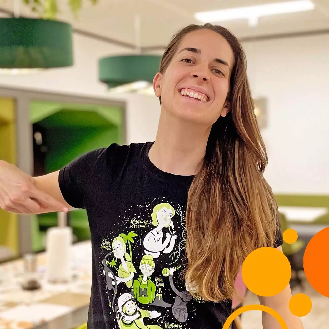
How to spot a multipotentialite: a case for being like a stem cell
Interview with Ana Pilar Rubio Gomez, Scientist in Cell Process Development at Orchard Therapeutics (UK)
A multipotentialite is someone with many intellectual and creative pursuits. They dive into one topic, resurfacing with skills and competencies. Then they discover a next interest which they pursue with the same elan. That doesn’t mean that they are unfocused. Quite the contrary. Each intellectual or creative foray is a path of intense, fast, and even fearless learning. What they learn, they expertly merge into a grander whole, no matter how disparate the concepts may seem. And the allure of new knowledge and experiences makes them thrive on diversity of situation, companionship, and challenges. Ana Rubio is a multipotentialite. She laughs when you tell her that but then retorts, “I like to think that I’m like a stem cell. I can become whatever.” Her reply is so fitting because if there is one thing that has served as a beacon throughout her career, it has been stem cells.“I’m not passionate about just cells,” she says. “I’m passionate about stem cells. They can become anything with no more than a few signals added to a petri dish, and that’s just crazy!”
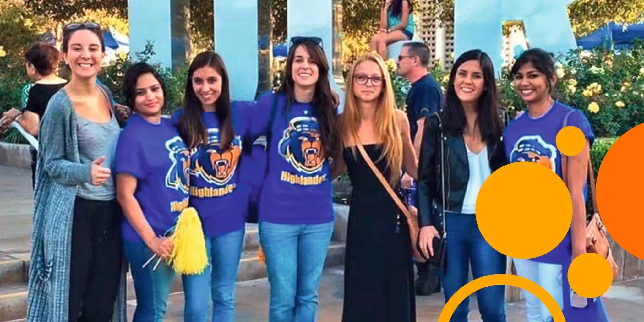
The first sign of a multipotentialite: idea synthesis
Ana is a Scientist at Orchard Therapeutics, a young and active company in the field of gene therapy using hematopoietic stem cells to potentially correct the causes of rare genetic diseases (affecting less than1% of the population). There, Ana bridges the gap between the initial development of a therapeutic strategy and realizing a fully manufacturable, feasible therapy product. “Our team works on making cell processing more efficient, robust, automated, and scaled. We find ways to reduce costs, shorten time in the cleanroom, and simplify processes to reduce the training burden on operators. For that, we need to understand the science, but the parameters we manipulate are geared toward getting the product through manufacturing and to the patient.”
When Ana describes her work responsibilities, it is clear why she enjoys her job. Ana is at home where several ideas intercept to solve concrete problems. Expertise among the various departments at Orchard is complementary, so each project is highly collaborative. Ana talks excitedly about brainstorming with colleagues and working out ways to resolve aspects of the manufacturing process. The interdependence of workflow steps makes it imperative to bear in mind and communicate about the entire system, from cell harvest to product infusion. Steeped in that diversity of know-how, Ana evaluates every change she makes to optimize a process in the context of manufacturability, time, cost, logistics, patient needs, and safety. “The Big Picture,” she calls it, and it’s a key mindset for her job. “Everyone in the company has the same purpose, and you work together toward that goal. While projects have unique challenges and your skills are tailored to tackle specific demands, you need to consider how your contribution impacts the endpoint.”
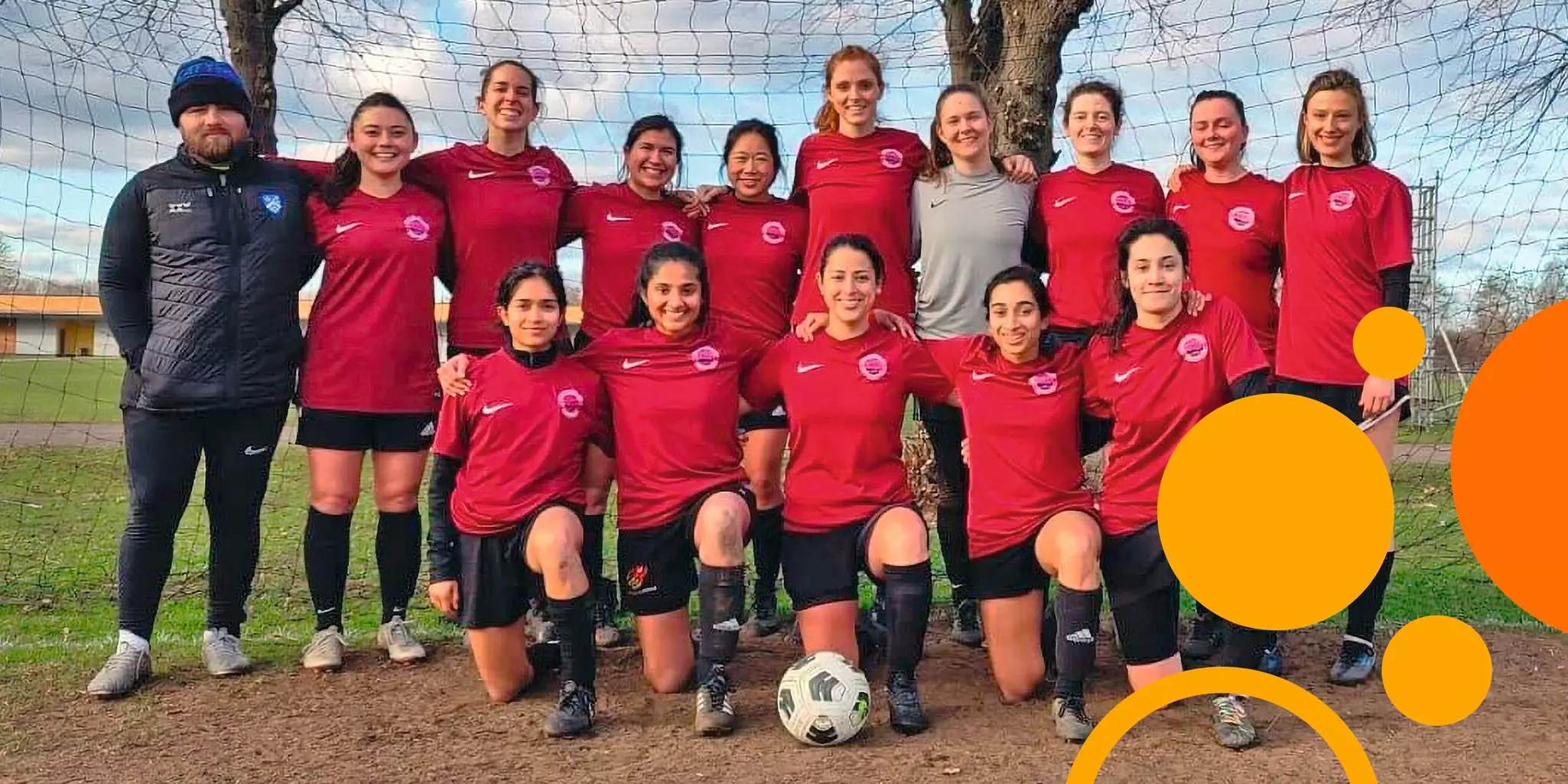
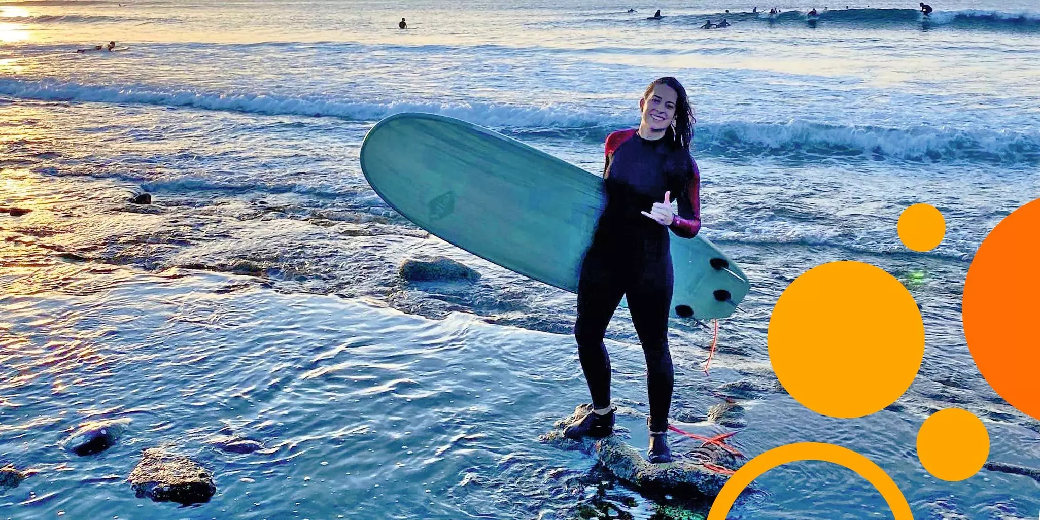
Embrace your (multi)potentiality
Although jazzed by her experience at Imperial College London, Ana knew that she did not want to go on to do a PhD. She wanted practical applications for her work. She sought brainstorming and exchanges with colleagues fueled by a common goal. So, she turned to industry, and she says that the last three years have been exceptional. She sees herself at the epicenter of what will someday be a seismic change in medicine. “It’s amazing how many companies are doing cell and gene therapies, but if you look closely, only a handful are at the point of approval, and there is a large gap between them and everyone else. I personally think the breakthrough will come when that gap is closed and when we transition to allogeneic strategies. Then you can multiply the number of patients you can treat with the same material. Then you can streamline and reduce manufacturing costs. Then these treatments become more accessible to everyone.”
For now, however, Ana found the type of teamwork she wanted, not only in terms of collaboration but also in terms of diversity of thought, background, and experiences. “That’s why I’m in London,” she says. “I love going to work and people telling me about their homes. Going to lunch and people bringing Tupperware with different foods.Those are tiny things that maybe people would not notice, but I like them because I learn new things every day, and not just about science. All these small details bring happiness to my workplace.”
Ana is one multipotentialite who’s found her place in the world.
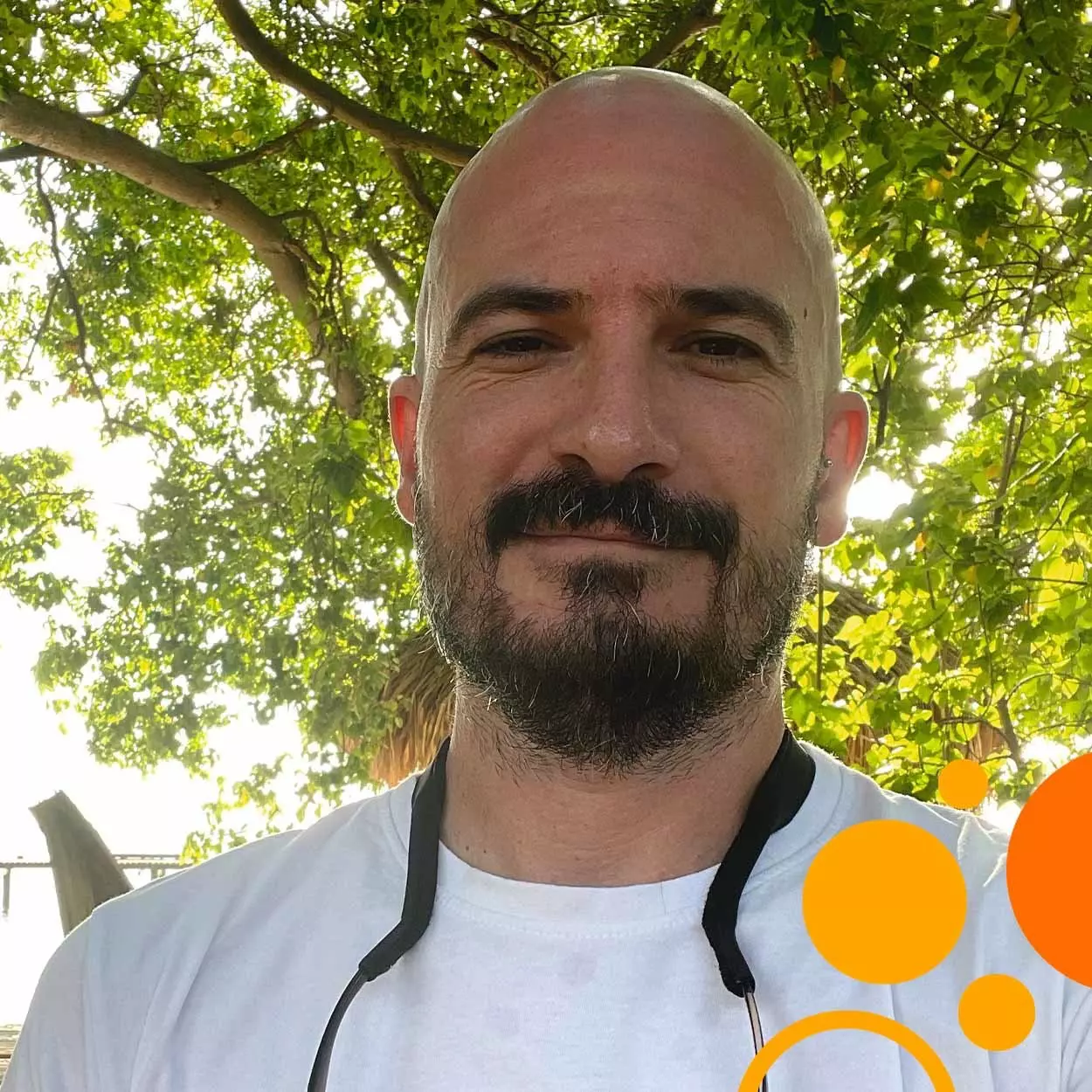
A microbiophile probes an invisible world of friends, foes, and manipulators
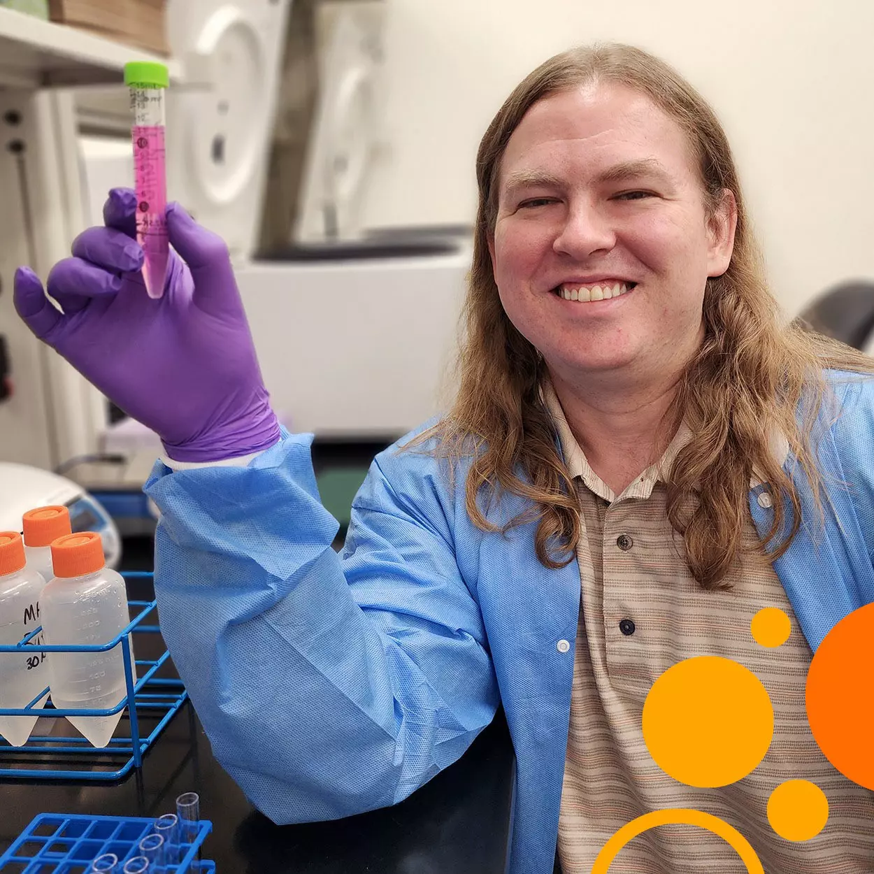
Interview with Dr. Javier del Campo, Group Leader at the Institute of Evolutionary Biology CSIC-UPF in Barcelona (ES) and Adjunct Professor at the Rosenstiel School of Marine, Atmospheric, and Earth Science at the University of Miami (US)
We all have a sense of what an individual is. You are distinct from your dog. But what about the microflora in your gut? Given their vital role in determining your health, might they be a part of you? And by functional extension – assuming your dog makes you smile and forces you to get out for fresh air – might your dog also be a part of you? If you’re giving this some thought, conversing with Dr. Javier del Campo would be an eye-opening experience. While the example with your dog might be a bit of a stretch, Javier explores similar questions in complex systems of organisms where the boundaries that define one individual from another are blurred. Engaged in diverse and intimate relationships, each member of these systems can drastically alter the nature, function, evolution, and survival of others. Javier is fascinated by the impact of these cross-species affairs and what they reveal about the almost-alien microscopic world.
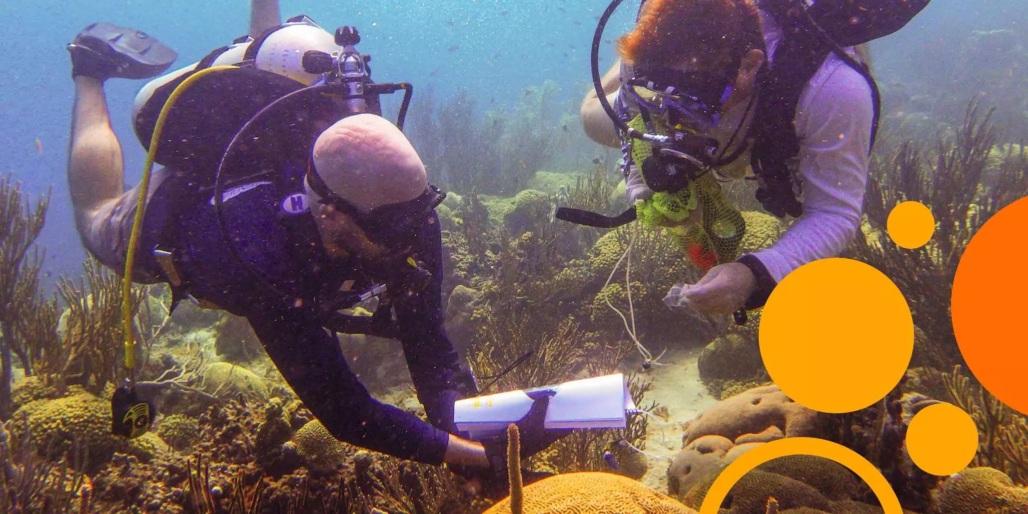
Boundless entities meet boundless lab
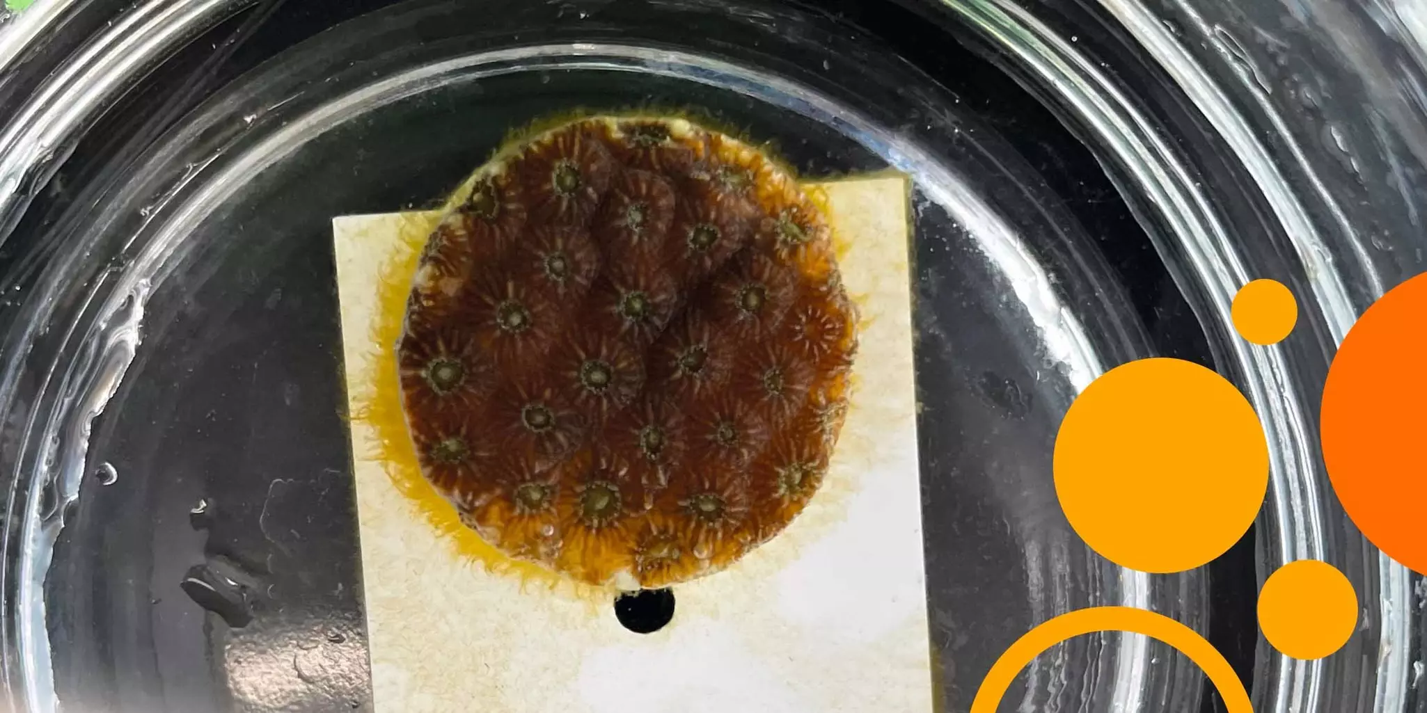
The systems Javier studies consist of corals and the organisms that live in them, on them, with them, and close to them. Javier explains that these assemblages of eukaryotic and prokaryotic partners – termed holobionts – are not defined by physical barriers. “In a coral, some microorganisms live within tissues and cells. Those are obvious members of the coral holobiont. But then you have the mucus that covers the coral. That mucus acts as a buffer with the external environment, and microbes live there too. Closer to the coral, the mucus microbial community is similar to that living in the tissue. As you move away from the coral, the microbial community changes in a matter of millimeters. It transitions to a composition that is more like communities in the surrounding water. There is no boundary where that transition suddenly happens. The mucus diffuses, and the community gradually becomes that of liters and liters of water around you.” Just so you don’t feel left out: you walk in a microbial cloud, a personal waft of biological hitch-hikers that likely extends (get this!) 90 centimeters from your body. So, where does a holobiont end? Javier takes a deep breath: “Holobiont is a definition because we need definitions. We put boundaries to what a holobiont is when there is no boundary in reality. The key is to have intimate relationships.”
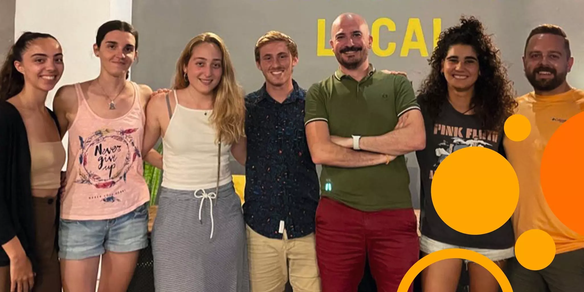
Minuscule but gargantuan: the microscopic world
The ubiquity and importance of microbes are no secret. We don’t see them but know that they move essential molecules from one biogeochemical phase to another, influencing earth-scale cycles. In fact, Javier and collaborators in Miami suspect that gut microbiota is responsible for producing the calcium carbonate pebbles that teleost fish poop out. That skill does not appear to reside in the physiology of the fish themselves. With those pebbles accounting for 3–15% of total oceanic carbonate, microbes may be heavy lifters in the workings of our planet and an essential node in carbon sequestration and ocean acidification models.
Leaving planetary dynamics behind, Javier draws your attention to the other end of the spectrum: the tiniest scales of life. “Microbes are everywhere and associated with every living being, even with other microbes that live at different scales. A microbe that is 200 microns in size can contain a plethora of smaller microbes!” And there, the cadence of Javier’s voice when talking about the minuscule divulges what drives his scientific curiosity. Microbes are his passion. He wants to know their world – how they perceive, respond, and modify it. In a holobiont, that translates into understanding how microbes build and rework relationships with their host over time and how, under certain circumstances, they disrupt its physiology.
On the Astros, astronauts, and the NK cells that live in them
Interview with Dr. Austin Bigley, Vice President and Head of Research and Development at Indapta Therapeutics (US)
“One of the things that happens in spaceflight is that people reactivate viruses, like cytomegalovirus. And it’s a weird biology, because you see the phenotypic shift associated with activated NK cells, but the cells won’t kill effectively.” Dr. Austin Bigley knows a thing or two about unexpected behavior in natural killer (NK) cells. Throughout his scientific career, he has watched NK cells respond to extreme conditions. He’s examined them in blood samples from astronauts on the International Space Station. He’s quantified outpours of NK cells into the bloodstream of subjects that are working out. And he’s documented a counterintuitive blunting of that exercise-driven effect by cytomegalovirus (CMV) infection, which normally activates NK cells. Today, Austin uses his intimate knowledge of these enigmatic cytotoxic lymphocytes at Indapta Therapeutics. As Vice President and Head of Research and Development, he is working to transform a unique population of NK cells – Fc epsilon RI gamma-deficient g-NK cells – into a powerful cancer treatment.
Like saying I'm in a big rock band
Austin chuckles when asked about his time working on a NASA-supported award to characterize the immune function of astronauts during spaceflight. “I’ve never found anybody who isn’t interested in that,” he says smiling. “You’re at a wedding and people may or may not be interested in your cancer work, but the NASA connection… It’s like saying, ‘yeah, I’m in a big rock band’ or ‘I play for the Astros’. Nobody’s not interested in that.”
In all fairness to admiring listeners, however, the project can only be described as awesome. Start with a small, elite study cohort, of which only a handful of subjects is actively going into space. Then, work out the logistics of getting fresh blood samples (yeah, you read right: fresh) from space to Houston. “They’d come back down to Kazakhstan, and then go to Scotland, and then get to Houston – all within 72 hours!” shares Austin. “We would get these samples in the middle of the night and would test them against their matched ground-based controls.” Finally, add that samples were taken not only before and after, but also during each astronaut’s stay on the ISS. That meant that one astronaut’s 90-day blood draw had to be timed with another astronaut’s return to Earth. And the returning astronaut also donated blood before embarking on the journey for their “return minus 1” data point.
In all, Austin’s project tested 6 astronauts in space for 6 months, 6 matched controls who stayed on Earth, and one very experienced long-hauler who was in space for a year. Every tested astronaut had impaired NK cell function at the midpoint of their stay compared to preflight levels and matched ground-based controls. While the effect was less marked in the experienced astronaut, it was still present, and this raises questions for longer-duration missions. “This is one of those things that needs more research. They need to figure out what’s going on for longer spaceflight, like the 9-month journey to Mars,” says Austin.
Curious who those very experienced astronauts may be? NASA keeps track here.

Of great minds and elegant science
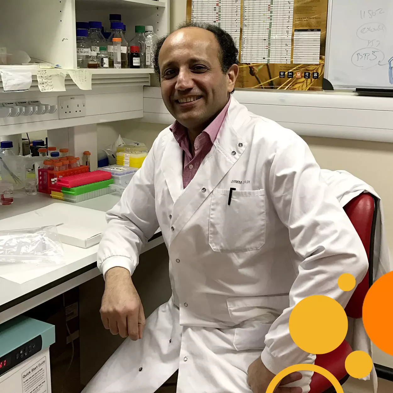
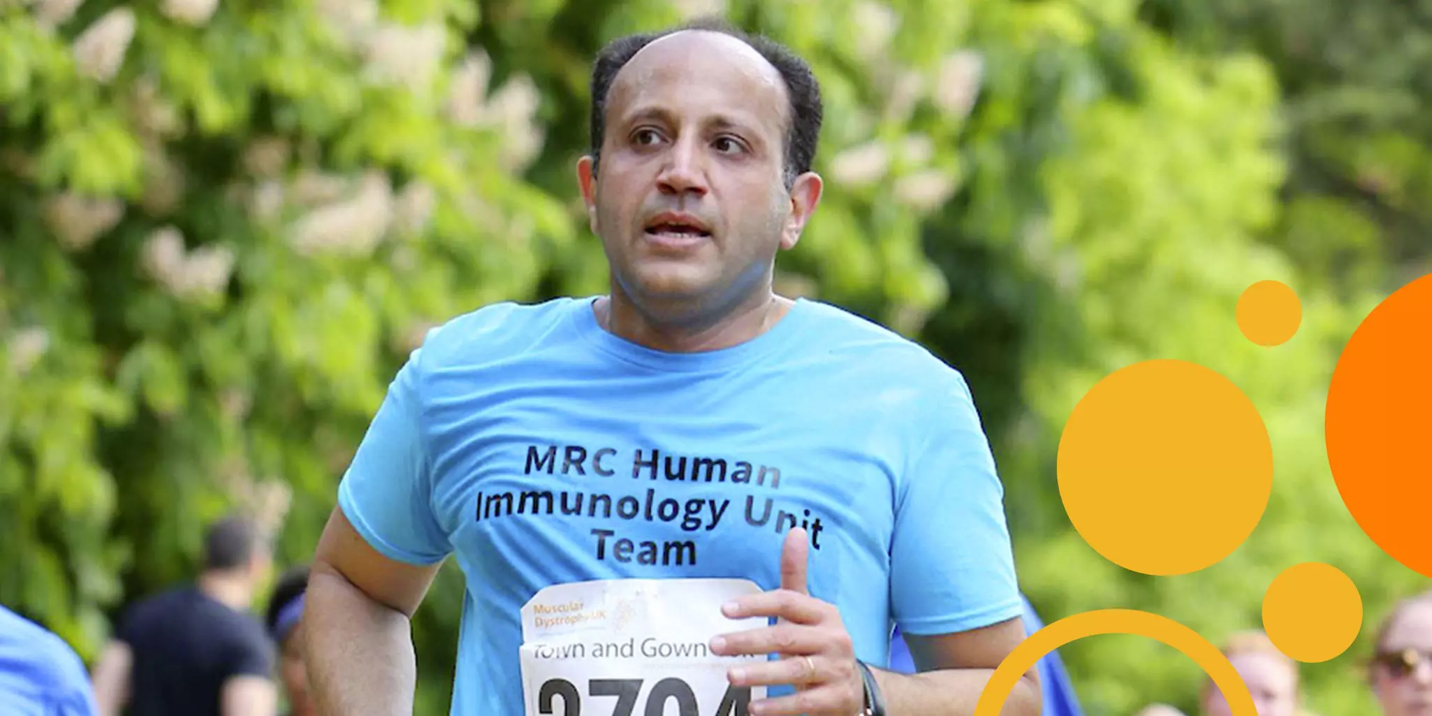
Interview with Dr. Ahmed Ahmed, Professor of Gynecological Oncology and Director of the Ovarian Cancer Cell Laboratory at the Weatherall Institute of Molecular Medicine (UK)
In ovarian cancer circles, he is recognized for his work on p53. Dr. Ahmed Ahmed’s groundbreaking paper in 2010 demonstrated that high-grade serous ovarian cancer is a p53 mutated tumor and marked the beginning of his systematic characterization of the origin and genetic drivers of the disease. His work has greatly improved the identification of ovarian cancer patients for treatment.
 Play video
A young doctor discovers his vocation in an intriguing paper
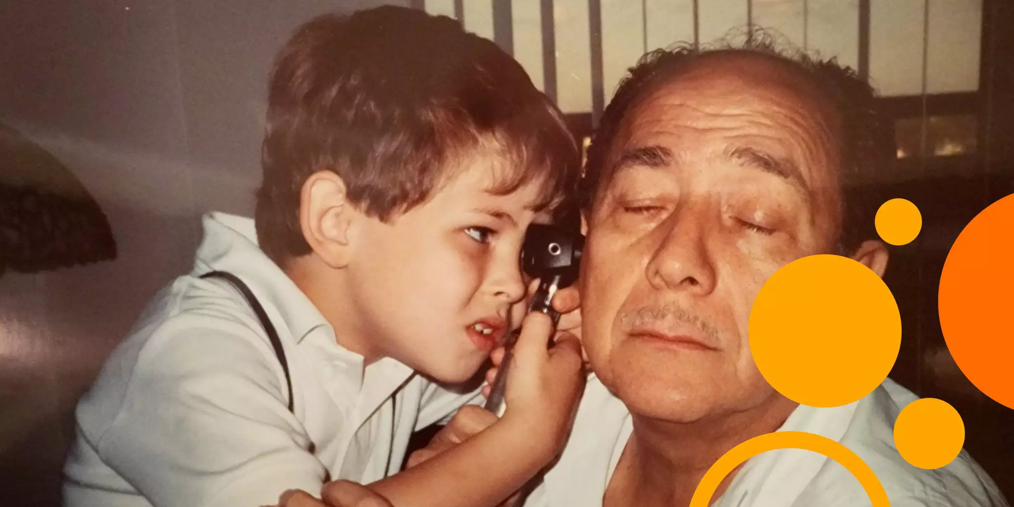
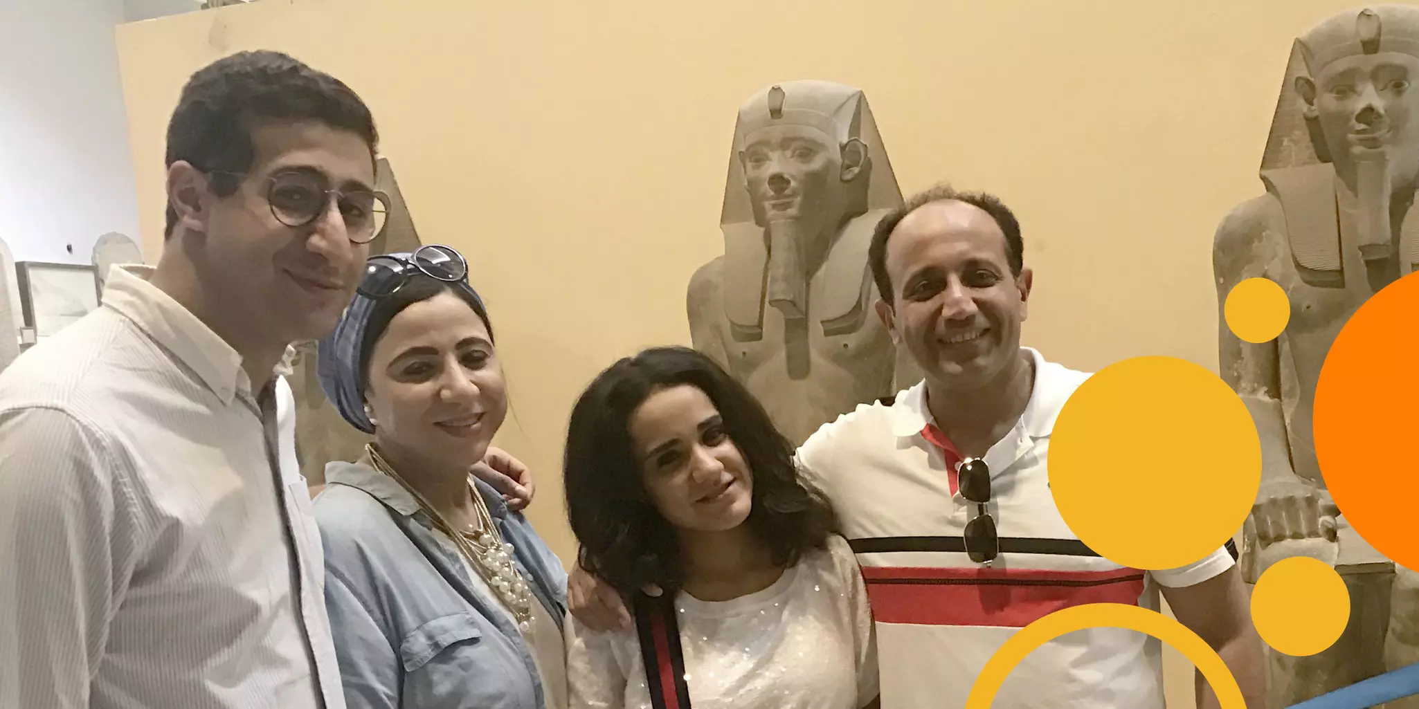
I've really always had a passion for trying to understand the how and why, and I naively thought I’d find all the answers in studying medicine. But actually, there were more questions than answers. My father is a retired surgeon. I think he was also very aware that there were questions to be answered; there were things to be made better. And that's what he encouraged me to look at from very early on. He always thought that me taking the route of discovery is more important to him than keeping me next to him in Egypt. I really owe it to him that he encouraged me to learn, think, and go wherever I need to go and stay as long as I need to stay to discover new things and try to improve things for patients.
When I started in medicine, I met my wife. Her father was a professor of biochemistry at the university where we both studied. He gave me a paper written by Professor Bob Bast where he made the discovery of CA125, which is the tumor marker that we now use for ovarian cancer. That paper was really fascinating to me as a student because it showed me how following simple, solid principles of science can lead to discoveries that make a huge impact. That really infused me to pursue a career in translational medicine – seeing how Bob had done his experiments, repeatedly looking at differences in antibody reactions between ovarian cancer and normal cell lines and finding at the 125th attempt an antibody that separates the two lines. They published these very nice observations and validated them in a large group of patients, showing that CA125 can discriminate between cancer and normal. And then it became routine! Every patient with an ovarian mass benefits from this discovery because patients where CA125 is elevated have priority for rapid surgery.
A very nice, really simple idea, but done very rigorously, lead to something that has helped a lot of people. That made the connection for me. It was clear that I needed to do research and for that I came to the UK. Then I took the opportunity to work for Bob Bast, which was a turning point in my career. Those two years were amazing for me. They helped me to develop as a scientist and gave me great exposure to science in the US, which I find fascinating and exciting.
The Oxford Classic: a radical shift in classifying ovarian cancers
Ovarian cancers have been notoriously difficult to classify in a way that supports better patient care. I thought, maybe the difficulties stem from trying to directly classify a highly heterogenous tumor. Instead, if we classify normal cells and then look for those subclasses in the tumor, we may be able to do a better job. So, we looked to subtype normal Fallopian tube cells using single-cell RNA sequencing, classify each cell into different molecular entities. We came up with 52 genes that could separate nicely ciliated cells and four subtypes of secretory cells. Then we thought, “if we now go and look at ovarian cancers, can we classify them into different types using the 52 genes?” And the answer was “yes, we can.” If this classification was meaningful, some of these subtypes would have an impact on how well a patient does. We called the classification system the Oxford Classic and tested it in stratifying patients with serous ovarian cancer. The Oxford Classic defined epithelial-to-mesenchymal transition-high (EMT-high) tumors that were consistently correlated with poor prognosis. In every single study that we looked at – we've now looked at nine different studies – we found that the EMT-high subtype, as classified by the Oxford Classic, indicates an up to 3-fold increase in risk of patients progressing or dying early.
The implications of using normal tissue to classify a tumor is fascinating and generalizable to other tissues. We want a phenotypic classification based on molecular background and one solid way to do this is to subclassify normal tissue and then look for those subclasses in the tumor. If you're able to find those connections, they will be solid connections because they are inherited from the normal tissue, and they stay there as the tumor develops.
What is the Oxford Classic?
The Oxford classifier of carcinoma of the ovary, or Oxford Classic for short, uses a panel of 52 genes to identify subtypes of serous ovarian cancer – the most common form of cancer of the ovary, which nevertheless has eluded classification that allows predicting prognosis. By stratifying patient populations according to the Oxford Classic, clinicians can better predict disease outcome and therapy developers gain additional insights about ways to target and eradicate cancerous cells.
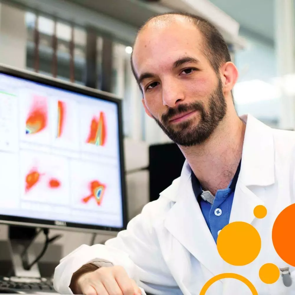
Unwavering. One man's quest to elevate lipids
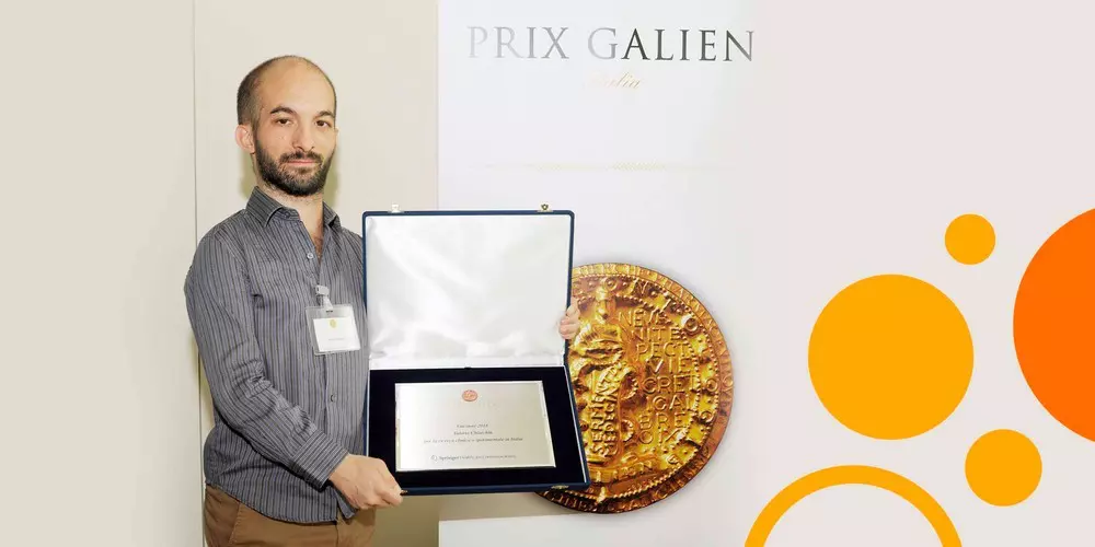
Interview with Dr. Valerio Chiurchiù, Neuroimmunologist at the Institute of Translational Pharmacology of Rome, CNR, and Santa Lucia Foundation Hospital (IT)
Breaking new ground in the sciences takes tenacity. Courage may start you down a less traveled research path, but Valerio Chiurchiù will tell you that stubborn persistence keeps you going. Determination is your best companion in uncharted waters. Valerio is an acclaimed researcher in neuroimmunology and pathophysiology. He is also an assistant professor in the Faculty of Medicine at the University of Rome but works primarily at the Institute of Translational Pharmacology of Rome, CNR, and at the Santa Lucia Foundation Hospital. There, Valerio heads the Laboratory of Resolution of Neuroinflammation.
DR. VALERIO CHIURCHIÙ
TITLE: PhD, Assistant Professor
INSTITUTE: Institute of Translational Pharmacology of Rome, CNR, and Santa Lucia Foundation Hospital (IT)
RESEARCH FOCUS: Immunomodulatory role of bioactive lipids in health and disease
QUOTE: “Sometimes in biochemistry, scientists do not see everything together. It is very compartmentalized. We should unify all this because in the end, these molecules talk to each other. A lipid will bind a protein, and that complex will go to DNA. So, in one step, we are talking about three different categories of molecules. This is missing in science now.”
“What I do embraces many fields of study,” Valerio describes. “I am an immunologist, a neuroscientist, but also a lipidologist. I don't know what that makes me. Maybe a neuroimmunologist in the field of lipids, something like that. What it means, however, is that I study the immunomodulatory role and the mechanisms of action of several bioactive lipids in health and disease, but mainly in the central nervous system.”
Valerio and a tightly knit community of lipid-studying scientists around the world are, in fact, leading a shift in our current perception of the who and how in inflammation. “For a long time,” says Valerio, “we thought that we have an inflammation, that inflammation becomes acute and you feel the symptoms in your body, and then all of a sudden, it disappears. Somehow your body manages to turn down all these processes, your symptoms disappear, and you get well. This is not the case. We don't resolve inflammation because all these inflammatory mediators just magically stop being produced by cells. Instead, at a specific time point, the cell undergoes a metabolic shift and starts to produce specialized lipid-based molecules to resolve the inflammation and re-establish homeostasis so that damage can be eliminated from the body and tissues can be repaired and function again.” These scientists have introduced a new set of molecular regulators of inflammation and, unexpectedly, they are hundreds of lipids.
Advocacy forged from personal strength
Advancing discoveries and ideas about the regulatory function of lipids is slow-going given the dominance of DNA and protein in the public mind and scientific discourse. Open any biology textbook and there’s a good chance that lipids are relegated to a structural role exemplified by the ubiquitous phospholipids of membranes. We rarely think of lipids as active agents of the cell, although we’ve known about the hormone-like eicosanoids (e.g., prostaglandins) since the first decades of the last century and about their binding receptors for over 40 years. Numerous other lipid families also carry out essential functions, like the endocannabinoids, sphingolipids, and ceramides. “We purposefully call them bioactive lipids,” explains Valerio. “Whenever the cell receives a stimulus, the first thing in its response is to use these lipids in the membrane, transform them into hundreds or thousands of molecules that then transport the signal into the cell, advising it how to behave or respond to any pathological or physiological stimulus.”
Valerio is single-minded in elevating lipids to the status they merit. That doggedness is very engrained. Already at the age of 6, he knew he would pursue the “science of the small,” as he puts it. Determined to become an internationally active scientist, a 16-year old Valerio moved to the UK for a couple years to become proficient in English. He learned about lipids during those years, but his return to Italy marked the beginning of his fascination. There, a physiology professor noticed him in class. Convinced of his talent, she invited him to join her lab. Valerio was only 19. “I was inside her lab studying and working every day. She believed in me so completely, that she trusted me to design and perform every experiment on my own since the beginning.” Unfortunately, his professor passed away prematurely, but she left an indelible mark. She was a lipidologist and it’s been lipids for Valerio ever since.
The value of understanding disease before it happens
Interview with Dr. Pablo Menéndez, ICREA Research Professor of Childhood Leukemia and Immunotherapy at the Josep Carreras Leukemia Research Institute (ES)
Dr. Pablo Menéndez is Campus Research Director at the Josep Carreras Leukemia Research Institute in Barcelona. He’s also an ICREA Research Professor, a title in recognition of his academic excellence. On the morning of our interview, he smiles over a hot cup of tea. He shares that later he will call his 92-year-old father and then have lunch with his kids. It’s Father’s Day in Spain.
Looking to the origin for answers
Where Pablo has made intriguing breakthroughs into understanding the natural history of leukemic cells is at the front end – their origin. Pablo and his team decode the mechanisms of hematopoiesis and leukemogenesis in the developing fetus. That’s right, in utero. Pablo explains his scientific pursuit in simple terms: “To get leukemia, you need at least two oncogenic hits. We know that the first one generally happens prenatally. The secondary hits are postnatal and can occur a few months or 15 years after birth, leading to overt leukemia. We call this latency. But what accounts for this difference in latency?”
This “two-hit” model is clearly illustrated in studies on monozygotic twins conducted by Pablo’s team and others. “We have experience with twins that develop leukemia at the same time or within a given timeframe, and we have cases where one twin is diagnosed with leukemia and the other is healthy. If we follow the healthy twin over time, we know that he or she will eventually develop leukemia,” says Pablo. “When you sequence the whole genome of these kids, you can learn which mutations and chromosomal damage are shared between the two. Those arose during very early stages of development.” These mutations are the first hit, or initiating mutations, in leukemogenesis. “Non-shared mutations or damage are produced afterwards when the monozygotic twins become independent individuals.” Patterns in these secondary mutations may correlate to latency and aggressiveness of leukemia.
Mechanistically connecting the initiating mutation with the secondary mutations that give rise to overt leukemia has been an active area of research in Pablo’s lab. For example, translocations that commonly occur in pediatric leukemia, like the ETV6-RUNX1 fusion, arise in utero. While they do not cause overt leukemia by themselves, they give rise to pre-leukemic progenitor cells that remain inconspicuous within bone marrow, outnumbered by normal progenitor cells. They are, however, especially susceptible to additional genetic insult that leads to leukemic transformation. Top contending triggers of those secondary mutations include infection and, as Pablo’s research suggests, inflammation. “It’s all about exposures; about accumulating mutations, accumulating risk. And if you look before the manifestation of symptoms, you can begin to understand many fundamental and clinical aspects of disease,” says Pablo. “These pre-leukemic cells are very interesting. They are able to escape many treatments and are easily confused with normal cells. They are also rare, so detecting them requires highly sensitive technology. Understanding them is very important.”
A disquiet with the unknown
Pablo’s fascination with the prenatal origin of pediatric leukemias began with an a-ha moment. “I was training in bone marrow transplantation and because my Ph.D. was very clinical, I wanted to step back and do more basic research,” Pablo explains. His doctoral work involved analyzing biopsies from adults undergoing bone marrow transplantation and he was dissatisfied with his understanding of molecular mechanisms regulating hematopoietic stem cells. “So, I moved to Canada to work on developmental hematopoiesis. During this period in Canada, I discovered that kids with leukemia are born with the mutations. So, because I had my background in bone marrow transplantation in adult leukemia patients, and then this basic expertise in how blood forms, I said, ‘Wow, these studies I’ve been doing would be very helpful to understand how these mutations occur during developmental hematopoiesis, prenatally and within the first weeks of life.’ And this may help to explain why kids are born with these mutations, and why some of them will eventually develop overt leukemia.”
Research idea in hand, Pablo approached world-renowned leukemia expert Sir Melvyn Greaves at The Institute of Cancer Research in the UK. “I proposed him to study these issues. He asked me, ‘please fly over, and let’s do it.’ So, I went to the UK to study pediatric leukemia but from a developmental angle, looking at how blood forms and what happens if the mechanisms of blood formation are impaired.”
Digging a bit deeper, the dissatisfaction with the unknown that took Pablo to Canada also drives his intellectual curiosity. “I think those fundamental questions need to be addressed. You have a disease and the first question you ask the doctor is ‘why me?’ The answer often is, ‘I don’t know. It’s nature, it’s a lottery.’ But no, it’s not a lottery. If we study epithelial tissue or mesenchymal tissues, eye development or brain development, there are many key mechanisms to understand why we get diseases; why these diseases in some cases have strong phenotypes, in some cases mild; why some cases are curable, and others have very bad prognosis. Everything has a biological basis.”
A Swedish lab at the epicenter of converging worlds and ideas
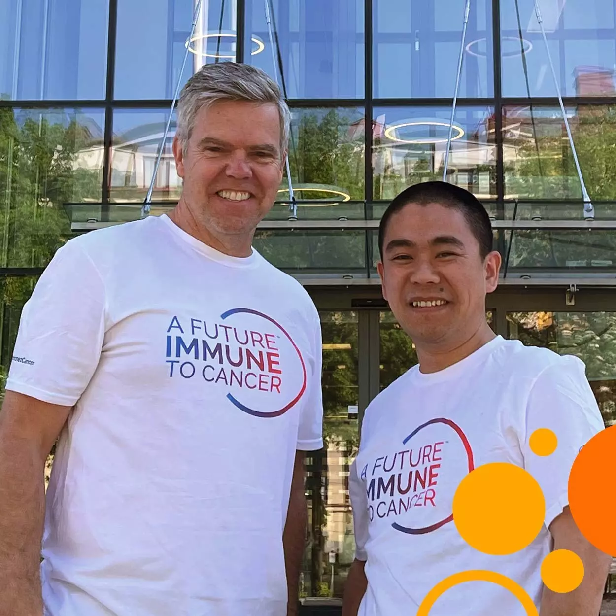
Interview with Dr. Magnus Essand, Professor of Gene Therapy, and Dr. Di Yu, Associate Professor of Immunotherapy at Uppsala University (SE)
The story of Magnus Essand and Di Yu is compelling in many ways. There’s the fact that they have been working together for nearly 15 years. There’s their approach in arming the immune system to not only attack cancerous cells but also obviate cancer’s escape from that offensive. And of course, their involvement in a publicly traded spin-off company to overcome the financial wasteland between lab and clinic. But what is striking about the storyline is its pattern of different life paths and ideas that converge into a collage of complementarity and opportunities.
Forests meet terracotta warriors
Magnus is Professor of Gene Therapy at Uppsala University. “Focused” and “driven” are words that come to mind when you hear Magnus speak. Di underscores that ambition with a little-known fact. “He is definitely a scientist. You know, he changed his last name to be more easily searched in PubMed!” Smiling, Magnus confirms the claim. “Yes. So, before my first publication my last name was Andersson, which is very common in Sweden. And if you searched PubMed, you would find too many people with that name. Essand comes from a lake in Norway, where my ancestors lived for a time in the past. So, I picked that name.”
The landscape around lake Esandsjøen is breathtaking.
Interestingly, it wasn’t always immunology for Magnus. He grew up in a small town near the Sweden-Norway border, where “most people were working in the forest industry,” he says. “And if you wanted to do something else, it should be something along the engineering track. So, I started to study engineering in high school – electrical engineering, actually. After finishing high school and doing my military service, I even worked for two years in programming before I started my university studies.” But Magnus wanted to see the world. His studies and research allowed him to do so – from Amsterdam and Paris to the National Cancer Institute in the US.
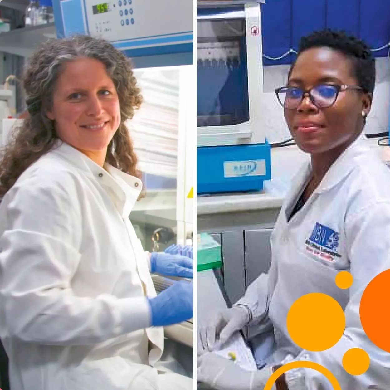
Science in Africa is science for the world. A clear message from Resty Nabeeta and Maike de la Roche.
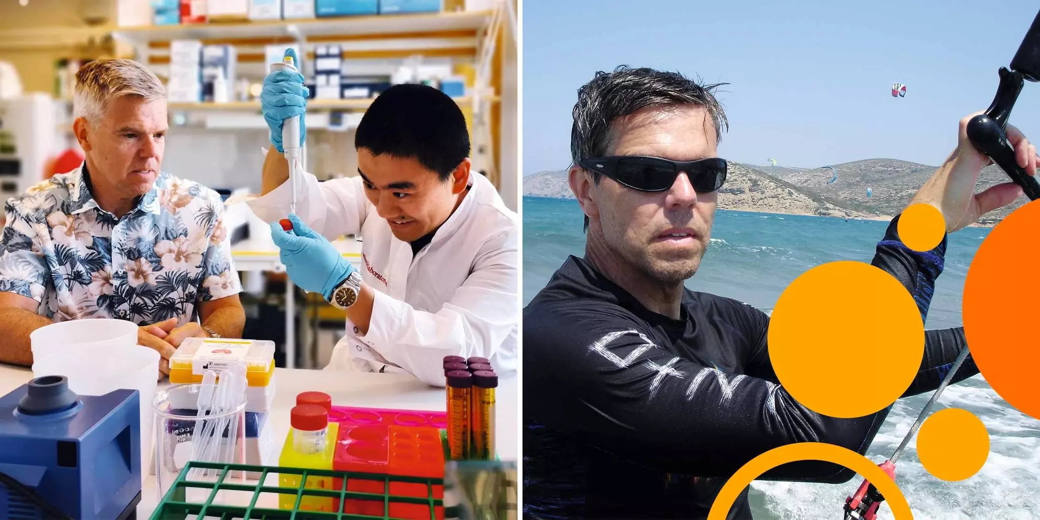
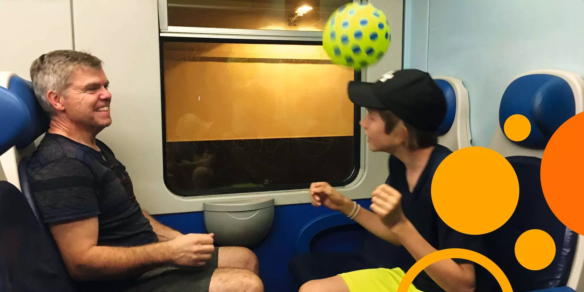
Interview with graduate student Resty Nabeeta from Makerere University, Kampala (UG), and group leader Dr. Maike de la Roche from the Cancer Research UK Cambridge Institute (UK)
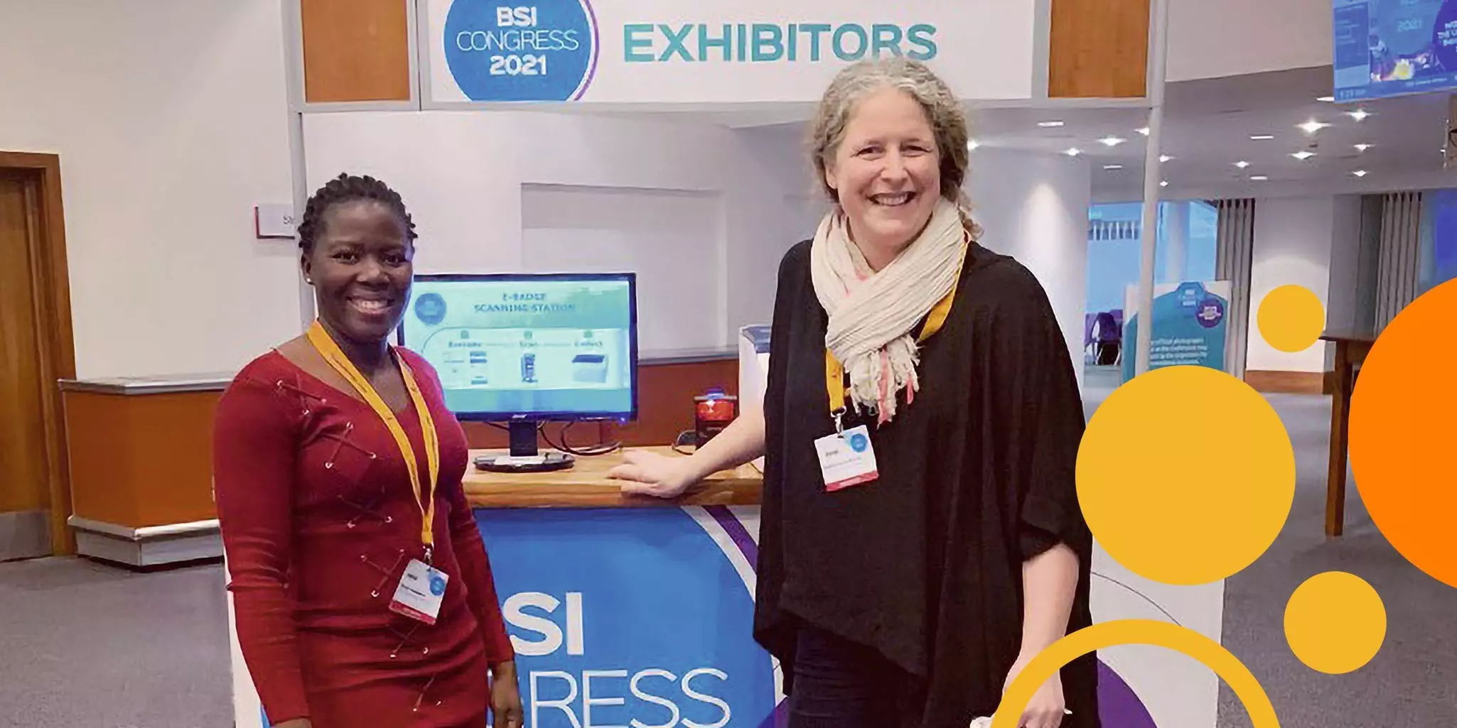
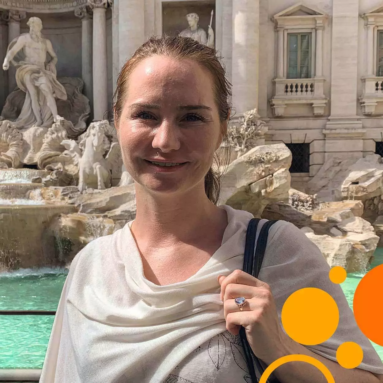
The world is becoming increasingly small. Activities in one corner of the planet influence, burden, empower, or wreak havoc in another. Human existence – in numbers, behavior, and impact – reverberates across natural and political boundaries, conquering distances with the incessant crisscrossing of container ships, flights, and Zoom calls. Oddly, how humans tackle the aftermath of globalization often remains localized, limited in reach, time, or inclusion. But if the very nature of society makes challenges global, then science must respond in kind. That impulse brought together Resty Nabeeta and Dr. Maike de la Roche on a gray morning in Edinburgh, just before heading to the buzzing poster halls and talks of the British Society of Immunology (BSI) Congress 2021.
Iben Lundgaard and the modern science lab:hip, focused, and empowered
Interview with Dr. Iben Lundgaard, Associate Professor at the Wallenberg Centre for Molecular Medicine, Lund University (SE)
“I never thought I would become a professor. When I was younger and at university, I thought of a professor as a gray-haired older male. That was my stereotype of a professor and I thought, ‘this is not for me; I will never be a professor.’ But now I am only one promotion away from becoming a professor.” Dr. Iben Lundgaard chuckles about that starting point to a career that led to an Associate Professorship at Lund University. In fact, Iben paints a stark contrast to the stereotypic image of an absentminded, out-of-touch, middle-aged academic. Iben is under 40 and female. She guides a productive research lab, insists on keeping science fun, and broadcasts their findings on social media. Add that Iben’s research focuses on a structure of the brain – the glymphatic system – that was first described just 8 years ago and you have the formula for today’s modern lab where tomorrow’s experts unfold their scientific curiosity.
Leading an expedition in uncharted territory
Iben did her postdoctoral research with Dr. Maiken Nedergaard at the University of Rochester in New York where she worked on the glymphatic system, a network of vessels that clears waste from the central nervous system. As she recounts it, the implications of its discovery were not clear to her at first. “I was first introduced to the glymphatic system during my interview with the Nedergaard lab when I was really jet-lagged. At that time, it hadn't been published yet. I felt like, ‘Okay, this is pretty cool, it could be big’. That day, I was mostly thinking about the system in connection with white matter diseases, which was related to my Ph.D. work. Later, I realized that it had wide implications for many different diseases.”
Watch a short video where Iben explains her work on the glymphatic system.
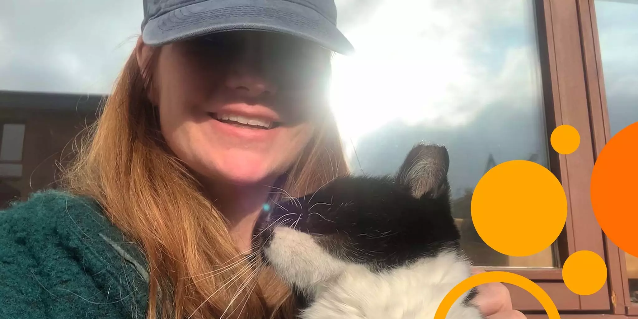
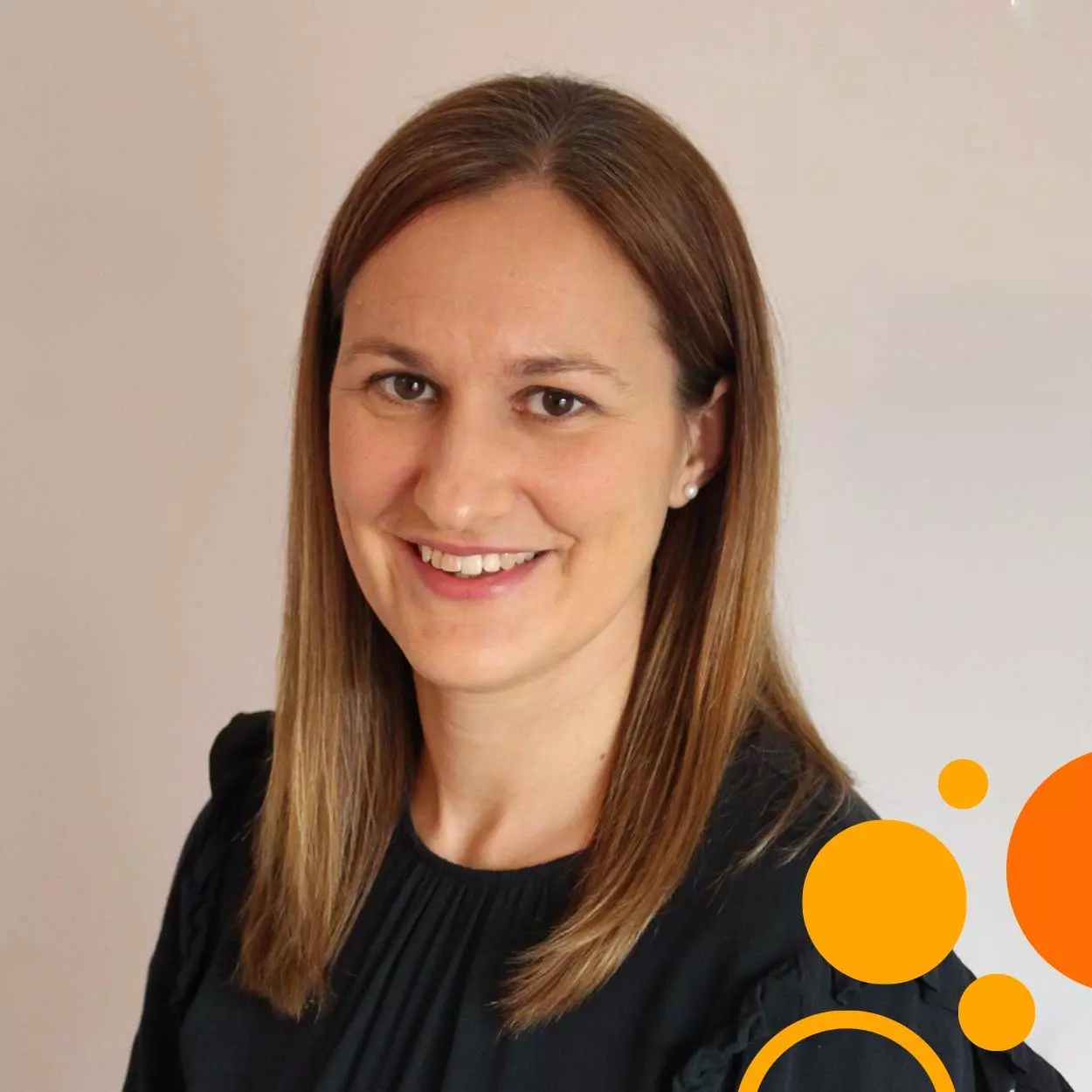
Rachel Abbott on seizing opportunities and pushing your own boundaries
Interview with Dr. Rachel Abbott, Head of TCR Pipeline and Dark Antigen Research at Enara Bio (UK)
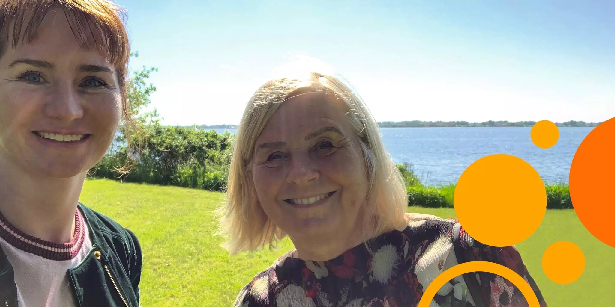
Rather early in life, Dr. Rachel Abbott realized that taking what others considered the safest route was not always the most interesting choice. In fact, by repeatedly deciding to take risks and learn something new, Rachel’s path has been an impressive progression through a spectrum of research areas and increased leadership. Today she is a young, successful pioneer in the biotech industry, heading up teams at Enara Bio to develop novel TCR-directed immunotherapies and discover unconventional cancer antigens.
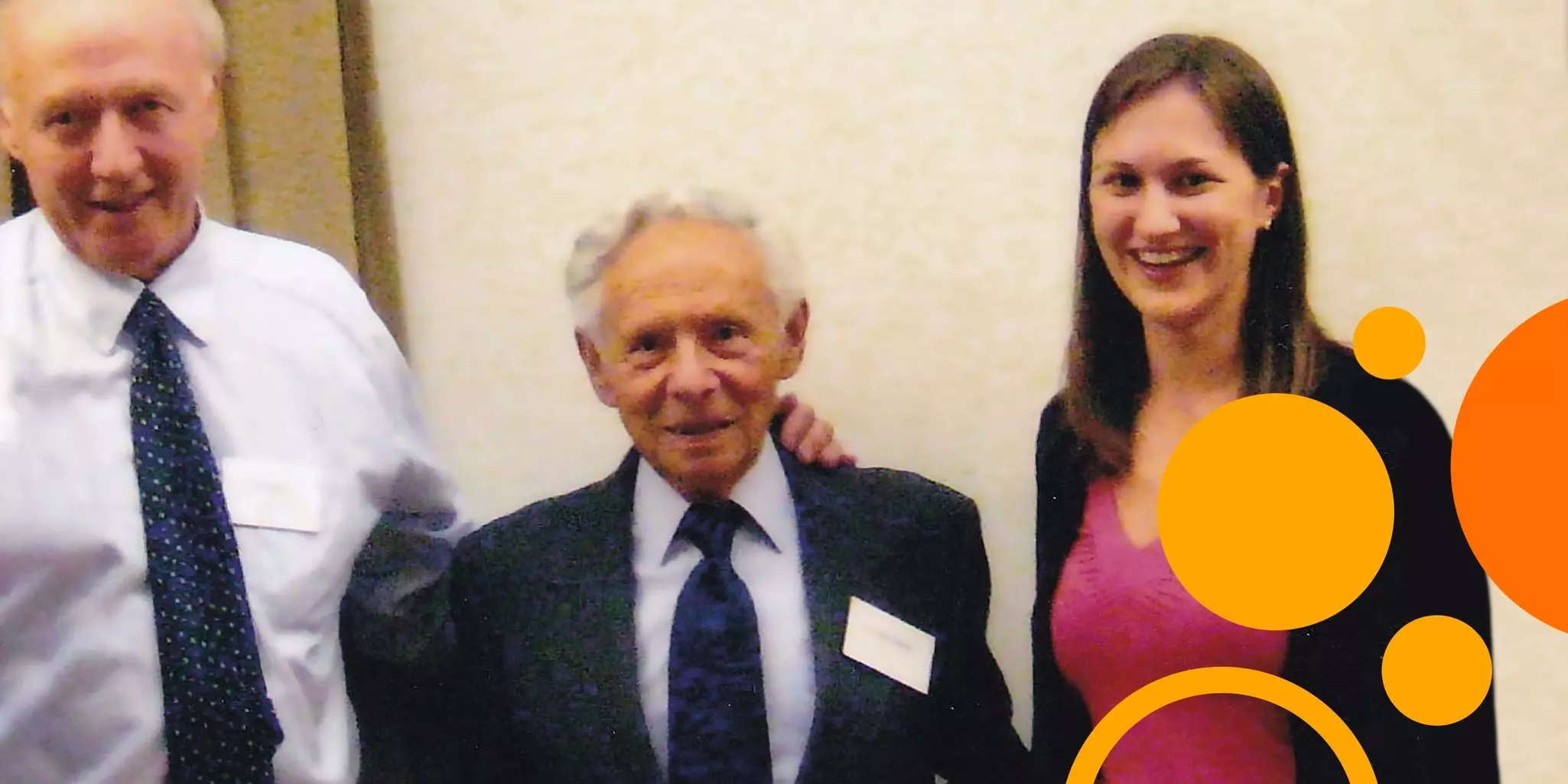
The Sohlberg-Björklund duet: orchestrating successful therapies with natural killer cells
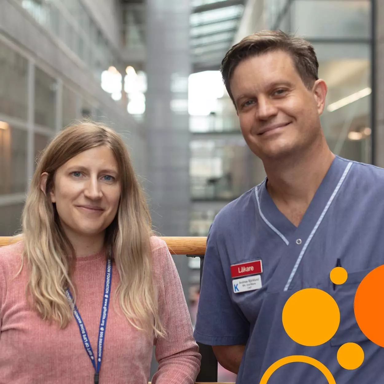
Interview with Dr. Ebba Sohlberg, project manager in the Center for Infectious Medicine at the Karolinska Institute and Dr. Andreas Björklund, clinician in the unit for Cell Therapy and Allogeneic Stem Cell Transplantation (CAST) of the Karolinska University Hospital, Stockholm (SE)
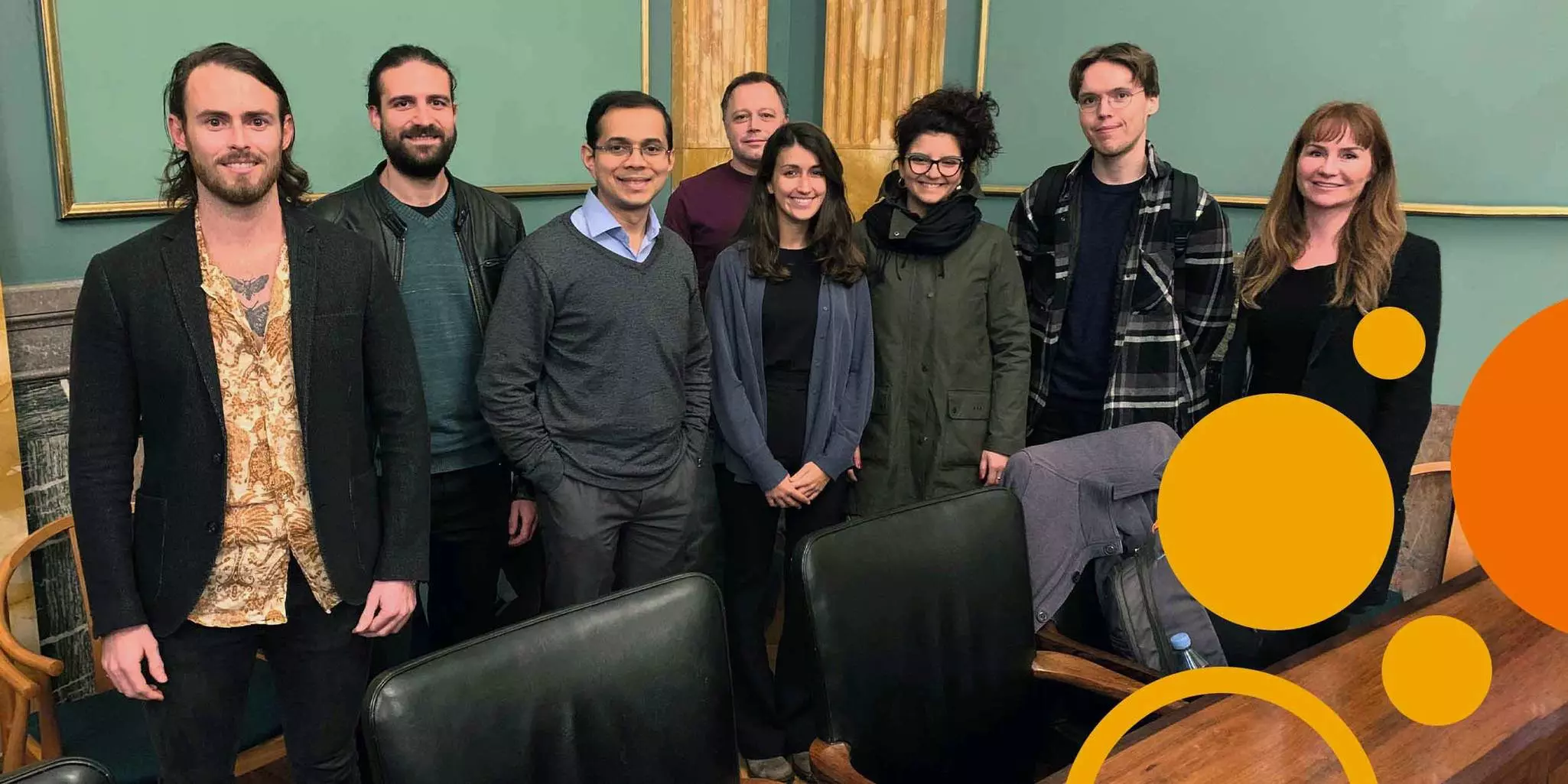
A new generation of NK cell therapies are under development at the Karolinska Institute and Karolinska University Hospital. Drs. Ebba Sohlberg and Andreas Björklund are part of a large team led by Professor Karl-Johan Malmberg that focuses on understanding the mechanisms underlying the diversification of human NK cell repertoires and implementing novel insights into NK cell biology in clinical trials for advanced cancer patients. Andreas was the PI for their recently closed clinical trial that successfully used natural killer (NK) cells to treat myelodysplastic syndrome (MDS) and acute myeloid leukemia (AML) patients. Ebba headed the immunomonitoring. In collaboration with various healthcare and industry partners, the team is now developing a new and exciting therapy based on ‘adaptive’ NK cells.
How did you end up where you are and how long have you been working together?
Andreas: I started as a PhD student in Prof. Malmberg’s group in 2005 and have been affiliated with the group in parallel to my clinical work at CAST ever since. In 2012, when our first NK cell therapy trial was launched, Karl-Johan moved to Oslo, and I was assigned the role of PI for the clinical study. When Ebba arrived in 2013, we worked together to decipher the correlations between the laboratory findings and the clinical outcomes. That analysis and the work of the entire research group led to a publication in Clinical Cancer Research in 2018. Since then, we have been the core of the KI [Karolinska Institute] group working on developing clinical study applications.
Ebba: When I was looking for a postdoc position, it was a natural step for me to come to the Karolinska Institute where NK cells were discovered. While we are finding out a lot about the inner workings of NK cells, our work nevertheless has the very concrete goal of treating patients. The infrastructure and community here offer unique opportunities in terms of translating our findings into improved patient care that are not available everywhere.
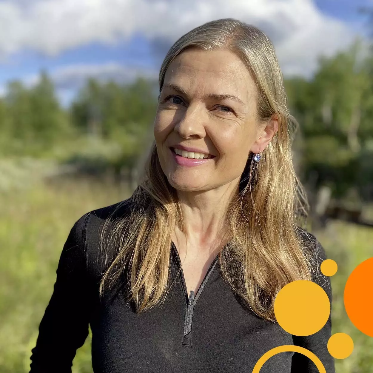
Leader, problem solver, challenge seeker. Johanna Olweus is a force to be reckoned with.
Interview with Dr. Johanna Olweus, Professor and Head of the Department of Cancer Immunology at Oslo University Hospital (NO)
From the start, Dr. Johanna Olweus puts you at ease in a conversation – she laughs easily; she gives every question careful consideration. In the wider world, she runs a prolific research group, heads an internationally renowned research department at Oslo University Hospital, and is a professor with teaching responsibilities at the University of Oslo. And yet, at this moment, she is 100% engaged in the intimate space of your exchange. She makes you hang on her every word as she describes the experiences, thoughts, and insights that shaped her career.
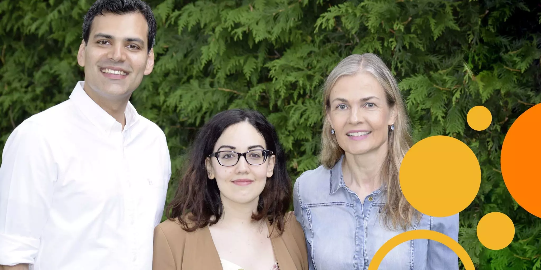
Advancing science, giving guidance, embracing humanity. A stroll with Paul Fairchild.
Interview with Dr. Paul Fairchild, Associate Professor in Medicine at the Sir William Dunn School of Pathology, Fellow and Tutor in Medicine at Trinity College, University of Oxford (UK)
On any given morning in Oxford, you may see pensive scholars on their way to a college, private hall, or department at the University of Oxford. One of them will be Dr. Paul Fairchild. For over 20 years, Paul has walked to and from work. Deep in thought, he mulls over problems, ideas, plans, and keeps himself "intellectually engaged with what we are doing, rather than answering infinite e-mails, which is more the norm, unfortunately."
Paul’s path as an immunologist did not begin in the natural sciences. Originally, his plan was to go into linguistics and learn multiple languages. But, then what? he wondered. Having changed his mind, he encountered immunology as an undergraduate and never looked back. “It made no sense to me whatsoever! So little was known about the immune system at that time. The T-cell receptor had not yet been elucidated. The MHC was taught to us as the complex that causes the rejection of allografts but if that was all you knew about it, it made no sense. Given that we’ve only done organ transplants for the last few years, how could something evolve to cause rejection?” he chuckles. “Nobody chose immunology because it was just so nonsensical.” Intrigued, he entered a field as it transitioned into a phase of exciting discoveries. He remembers how “during my PhD, the crystal structure of MHC class I was published, showing that it actually had this substance bound in a groove, which people assumed might be a foreign peptide. That was the first that was really known about what MHC did at all, and since then things have begun to slot into place.”
It was also during his PhD work with Professor Jon Austyn that Paul was introduced to dendritic cells and his interest in them has not waned. Paul was among the first to describe the antigen-presenting role of dendritic cells during clonal deletion in the thymus and he has worked throughout his career on questions of immune tolerance in the context of autoimmune disease and allograft rejection. Today, his research lab uses knowledge about what tips the immune system toward response or tolerance to figure out how to coax it back. “Even now, 30 years after understanding the way the immune system works, we still have not really got to that point of actually inducing tolerance clinically. So, it's still quite a long way off. And that's really because trying to reestablish tolerance in a primed immune system is exceptionally difficult to do” says Paul. The same but opposite inertia holds true for immunotherapies, he argues. “All tumor antigens are effectively self proteins. Some level of tolerance has been established to them. So, we're trying to break through that barrier of tolerance to trigger an immune response against what is effectively a self component.”
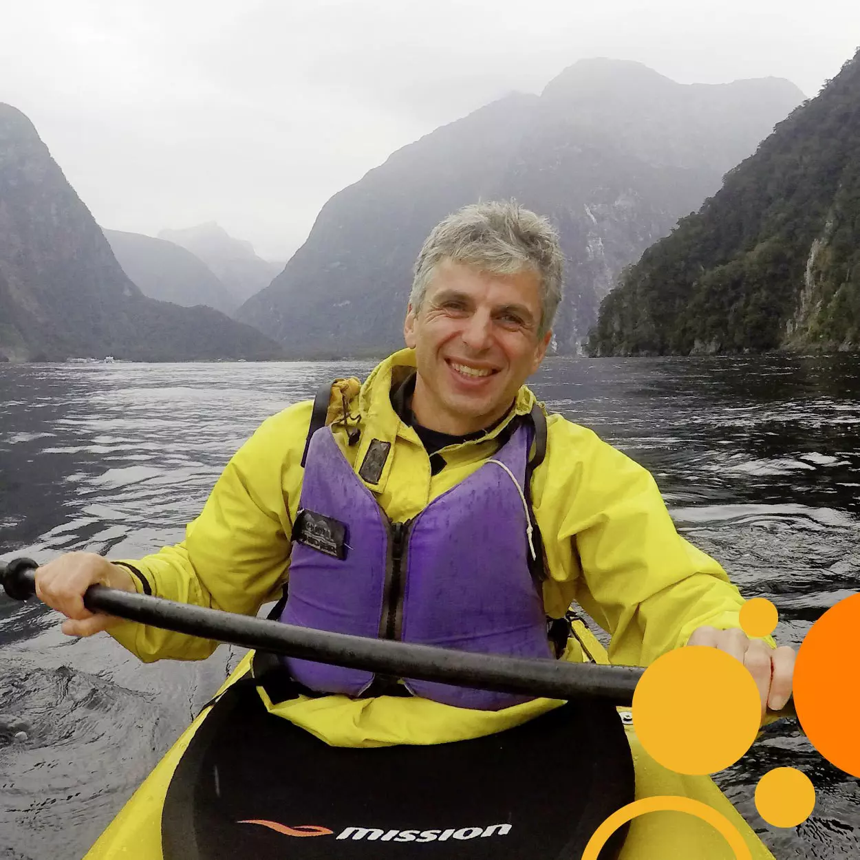
Paul Klenerman and the fine art of serendipity surfing
Interview with Dr. Paul Klenerman, Sidney Truelove Professor of Gastroenterology at the Nuffield Department of Medicine, University of Oxford (UK)
Google Paul Klenerman and you’ll stumble upon a Wikipedia page listing past accomplishments of a young fencing champion. What he does today is splashed across numerous other websites and videos with terms like University of Oxford, viral immunology, T-cell responses, HIV, hepatitis C, host-virus interactions.
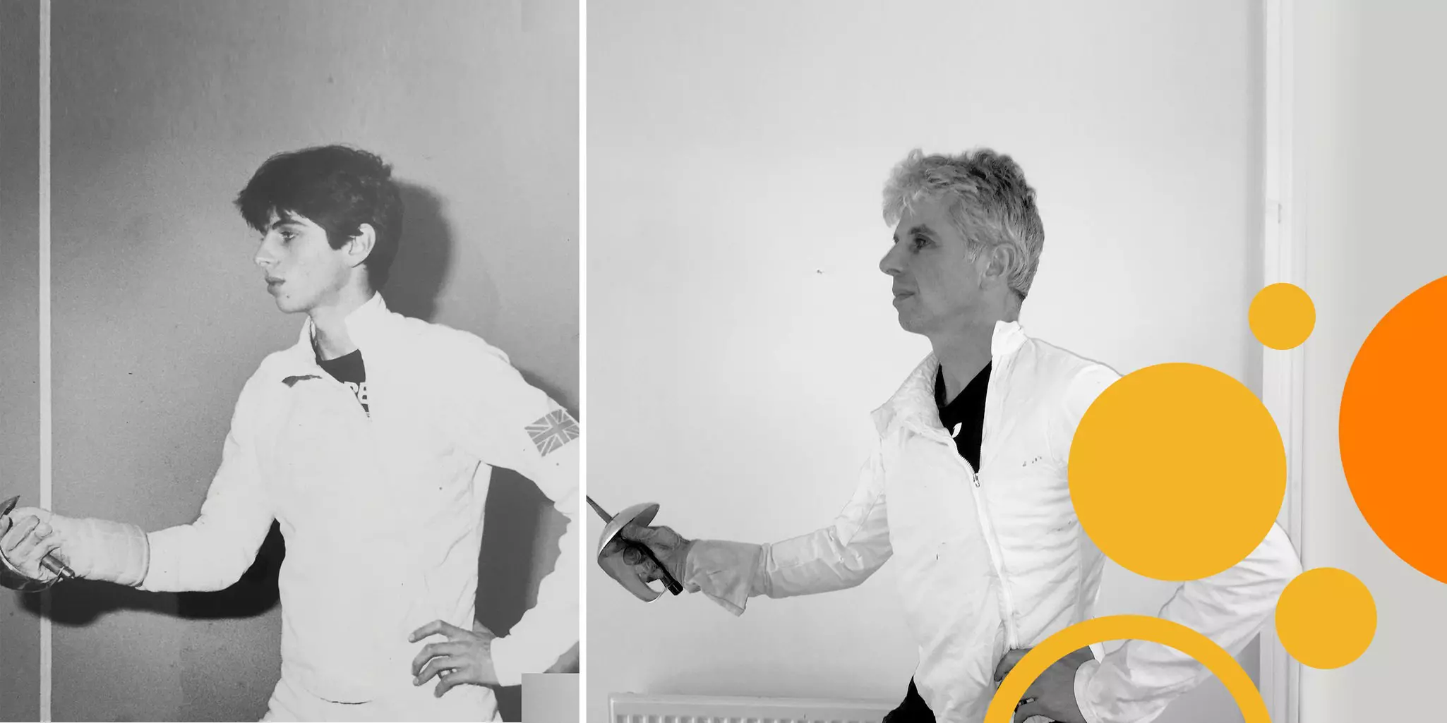
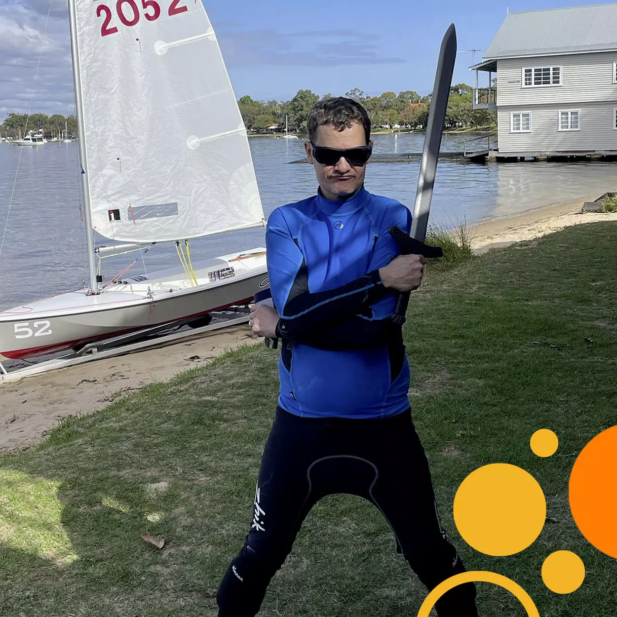
A far cry from “Little Tortoise". Jonas Nilsson in four acts.
Interview with Dr. Jonas Nilsson, Inaugural Perkins Chair of Melanoma Discovery at the Harry Perkins Institute of Medical Research (AU) and Professor in Experimental Cancer Surgery at the University of Gothenburg (SE)
Conversations with some people take you on an unexpected journey of unique perspectives and tantalizing insights. That’s the case with Dr. Jonas Nilsson. Last November, he packed up the family and moved across the globe from Sweden to Western Australia on a personal and professional expedition. He isn’t trekking through the Australian bush. Instead, he is clearing a path for clinical melanoma research out of Perth. In the process, he’s seized the opportunity to leave a positive mark, regardless of where life takes him next. Taking chances runs deep in his career and has prevented stagnation on more than one occasion.
Act 1: the past
“Actually, I fell into research during basic studies,” Jonas recounts. “I got really interested in a project that a professor had at the university I attended in Umeå in the north of Sweden, trying to understand what these small molecules called polyamines do. At that time, we were cloning genes in the lab. This was a bit before the human and mouse genomes were known, so we had to do these things manually. I would screen libraries full of genes and identify those that we wanted to examine. I cloned a cDNA that encodes a protein that regulates the stability of a key enzyme in the synthesis of polyamines called ornithine decarboxylase or ODC. As it turned out, that gene is growth regulated. So, when you stimulate cells to grow, the protein encoded by this gene can activate ODC to produce polyamines needed for cancer cells to divide. That was the link to cancer.”
The clone Jonas developed was the ticket to his next adventure. It caught the attention of Dr. John Cleveland, then at St. Jude Children's Research Hospital in the US, who was working on the well-known oncogenic transcription factor Myc. “He wanted to get this cDNA that I had cloned,” says Jonas smiling. “I had just finished my PhD, so I just said: ‘you can get the clone if I can go with the clone’ and that is how I moved with the clone to do my postdoc in the US.”
Jonas and his wife Lisa Nilsson were the first to dissect the downstream connection between Myc and ODC, revealing its importance in tumorigenesis. “We showed that even though inhibiting ODC to halt a growing cancer is not feasible, if you trigger polyamine inhibition before cancer develops as a chemoprevention, then it is quite powerful because elevated polyamine synthesis is required for the transformation event,” Jonas explains. “At the time, we hypothesized about genetic mechanisms but now I think it has to do with tumor-promoting inflammation, which is probably a major function of pre-cancerous elevation of ODC.”
The work granted Jonas an assistant professorship at his alma mater in Umeå, where Jonas began thinking of doing so-called patient-derived xenograft (PDX) mouse models. However, he was struggling to gain access to patient samples. That would change through a chance encounter with Professor Peter Naredi, a surgeon. “We were talking in a room full of people and it seemed like the two of us were the only ones talking. We had a discussion basically in front of everyone watching us like we were crazy. We decided we had to work together.” Again, Jonas jumped at the opportunity and with access to samples, he and Lisa successfully transplanted colon cancer tissue into a mouse.
Urged by the surgeon to try his luck for a position in a clinical department, Jonas sought openings and noticed the newly established Sahlgrenska Center for Cancer Research (SCC) at the University of Gothenburg. Jonas and the PDX models were a perfect fit. Enthusiasm for his work at the SCC lead to a shift in focus to melanoma, with surgeons collecting patient samples for a biobank study and the establishment of novel PDX models. Jonas and Lisa teamed up with Roger Olofsson Bagge and Lars Ny to establish the Sahlgrenska Translational Melanoma Group (SATMEG). Using biopsies from metastatic uveal melanoma patients undergoing liver perfusion, they have also characterized the genetics of the disease via whole-genome sequencing. The list of his achievements goes on. In fact, Jonas contributed in different ways to 5 clinical trials, including the recently published PEMDAC study for patients with metastatic uveal melanoma. “Lisa and I had a mantra for many years that our goal in research was for Lars to start another clinical trial because that was the best thing we could do for patients.”
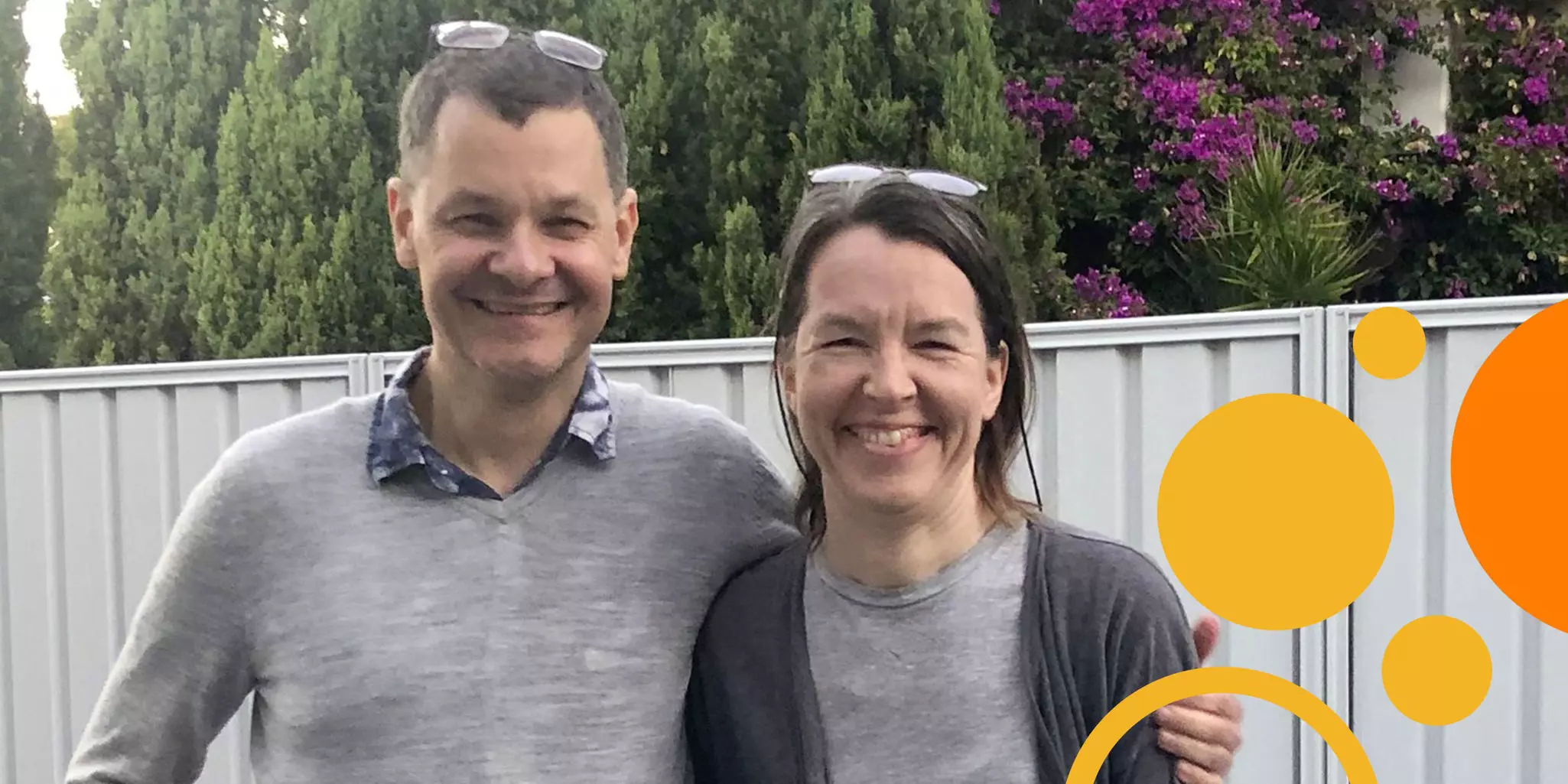
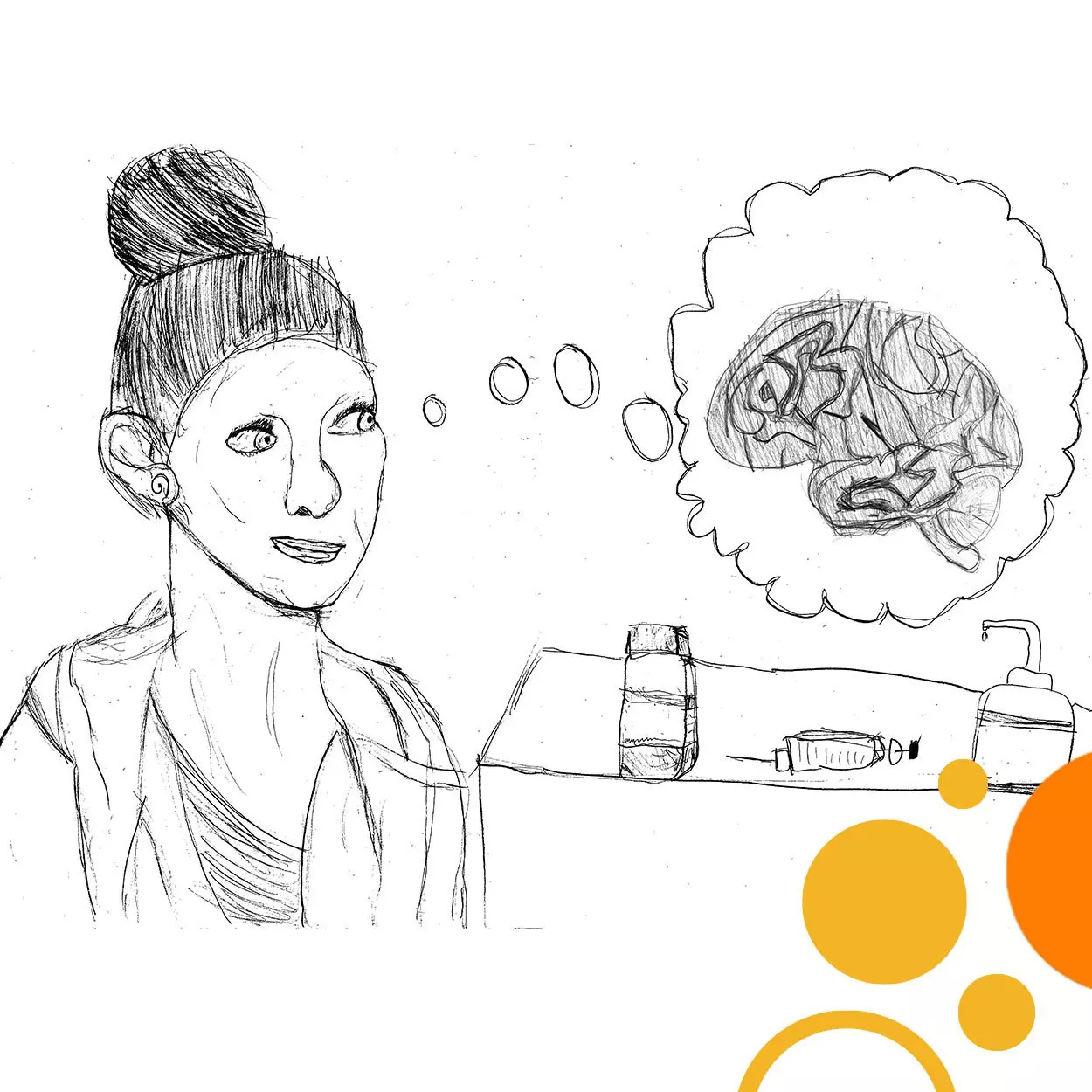
Stem cell wrangler by day, therapy maker… also by day. This is Agnete Kirkeby.
Interview with Dr. Agnete Kirkeby, Associate Professor at the University of Copenhagen (DK) and Lund University (SE)
“My mom helps making new medicines.” As simple as that sounds, Dr. Agnete Kirkeby’s ten-year-old daughter describes her mother’s work perfectly. Not only what she does, but also the reason that sustains her tenacity in a field that demands patience and precision like few others.
The privilege to apply your research
Creating novel but also “economically and efficaciously competitive therapies”, as Agnete points out, dictates her research approach. “Few of us have the privilege to actually go and apply our research to patients in the clinic. To me, the prospect of doing that is a huge driving force. Opening my eyes to this possibility – that it’s not just something you talk about but a real possibility if you decide to go that way – has been very important in the way I run research today. Every time I plan a research project, it is with that aim in mind.”
Thus, a research program in Agnete’s group begins with a clinical need where current treatments do not solve the underlying problem and where cell replacement makes sense. “The diseases we are looking at here involve small regions of the brain where our therapy can have a more targeted impact. In Parkinson’s disease, we know that the motoric symptoms are caused mainly by a small area of dopaminergic neurons that are lost. We go in and replace exactly those neurons. Similarly for Parkinson’s disease associated with dementia, another small cluster of cholinergic neurons in the basal forebrain is lost and we are looking into replacing those to regain functionality. So, we are looking at those diseases where we know specific subtypes of neurons are being lost in a rather small region but with a huge impact on symptoms.”
Other efforts in her lab are developing protocols to generate neuron subtypes of the incredibly diverse populations that constitute the hypothalamus. “We have several projects where we are generating hypothalamic neurons. One is generating hypocretin-secreting neurons with the aim of treating narcolepsy and another is working on hypothalamic neurons involved in appetite and metabolic regulation to tackle critical questions about the recently demonstrated central involvement of hypothalamic dysregulation in obesity and diabetes.” Through experience, the teams have learned that hypothalamic cells require finesse in fine-tuning developmental steps to derive the right cell types and it is possible that the inherent versatility of hypothalamic progenitor cells limits the purity of the outcome. That requirement for precision, Agnete advises, is agnostic of cell type because “when you work with stem cells, they are never forgiving. If you make a tiny mistake, you’re immediately going to be punished for it because you are not going to get what you were aiming for.”