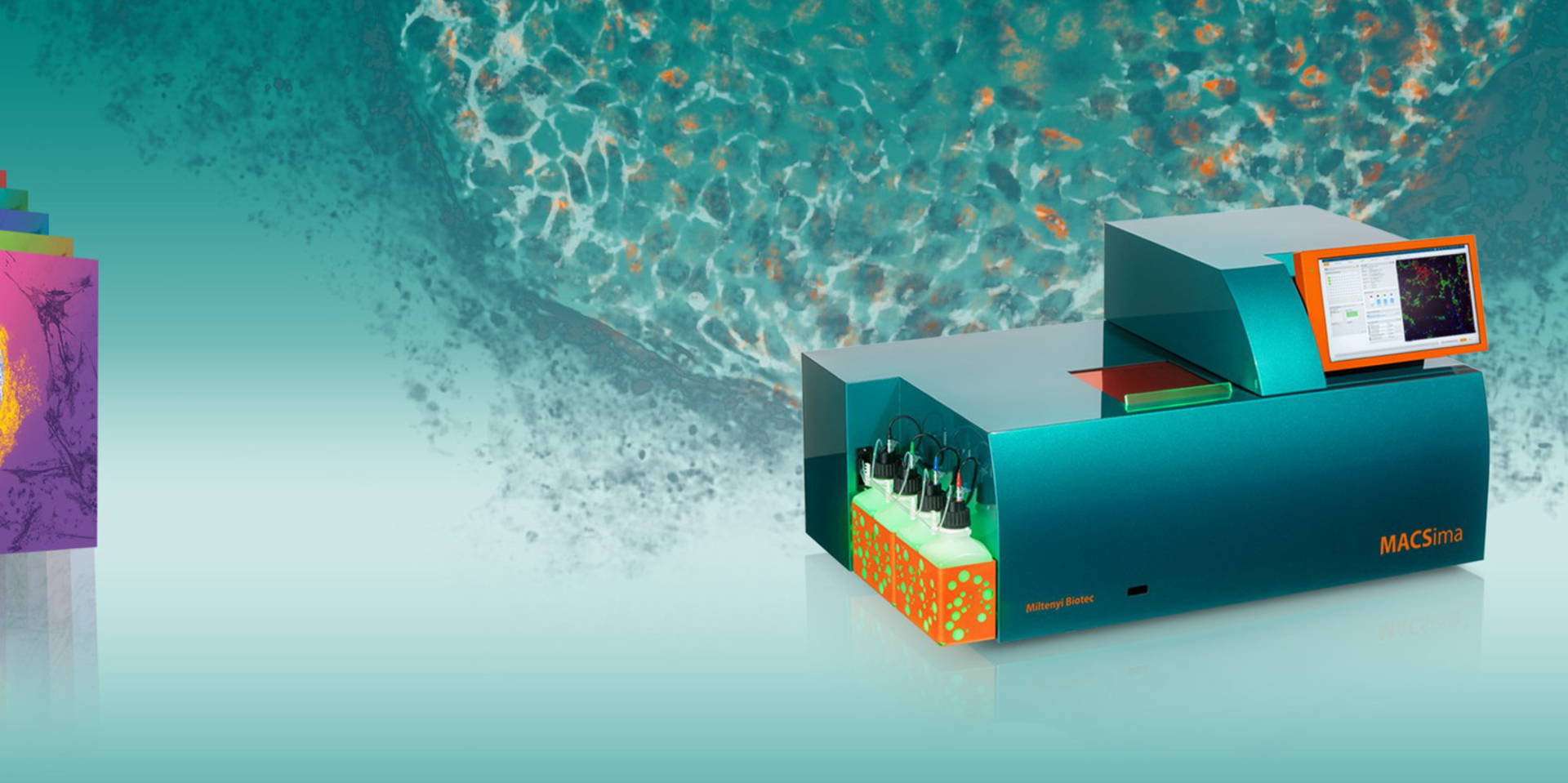
MACSima™ Platform

Unlock the next level in immuno-oncology research

Immuno-oncology (IO) as a research field has grown tremendously in recent years as researchers the world over try to understand and manipulate the immune response within the tumor microenvironment in the continuing fight against cancer.

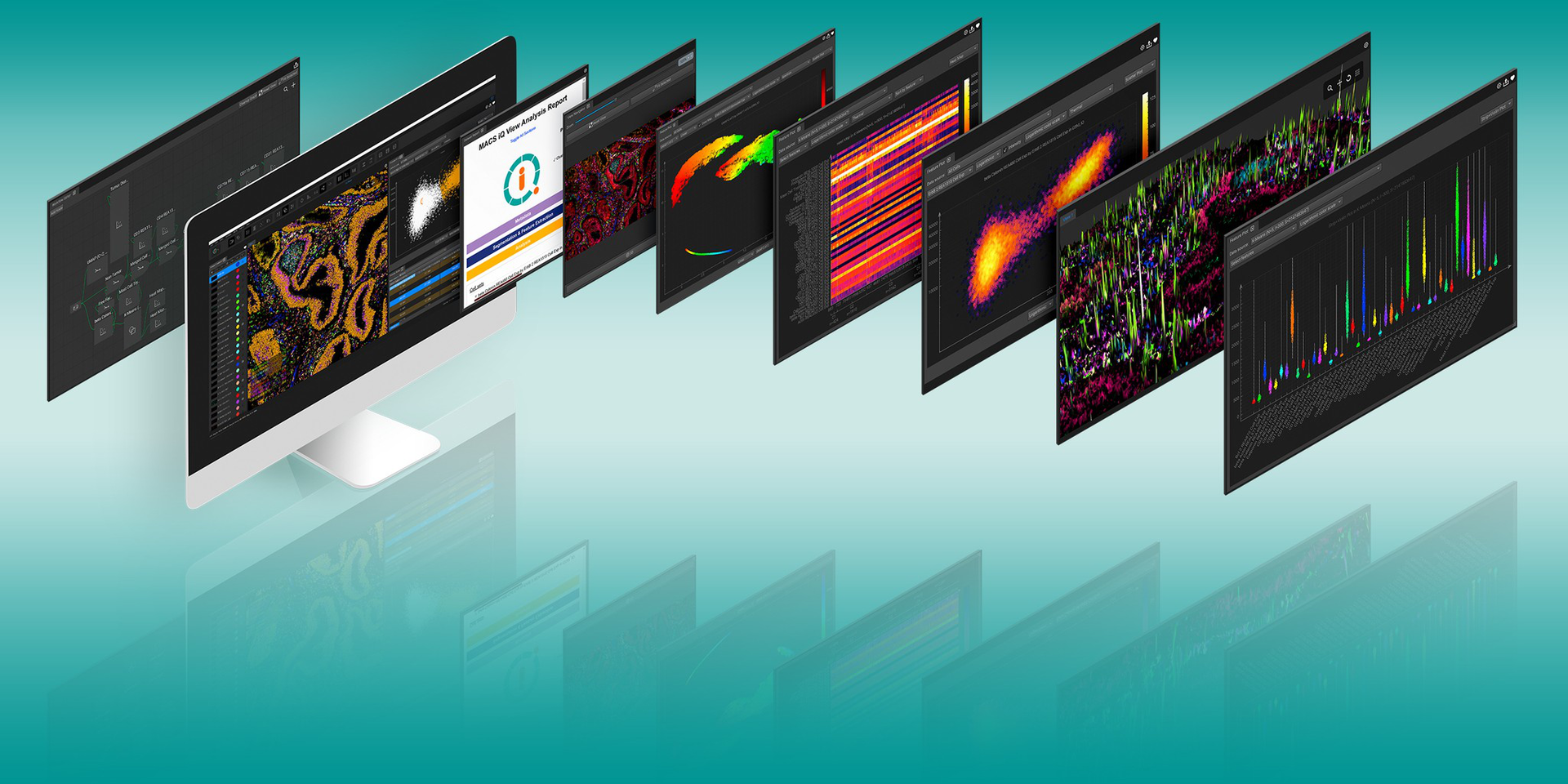
The deep phenotyping of tumor cells in the tumor microenvironment can help define new targets to be used in promising IO therapies. Current tools demand a tradeoff between spatial information and the number of markers that can be analyzed. With the MACSima Platform, no such sacrifice is necessary: hundreds of markers on a single sample can be stained in an automated manner, while preserving tissue integrity and capturing the spatial context, opening new possibilities for advances in IO research.

In action in IO
Hot off the press

Predicting immunotherapy response in lung cancer

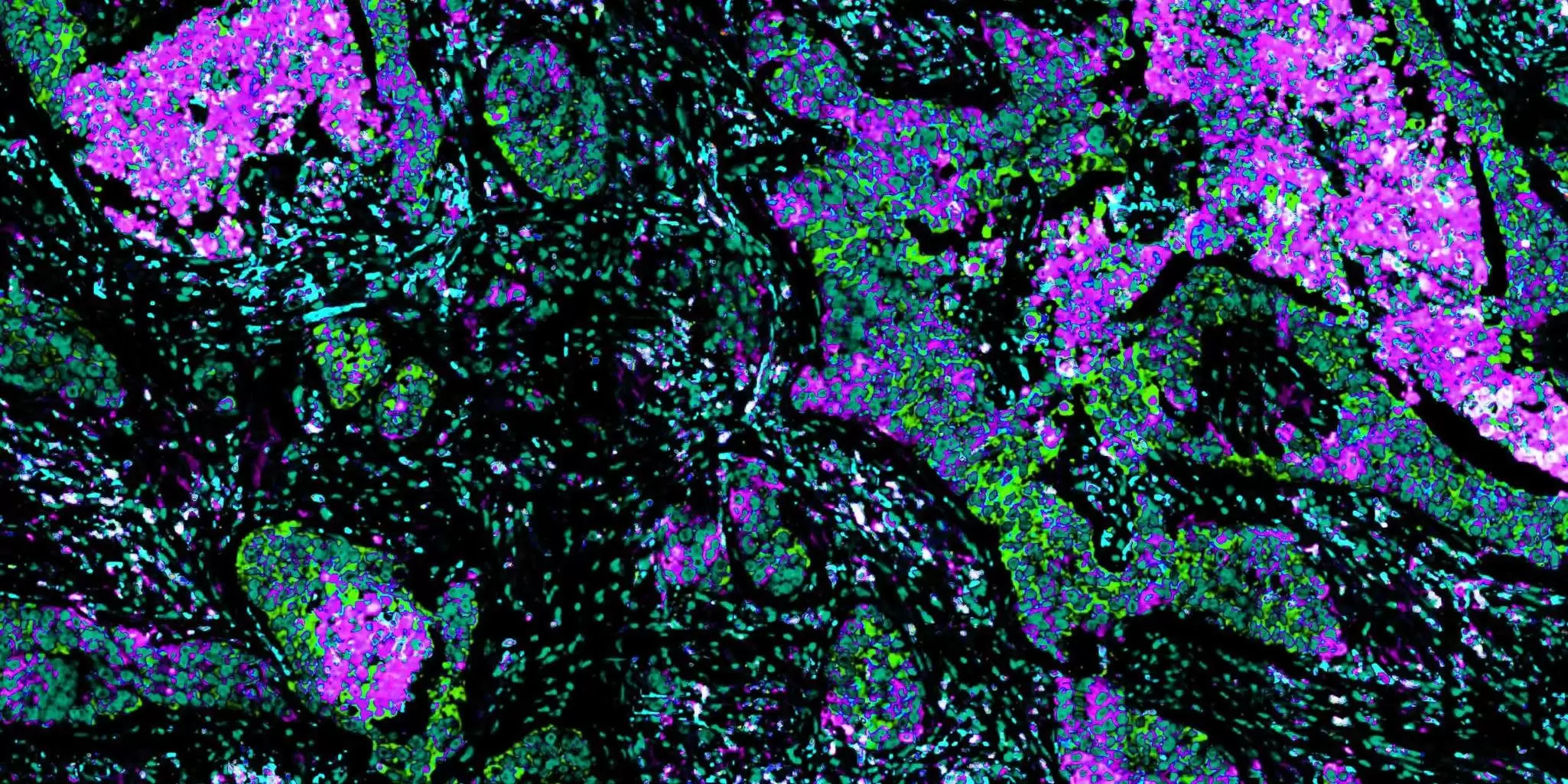
In their recent paper published in Nature Commmunications, Clemens Hinterleitner et al. discovered that platelets expressing PD-L1 (pPD-L1) interact with lung cancer cells and that the expression level of PD-L1 can predict responses in immunotherapy for non-small cell lung cancer (NSCLC). Using the MACSima Platform they confirmed that NSCLC samples with high pPD-L1 levels show lower numbers of T cells in the tumor microenvironment and less infiltrating T cells.

Read the paper
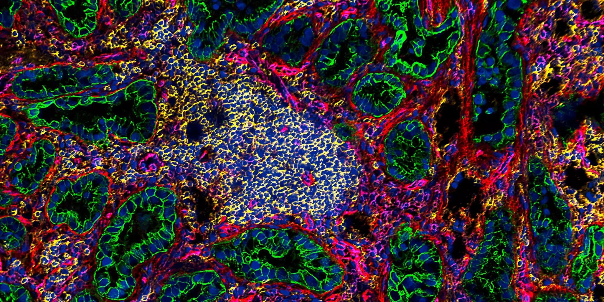
Microscopy image of pancreas carcinoma imaged with the MACSima system
Publication in Nature Communications

Discovery of CAR target candidates in pancreatic carcinoma

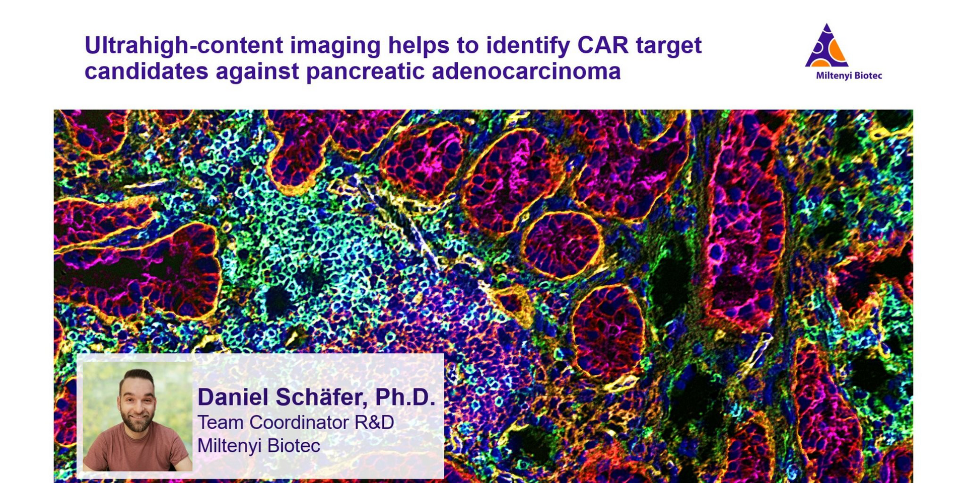
A new publication in Nature Communications reveals target candidates for CAR T cell–based immunotherapy of pancreatic adenocarcinoma. A research team from across Germany, led by Miltenyi Biotec, used a smart combination of data mining, flow cytometry, and ultrahigh-content analysis with the MACSima Platform to bypass a major roadblock preventing potential cellular immunotherapy: the lack of suitable tumor-specific antigens. Check out the full story at Nature Communications.

Read the paper
Peer-reviewed publication

New potential target pair for cell therapy of ovarian carcinoma

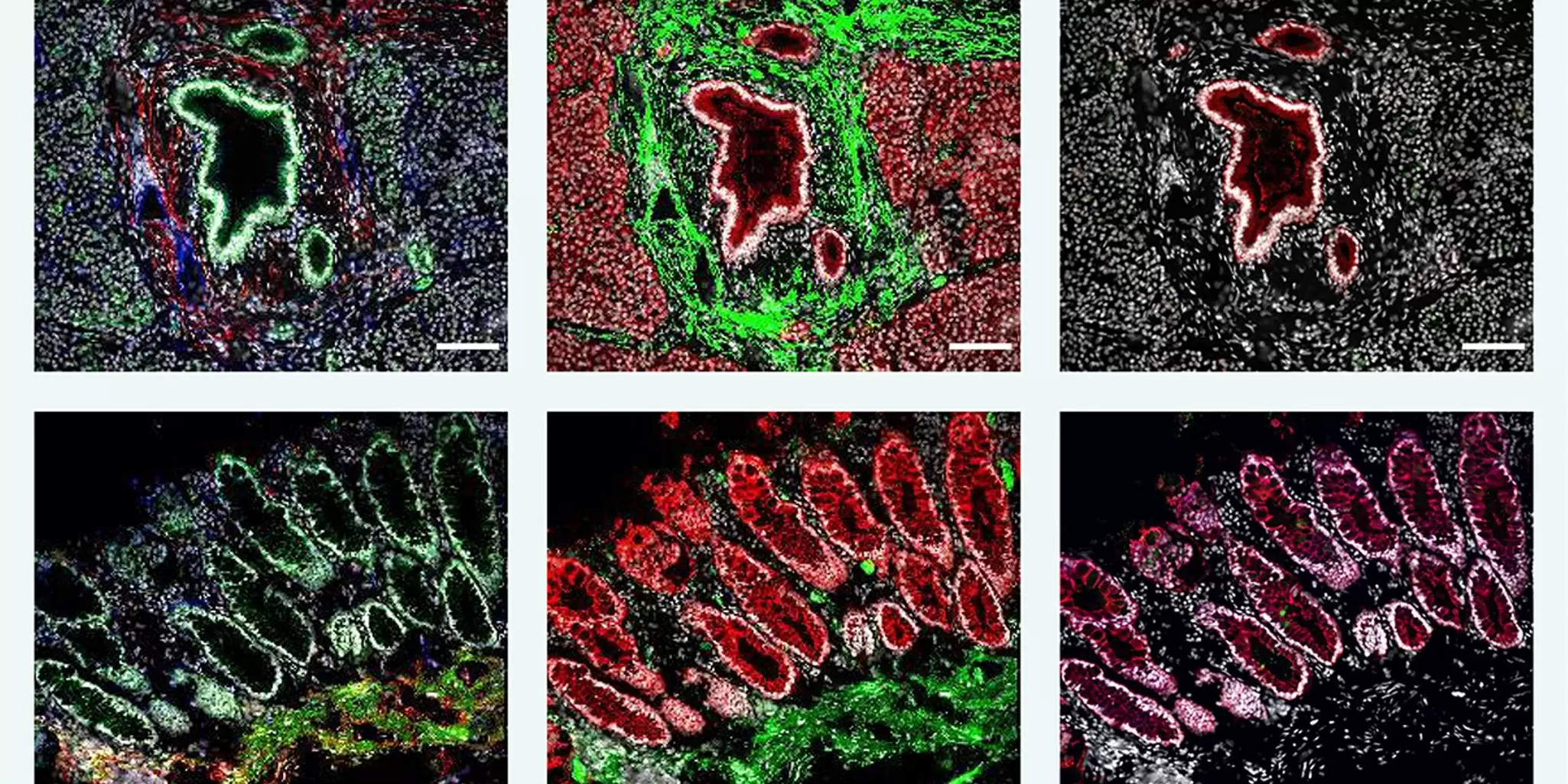
Ali Kinkhabwala et al. used MICS (MACSima Imaging Cyclic Staining) technology to analyze human glioblastoma, ovarian and pancreatic carcinoma, as well as 16 healthy tissues. Showing that EPCAM and THY1 are co-expressed only on ovarian cancer cells, but not on healthy tissue, they identified a new potential target pair for CAR T cell–based therapy. Validation with CAR T cells showed efficient killing of double-positive cells only, suggesting a reduced toxicity of CAR T cells. Kinkhabwala, A. et al. (2022) Sci. Rep. 12: 1911. Licensed under a Creative Commons Attribution 4.0 International License (CC BY 4.0).

Read the paper

See how it works

Video

Powerful and automated: Mastering spatial biology with the MACSima™ Platform

Learn how the MACSima System can stain hundreds of markers on a single sample, and stain multiple samples at a time, all in an entirely automated fashion. See more than ever before: Unlock new insights and advance your research further, faster!

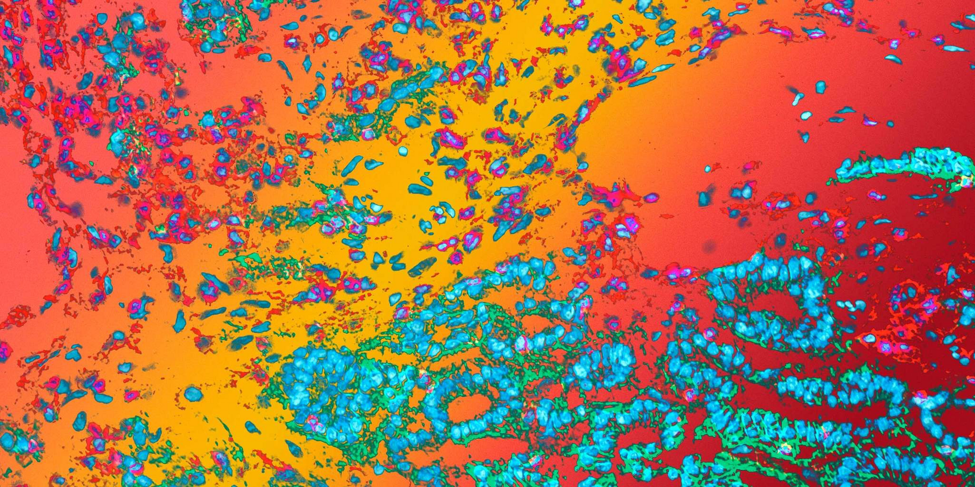
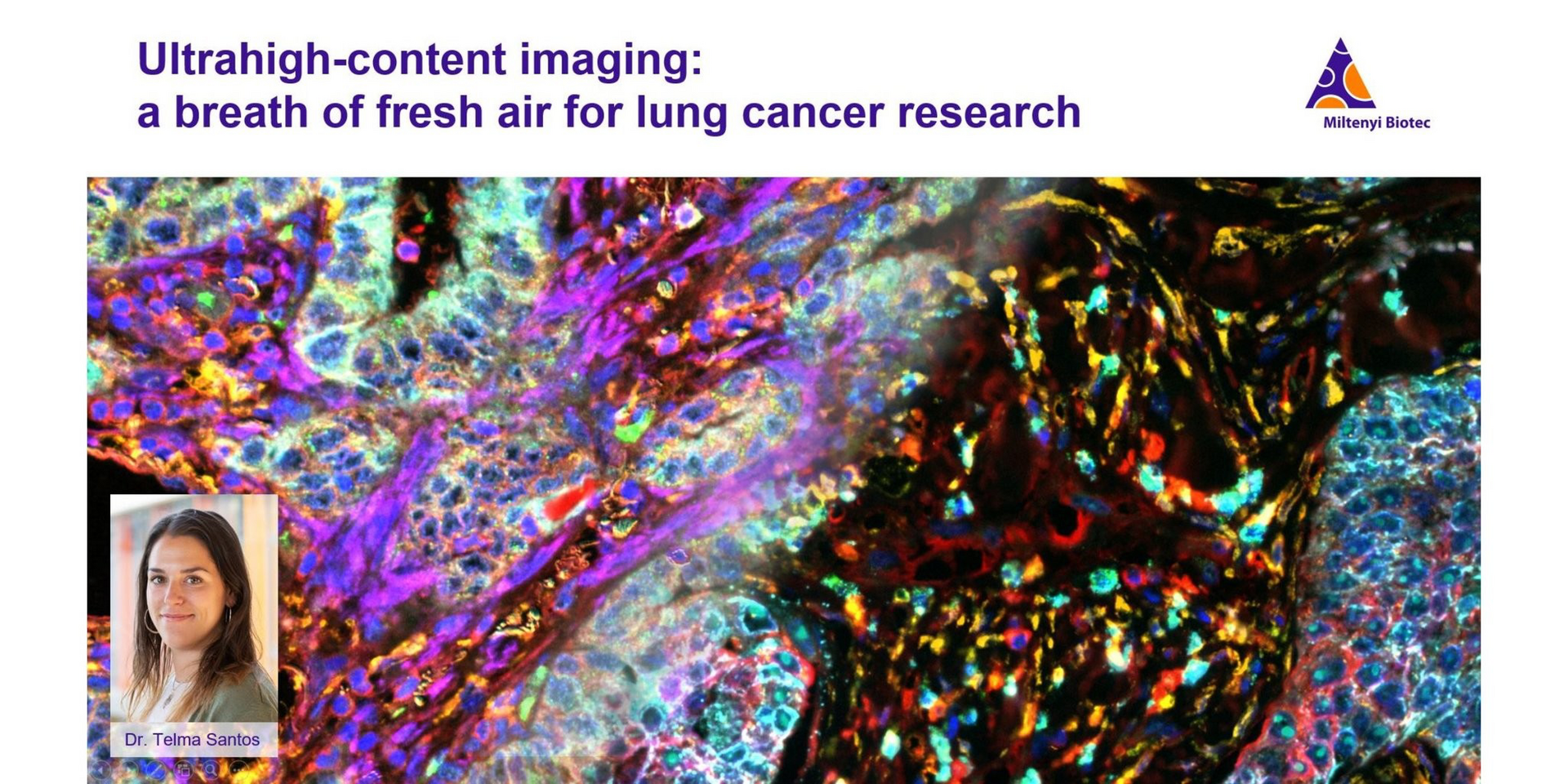
Try for yourself with our Art Tool:
Select the tissue type of your interest and up to six markers to create your own image with data taken from real-life samples.

Selected antibodies (tap to toggle on and off)

Choose antibodies (max. 6 simultaneously)
CCR6
CCR7
CD3
CD4
CD5
CD7
CD8
CD9
CD14
CD15
CD19
CD20
CD22
CD24
CD27
CD28
CD31
CD34
CD38
CD40
CD45
CD45RA
CD45RO
CD52
CD53
CD58
CD64
CD69
CD79b
CD80
CD83
CD90
CD134
CD137
CD163
CD244
CD278
CEACAM5
CLEC10A
CX3CR1
CXCR3
DAPI
DPP4
ENTPD1
EpCAM
FasR
HLA-DR
IGSF2
IL2RA
Endoglin
IgM
Integrin a2
Integrin a5
Integrin a6
Ki67
LAG3
LAMP1
NCAM
NT5E
Neprilysin
PD-1
Syndecan 1
TfR1
Select any antibody to start

Get further information on the MACSima Platform