Advance your spatial biology with integrated 3D to 2D tissue profiling

Advance your spatial biology with integrated 3D to 2D tissue profiling
To gain a deeper understanding of tissue complexity we present a unified spatial biology workflow that enables whole-organ 3D imaging and high-plex molecular spatial analysis from the same intact sample.
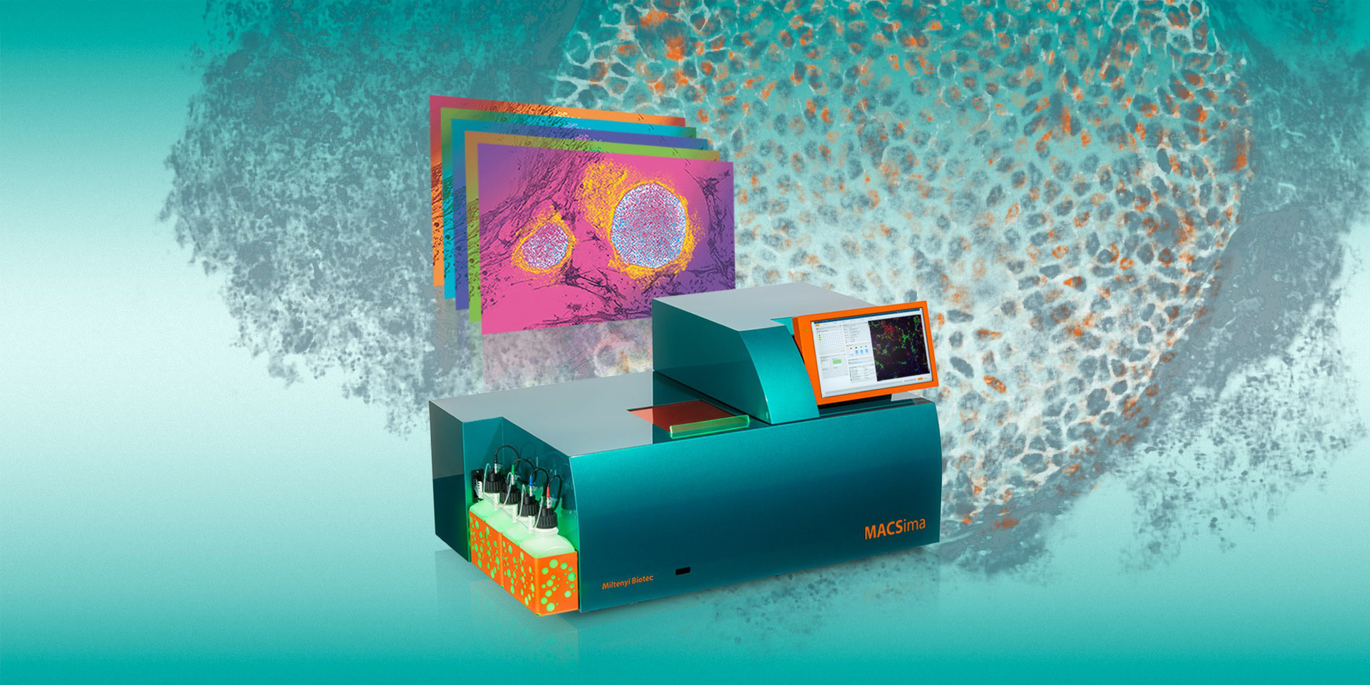
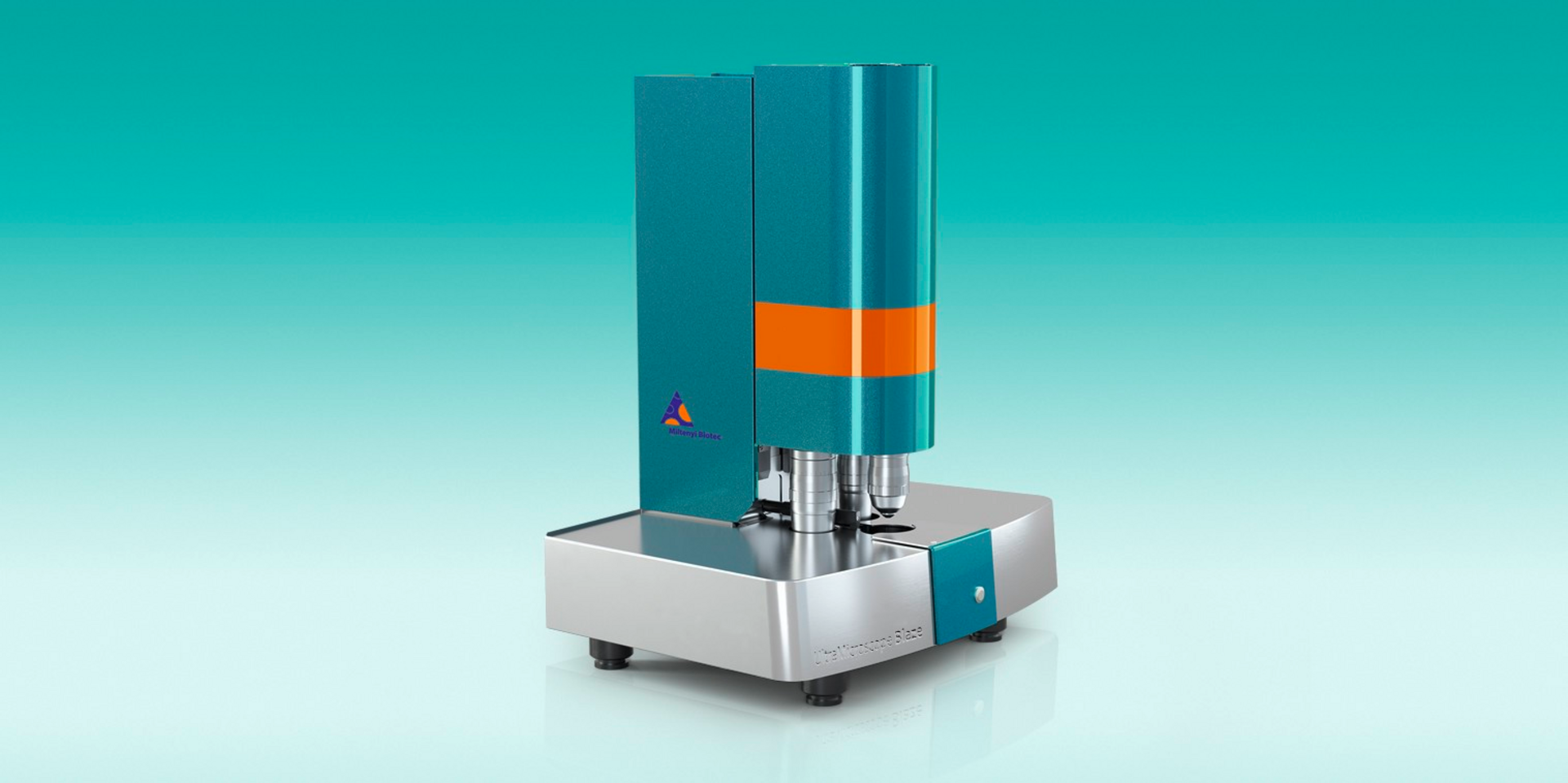
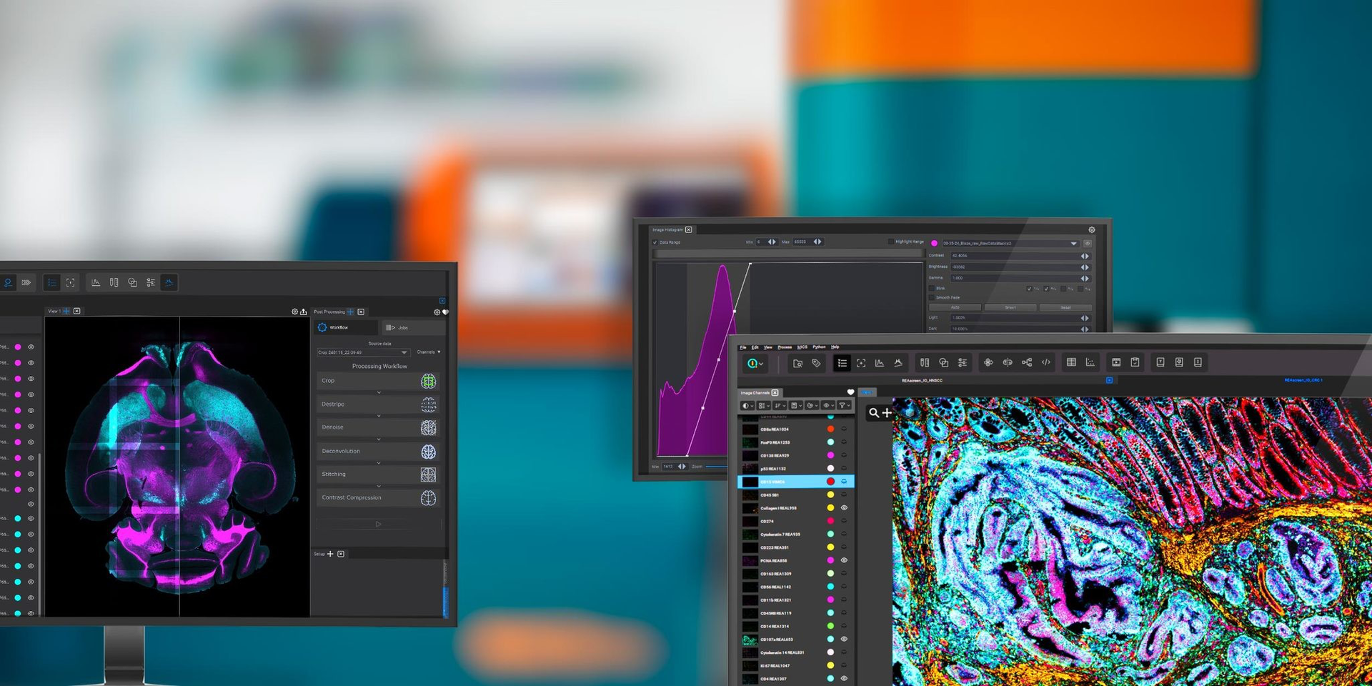
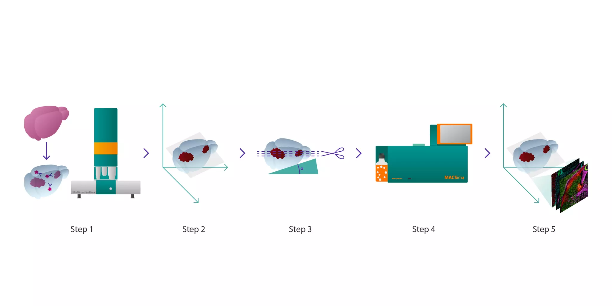
This integrated approach combines light sheet fluorescence microscopy using the UltraMicroscope Blaze™ with multiplexed cyclic immunofluorescence via MACSima® Imaging Cyclic Staining (MICS). It allows researchers to perform volumetric imaging of optically cleared tissues, identify anatomically precise regions of interest, and conduct single-cell level multiplexed proteomic profiling with high spatial fidelity.
Crucially, the workflow preserves both tissue morphology and epitope integrity throughout the entire process, from 3D immunolabeling and optical clearing to sectioning and multiplex staining, ensuring accurate coregistration between macroscopic structures and molecular details.
By bridging whole-tissue 3D imaging with cellular-resolution multiplex analysis, this workflow enables multiscale investigation of tissue microenvironments. Tailored for translational and preclinical research in oncology, neurobiology, immunopathology, and beyond, this integrated approach empowers scientists to explore complex biological systems with high spatial accuracy and deep molecular insight.


Glioblastoma analysis in a preclinical mouse model
Background:
Glioblastoma is a highly invasive brain tumor with complex spatial heterogeneity that presents significant challenges in understanding tumor microenvironments and treatment response. Traditional 2D analysis methods often miss rare or regionally restricted cell populations crucial to disease progression.
Methods:
Using a glioblastoma mouse model, researchers implemented a novel workflow that integrates whole-brain 3D-immunofluorescence imaging with high-plex proteomic analysis, enabling multi-scale investigation of the same specimen. This allowed them to overcome the limitations of thin-section analysis and gain more comprehensive insights into glioblastoma architecture, immune cell infiltration, and spatial biomarker distribution within intact tissues.
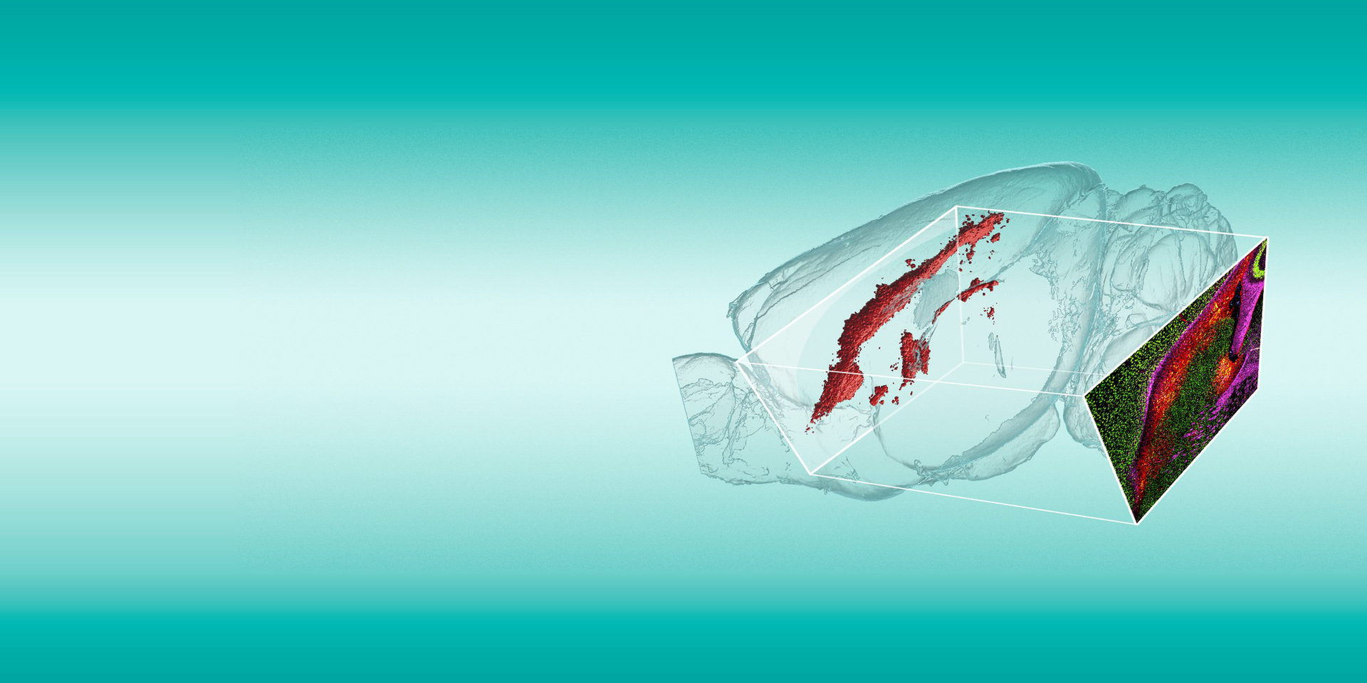
Whole mouse brain hemispheres were optically cleared and imaged using the UltraMicroscope Blaze™, providing detailed 3D maps of tumor location. Using the 3D map of the brain, a region of interest (ROI) was identified, specifically sectioned from the specimen, and analyzed on the MACSima® Platform using multiplexing cyclic immunofluorescence for 108 markers at subcellular resolution.
Results:
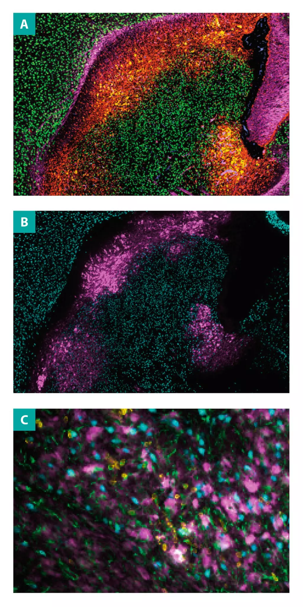
The workflow enabled high-fidelity spatial registration between 3D tumor structures and 2D proteomic profiles. High-resolution 3D imaging of optically cleared mouse brain was performed using the UltraMicroscope Blaze to visualize tumor cell distribution (red) in the intact tissue context (see video). To accurately section along the tumor plane, precise cryosectioning through the tumor area was performed, preserving structural integrity and enabling downstream molecular analysis using MACSima Imaging Cyclic Staining (MICS) technology. The selected tissue section was successfully stained with 108 different Fluorescein isothiocyanate (FITC)-, Phycoerythrin (PE)-, and Vio® 780–conjugated antibodies. Figure A shows staining of the mouse brain hemisphere tissue sections after 3D imaging with antibodies against nestin in red, CD271 (REA648) in yellow, CD146 in blue, NeuN in green, and neurofilament in magenta. Figure B shows NeuN in teal and EGFR (REA939) in magenta. Figure C is the enlarged view from figure B and shows the GFP staining from the 3D imaging in magenta, the MICS staining NeuN in blue, CD45 (REA737) in yellow, and GFAP (REA335) in green.
Tissue morphology and epitope integrity were preserved throughout, allowing detection of marker colocalization and retrospective ROI tracking.
Future perspectives:
This integrative 3D–2D workflow provides a powerful tool for preclinical oncology research. It supports biomarker discovery, targeted therapy development, and deeper understanding of glioblastoma spatial dynamics. The adaptability of the workflow also opens doors for investigating other diseases requiring spatial precision, such as neurodegeneration and immunopathology.