TH1, TH2, and TH17 polarization of naive CD4+ mouse T cells
A complete workflow for cell preparation, isolation, polarization, and analysis
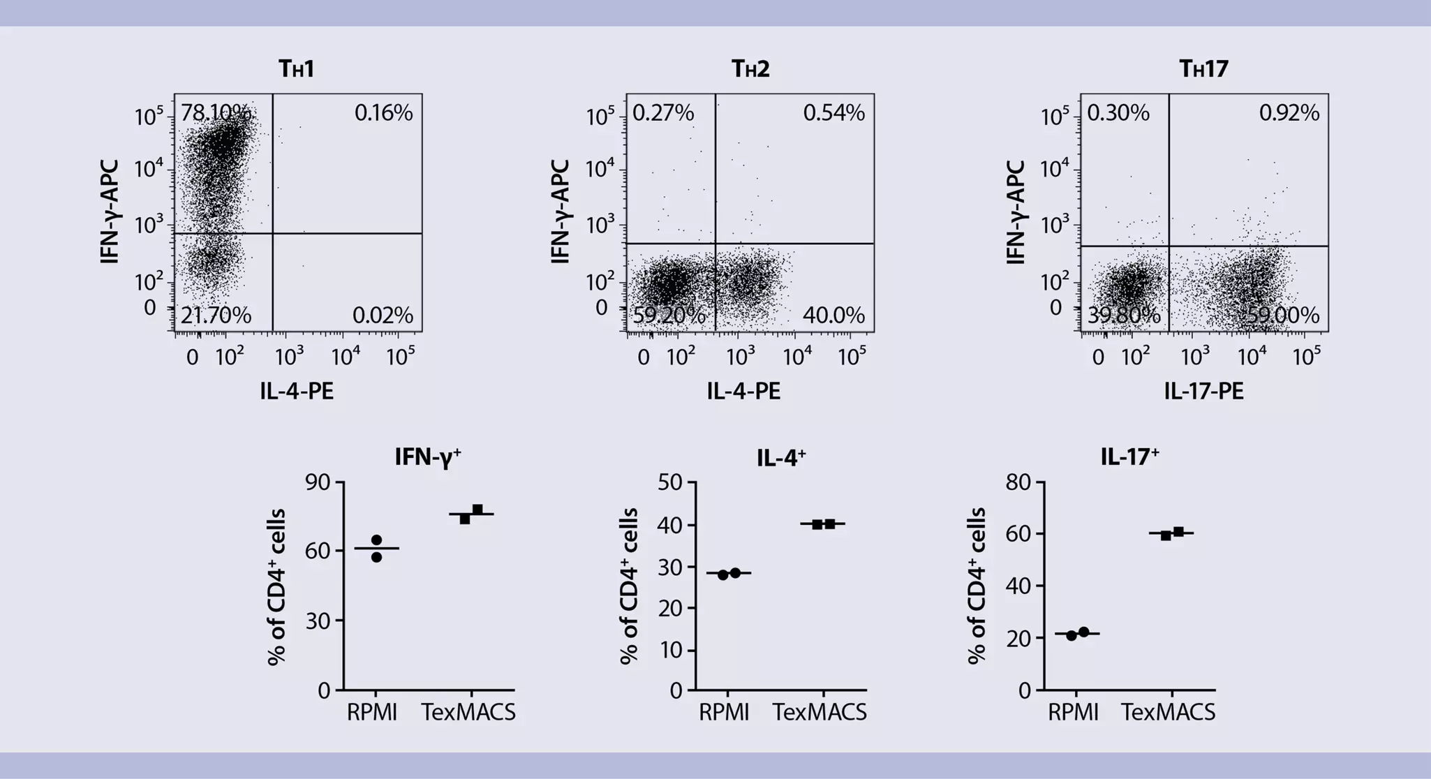
This application protocol describes a complete workflow for reliable and efficient mouse TH cell differentiation, starting with single-cell preparation, followed by isolation of naive CD4+ T cells and in vitro activation and differentiation, through to comprehensive cell analysis. It demonstrates that in vitro TH cell differentiation in the presence of TexMACS™ Medium leads to a higher expression level of the characteristic effector cytokines in the various TH subsets compared to RPMI 1640.
Protocol
TH1, TH2, and TH17 polarization of naive CD4+ mouse T cells
5 min
Preparation of a single cell suspension from mouse spleen
18 min
Isolation of naive CD4+ T cells
6 d
TH1, TH2, or TH17 polarization of naive CD4+ T cells
20 min
Removal of MACSiBead™ Particles
5 h
Intranuclear staining of lineage-specific transcription factors
2 h
Restimulation of polarized T cells and brefeldin A treatment
1.5 h
Intracellular staining of lineage-specific effector cytokines
Flow cytometric analysis
Protocol

This protocol includes detailed instructions for preparing necessary reagents and materials and then carrying out each step.
PBE buffer (standard wash and dilution buffer)
Prepare a solution of PBS, pH 7.2, 2 mM EDTA, and 0.5% BSA by diluting MACS® BSA Stock Solution 1:20 with autoMACS® Rinsing Solution.
Preparation of a single cell suspension from mouse spleen
- For details on the use of the gentleMACS™ Dissociators, refer to the gentleMACS Dissociator user manuals.
- For cell culture experiments subsequent to tissue dissociation, perform all steps under sterile conditions.
- The weight of one mouse spleen amounts to 80–120 mg (female BALB/c mouse, 6–7 weeks old).
- Transfer mouse spleens into a gentleMACS C Tube containing the following amount of PBE buffer:
1–2 mouse spleens: 3 mL
3–4 mouse spleens: 6 mL
5–6 mouse spleens: 9 mL - Close the C Tube tightly and attach it upside down onto the sleeve of the gentleMACS Dissociator.
▲ Note: Close the C Tube tightly beyond the first resistance.
▲ Note: Ensure that the sample material is located in the area of the rotor/stator. - Choose and run one of the following gentleMACS Programs:
1–2 mouse spleens: m_spleen_01
3–6 mouse spleens: m_spleen_04 - After termination of the program, detach the C Tube from the gentleMACS Dissociator.
- (Optional) Perform a short centrifugation step to collect the sample material at the bottom of the tube.
- Resuspend the sample and apply the cell suspension to a MACS SmartStrainer (30 μm) placed on a 15 mL tube (1–2 mouse spleens) or to an appropriate cell strainer placed on a 50 mL tube (3–6 mouse spleens). Wash cell strainer with 5 mL of PBE buffer.
- Discard cell strainer and centrifuge cell suspension at 300×g for 10 minutes at room temperature. Aspirate supernatant completely.
- Resuspend cells in PBE buffer to the required volume for further applications. For example, resuspend cells in 10 mL of PBE buffer for magnetic labeling.
▲Note: Process cells immediately.
Magnetic labeling
- Prepare cells and determine cell number.
- Centrifuge cell suspension at 300×g for 10 minutes. Aspirate supernatant completely.
- Resuspend cell pellet in 400 µL of PBE buffer per 1×10⁸ total cells.
- Add 100 µL of Naive CD4+ T Cell Biotin-Antibody Cocktail, mouse per 1×10⁸ total cells.
- Mix well and incubate for 5 minutes in the refrigerator (+2 to +8 °C).
- Add 200 µL of PBE buffer per 1×10⁸ total cells.
- Add 200 µL of Anti-Biotin MicroBeads per 1×10⁸ total cells.
- Add 100 µL of CD44 MicroBeads per 1×10⁸ total cells.
- Mix well and incubate for 10 minutes in the refrigerator (+2 to +8 °C).
- (Optional) For highest recovery, wash cells by adding 1–2 mL of PBE buffer per 1×10⁸ total cells and centrifuge at 300×g for 10 minutes. Aspirate supernatant completely.
- Resuspend up to 1×10⁸ cells in 500 µL of PBE buffer.
- Proceed to section Magnetic separation.
Magnetic separation
- Always wait until the column reservoir is empty before proceeding to the next step.
- Choose an LS Column and a suitable MACS Separator.
- Place LS Column in the magnetic field of a suitable MACS Separator. For details refer to the respective MACS Column data sheet.
- Prepare column by rinsing with 3 mL of PBE buffer.
- Apply the cell suspension onto the column. Collect flow-through containing unlabeled cells, representing enriched naive CD4+ T cells.
- Wash column with 3 mL of PBE buffer. Collect unlabeled cells that pass through, representing enriched naive CD4+ T cells, and combine with the effluent from step 3.
▲ Note: (Optional) If flow cytometric analysis of the freshly isolated naive CD4+ T cells is desired, take an aliquot (up to 1×10⁶ cells) and proceed to section Flow cytometry analysis of freshly isolated naive CD4+ T cells. - (Optional) Remove column from the separator and place it on a suitable collection tube. Pipette 5 mL of PBE buffer onto the column. Immediately flush out the magnetically labeled non-naive CD4+ T cells by firmly pushing the plunger into the column.
(Optional) Flow cytometric analysis of freshly isolated naive CD4+ T cells
- The recommended antibody dilution for labeling of cells and subsequent analysis by flow cytometry is 1:50 for up to 1×10⁶ cells/50 µL of PBE buffer.
- Volumes given below are for up to 1×10⁶ nucleated cells. When working with fewer than 1×10⁶ cells, use the same volumes as indicated. When working with higher cell numbers, scale up all reagent volumes and total volumes accordingly (e.g. for 2×10⁶ nucleated cells, use twice the volume of all indicated reagent volumes and total volumes).
- Determine cell number.
- Centrifuge cell suspension at 300×g for 10 minutes. Aspirate supernatant completely.
- Resuspend up to 1×10⁶ nucleated cells per 45 µL of PBE buffer.
- Add the following antibodies according to manufacturer's recommendations:
CD45 Antibody, anti-mouse, VioGreen™, REAfinity™
CD4 Antibody, anti-mouse, VioBlue®, REAfinity
CD62L Antibody, anti-mouse, PE, REAfinity
CD3ε Antibody, anti-mouse, APC-Vio® 770, REAfinity
CD44 Antibody, anti-mouse, FITC, REAfinity - Mix well and incubate for 10 minutes in the dark in the refrigerator (+2 to +8 °C).
▲ Note: Higher temperatures and/or longer incubation times may lead to non-specific cell labeling. Working on ice requires increased incubation times. - Wash cells by adding 1−2 mL of PBE buffer and centrifuge at 300×g for 10 minutes. Aspirate supernatant completely.
- Resuspend cell pellet in a suitable amount of PBE buffer (e.g. 500 µL) for analysis by, e.g., flow cytometry.
- (Optional) Add Propidium Iodide Solution to a final concentration of 1 µL per mL (e.g. 0.5 µL in 500 µL) to the cell suspension if exclusion of dead cells via fluorescence is desired.
Things to prepare in advance
Supplemented T cell medium
TexMACS Medium supplemented with FBS (final concentration 10%), 2-mercaptoethanol (final concentration 0.01 mM), and 100× penicillin/streptomycin stock solution (final concentration 1%).
- Medium referred to as fully supplemented T cell medium (see section Polarization of naive T cells) consists of supplemented T cell medium (TexMACS Medium including 10% FBS, 0.01 mM 2-mercaptoethanol, and 1% penicillin/streptomycin stock solution) with the addition of cytokines and antibodies from CytoBox Th1, mouse, CytoBox Th2, mouse, or CytoBox Th17, mouse.
- Addition of penicillin/streptomycin to the T cell medium is optional.
Loading of Anti-Biotin MACSiBead™ Particles
- Resuspend Anti-Biotin MACSiBead Particles thoroughly by vortexing before use to obtain a homogenous suspension.
- Anti-Biotin MACSiBead Particles are supplied without preservative. Remove aliquots under aseptic conditions.
- It is recommended to load Anti-Biotin MACSiBead Particles in batches of 1×10⁸ Anti-Biotin MACSiBead Particles. Loaded Anti-Biotin MACSiBead Particles are stable for up to 4 months when stored at +2 to +8 °C.
- Pipette 100 μL of CD3ε Antibody, Biotin and 100 μL CD28 Antibody, Biotin into a sealable 2 mL tube and mix well.
▲ Note: This staining cocktail with a final antibody concentration of 10 μg antibody per 1 mL loaded Anti-Biotin MACSiBead Particles is optimized for achieving maximum T cell activation. - Add 300 μL of PBE buffer and mix well.
- Resuspend Anti-Biotin MACSiBead Particles thoroughly by vortexing.
- Add 500 μL Anti-Biotin MACSiBead Particles (1×10⁸ Anti-Biotin MACSiBead Particles) to the staining cocktail.
▲ Note: Anti-Biotin MACSiBead Particles can be loaded in a flexible manner with biotinylated antibodies or ligands other than those supplied. If desired, add other biotinylated antibodies or ligands at appropriate concentrations and adjust with buffer to a total volume of 1 mL accordingly. - Incubate for 2 hours at +2 to +8 °C under constant, gentle rotation by using the MACSmix™ Tube Rotator at approximately 4 rpm (slowest permanent run program).
- The loaded Anti-Biotin MACSiBead Particles (1×10⁸ Anti-Biotin MACSiBead Particles/mL) are now ready to use. Do not remove the loaded Anti-Biotin MACSiBead Particles from the antibody mix. Store at +2 to +8 °C for up to 4 months.
Calculation of cytokine concentration
In order to obtain maximal reproducibility for TH cell differentiation experiments, it is recommended to always add recombinant cytokines at a defined unit dose in U/mL. To calculate the cell culture concentration in ng/mL corresponding to the concentration in U/mL, apply the following formula:
Final culture concentration [ng/mL] = (60 U/mL)/(biological activity [U/mg]*) × (106)
* Refer to the corresponding data sheet or CoA to obtain the biological activity.
Plate sizes for in vitro T cell polarization
For T cell polarization, the cells should be resuspended in culture medium at 1×10⁶ cells/mL. Plate the cells at a density of 1×10⁶ cells/cm2. Both the dilution and the cell density are important to assure optimal stimulation and cell growth. The following table lists culture plate sizes suitable for different cell numbers. It also indicates the appropriate amount of medium to add.
Table 1: Plate sizes for in vitro T cell polarization.
| Total cell number | Medium volume to add | Culture plate | Well diameter |
| 2.5×10⁵ | 0.25 mL | 96 well | 0.64 cm |
| 1×10⁶ | 1.00 mL | 48 well | 1.13 cm |
| 2×10⁶ | 2.00 mL | 24 well | 1.60 cm |
| 4×10⁶ | 4.00 mL | 12 well | 2.26 cm |
| 1×10⁷ | 10.00 mL | 6 well | 3.50 cm |
Polarization of naive T cells
Prepare fully supplemented T cell medium by adding cytokines and antibodies to the supplemented TexMACS Medium (see section Things to prepare in advance) as follows:
For TH1 cell polarization (CytoBox Th1, mouse):
- 10 ng/mL (60 U/mL) Mouse IL-12, research grade
- 10 ng/mL (50 U/mL) Mouse IL-2 IS, premium grade
- 10 µg/mL IL-4 Antibody, anti-mouse, pure-functional grade
- 10 ng/mL (200 U/mL) Mouse IL-4, premium grade
- 10 ng/mL (50 U/mL) Mouse IL-2 IS, premium grade
- 10 µg/mL IFN-γ Antibody, anti-mouse, pure-functional grade
- 20 ng/mL (10.000 U/mL) Mouse IL-6, premium grade
- 10 ng/mL (40 U/mL) Mouse IL-23, research grade
- 10 ng/mL (8400 U/mL) Mouse IL-1β, premium grade
- 2 ng/mL (10 U/mL) Human TGF-β1, premium grade
- 10 µg/mL IL-4 Antibody, anti-mouse, pure-functional grade
- 10 µg/mL IFN-γ Antibody, anti-mouse, pure-functional grade
- 10 µg/mL IL-2 Antibody, anti-mouse, pure-functional grade
▲ Note: Refer to section Things to prepare in advance for conversion from U/mL to ng/mL.
- Resuspend loaded Anti-Biotin MACSiBead Particles thoroughly and transfer, for example, 40 μL (4×10⁶ loaded Anti-Biotin MACSiBead Particles) per 2×10⁶ cells (bead to cell ratio 2:1) into a suitable tube.
▲ Note: If unloaded MACSiBead Particles shall be used for negative control experiments, replace loaded Anti-Biotin MACSiBead Particles with unloaded Anti-Biotin MACSiBead Particles. - Add 1 mL of culture medium to the loaded Anti-Biotin MACSiBead Particles and centrifuge at 300×g for 5 minutes.
- Aspirate supernatant and resuspend loaded Anti-Biotin MACSiBead Particles in, e.g., 1 mL of fully supplemented T cell medium including the cytokines and antibodies (from CytoBox) to a final concentration of 4×10⁶ Anti-Biotin MACSiBead Particles per mL.
- Determine cell number of freshly isolated mouse naive CD4+ T cells from chapter 2. Isolation of naive CD4+ T cells.
- Centrifuge cell suspension at 300×g for 10 minutes. Aspirate supernatant completely.
- Resuspend cells at a density of 2×10⁶ cells per mL in fully supplemented T cell medium including cytokines and antibodies (from CytoBox).
- Transfer the desired amount of cells and the loaded Anti-Biotin MACSiBead Particles at a ratio of 1:1 to a suitable cell culture vessel to a final density of 1×10⁶ cells per mL per cm2 with two loaded Anti-Biotin MACSiBead Particles per cell (e.g. 2.5×10⁵ cells in 125 µL medium + 125 µL loaded Anti-Biotin MACSiBeads Particles per well of a 96-well plate).
▲ Note: Depending on the cell number to be cultivated and differentiated per well, 6-, 12-, 24- or 48-well plates might be used. Please upscale reagents accordingly and refer to "Plate sizes for in vitro T cell polarization" to choose the appropriate cell culture dish. - Incubate at +37 °C and 5–10% CO2 for up to 6 days.
▲ Note: Inspect cultures daily and add fresh fully supplemented TexMACS Medium if required. - At day 2, gently pipette culture up and down to break up all cell clumps.
- Split the cell culture every two days 1:4 or 1:2, depending on the proliferation of cells, and add fresh fully supplemented TexMACS Medium.
- After 6 days of cultivation, polarized TH1, TH2, or TH17 cells can be further processed for downstream analysis. Refer to "Intranuclear staining of lineage-specific transcription factors" or "Intracellular staining of lineage-specific effector cytokines". T cells require a restimulation for further expansion or analysis of cytokine expression. See "Restimulation of polarized T cells and brefeldin A treatment" for details.
For further flow cytometric analysis, it is recommended to remove the MACSiBead Particles from the cell suspension.
- Harvest cells and pool different wells of the same condition.
- Wash empty wells with cold PBE buffer to rinse out the remaining cells on the plate.
- Determine cell number.
- Wash cells with cold PBE buffer.
- Resuspend cells in PBE buffer at a density of up to 2×10⁷ cells/mL and vortex thoroughly.
- Place the tube in the magnetic field of the MACSiMAG™ Separator.
- Allow the MACSiBead Particles to adhere to the wall of the tube for 4 minutes.
- Keeping the tube in the magnet, carefully remove the supernatant containing the MACSiBead-depleted cells and place in a new tube.
- Remove the tube from the separator and add PBE buffer to the same volume as before.
- Vortex sample, replace tube in the MACSiMAG Separator, and repeat steps 7–8.
- Collected cells can now be further processed as required.
Things to prepare in advance
Fixation and Permeabilization Solution (FoxP3 Staining Buffer Set)
To achieve the appropriate working concentration for safe fixation and permeabilization of cells, Fixation/Permeabilization Solution 1 must be diluted 1:4 with Fixation/Permeabilization Solution 2 (i.e. for 1×10⁶ cells use 0.25 mL of Fixation/Permeabilization Solution 1 plus 0.75 mL of Fixation/Permeabilization Solution 2).
Permeabilization Buffer (FoxP3 Staining Buffer Set)
To achieve the appropriate working concentration for safe permeabilization of cells, 10× Permeabilization Buffer must be diluted 1:10 with deionized or distilled water before use (i.e. 1 mL of 10× Permeabilization Buffer plus 9 mL of deionized/distilled water).
▲ Note: Before preparing the dilution, make sure that the buffer does not contain any precipitates.
Transcription factor antibodies
- Analysis of TH1 cells (e.g. T-bet; T-bet Antibody, anti-human/mouse, PE, REAfinity)
- Analysis of TH2 cells (e.g. T-bet and Gata3; T-bet Antibody, anti-human/mouse, PE and Gata3 Antibody, anti-human/mouse, APC, REAfinity)
- Analysis of TH17 cells (e.g. RORγ (t); RORγ (t) Antibody, anti-human/mouse, APC, REAfinity)
Notes:
- The recommended antibody dilution for all above-mentioned antibodies is 1:50 for up to 1×10⁶ cells/100 μL of buffer for labeling of cells for analysis by flow cytometry.
- Use freshly prepared Fixation and Permeabilization Solution and Permeabilization Buffer from FoxP3 Staining Buffer Set for cell fixation and permeabilization.
Intranuclear staining of lineage-specific transcription factors
- Determine cell number.
- Centrifuge cell suspension at 300×g for 10 minutes. Aspirate supernatant completely.
- Resuspend up to 1×10⁶ nucleated cells in 1 mL of cold, freshly prepared Fixation/Permeabilization Solution.
- Mix well and incubate for 30 minutes in the dark in the refrigerator (+2 to +8 °C).
- Wash cells by adding 1−2 mL of cold buffer per 1×10⁶ cells and centrifuge at 300×g for 5 minutes at +2 to +8 °C. Aspirate supernatant completely.
- Wash cells by adding 1−2 mL of cold 1× Permeabilization Buffer per 1×10⁶ cells and centrifuge at 300×g for 5 minutes at +2 to +8 °C. Aspirate supernatant completely.
- Resuspend up to 1×10⁶ nucleated cells in 100 μL of cold 1× Permeabilization Buffer.
▲ Note: For staining with several antibodies in this step, reduce the volume of 1× Permeabilization Buffer accordingly. For efficient permeabilization, the volume of 1× Permeabilization Buffer should be at least 30% of the overall staining volume. - Add 2 μL of each needed transcription factor antibody.
- Mix well and incubate for 30 minutes in the dark in the refrigerator (+2 to +8 °C).
- Wash cells by adding 1−2 mL of cold 1× Permeabilization Buffer per 1×10⁶ cells and centrifuge at 300×g for 5 minutes at +2 to +8 °C. Aspirate supernatant completely.
- Resuspend cell pellet in a suitable amount of buffer for analysis by flow cytometry or fluorescence microscopy.
▲ Note: Due to fixation and permeabilization, cells are smaller than viable cells. Thus, FSC/SSC settings of the flow cytometer may need to be adjusted.
- Add 20 ng/mL PMA and 1 µg/mL Ionomycin to cells and incubate for 3 hours at +37 °C.
- Add brefeldin A to a final concentration of 2 µg/mL to the cells and incubate for another 2 hours at +37 °C.
- Harvest cells, determine cell number, and proceed to next step.
- For analysis of TH1 and TH2 cells, use, e.g., IFN-γ and IL-4; IFN-γ Antibody, anti-mouse, APC, REAfinity and IL-4 Antibody, anti-mouse, PE, REAfinity.
- For analysis of TH17 cells, use e.g., IFN-γ and IL-17; IFN-γ Antibody, anti-mouse, APC, REAfinity and IL-17 Antibody, anti-mouse, PE, REAfinity).
- The recommended antibody dilution of the above-mentioned antibodies for cell labeling and subsequent flow cytometry analysis is 1:50 for up to 1×10⁶ cells/50 μL of PBE buffer.
- Volumes given below are for up to 1×10⁶ nucleated cells. When working with fewer than 1×10⁶ cells, use the same volumes as indicated. When working with higher cell numbers, scale up all reagent volumes and total volumes accordingly (e.g. for 2×10⁶ nucleated cells, use twice the volume of all indicated reagent volumes and total volumes).
- T cells require a restimulation prior to analysis of cytokine expression.
- After successful restimulation and brefeldin A treatment, intranuclear transcription factor and intracellular cytokine staining can be combined for certain cytokine/transcription factor combinations. In this case, please follow the protocol steps "Restimulation of polarized cells and brefeldin A treatment", "Intracellular staining of lineage-specific effector cytokines", and "Intranuclear staining of lineage-specific transcription factors" in this order.
- IL-4 staining cannot be combined with intranuclear staining using the FoxP3 Staining Buffer Set.
- Use Inside Fix and Inside Perm from Inside Stain Kit for cell permeabilization and fixation.
- Wash up to 1×10⁶ cells by adding 1–2 mL of PBE buffer and centrifuge at 300×g for 10 minutes. Aspirate supernatant completely.
- (Optional) Stain cell surface antigens that are sensitive to fixation with appropriate antibodies according to the manufacturer's recommendations.
- Wash cells by adding 1–2 mL of PBE buffer and centrifuge at 300×g for 10 minutes. Aspirate supernatant completely.
- Resuspend up to 1×10⁶ cells in 250 μL of PBE buffer.
- Add 250 μL of Inside Fix (from the Inside Stain Kit). Mix well and incubate for 20 minutes in the dark at room temperature.
- Centrifuge at 300×g for 5 minutes. Aspirate supernatant carefully.
- Wash cells by adding 1 mL of PBE buffer and centrifuge at 300×g for 5 minutes. Aspirate supernatant carefully.
▲ Note: Fixed cells may be stored in azide-containing buffer at +2 to +8 °C for up to one week. - (Optional) Stain cell surface antigens that are sensitive to permeabilization with appropriate antibodies.
- Wash cells by adding 1–2 mL of PBE buffer and centrifuge at 300×g for 10 minutes. Aspirate supernatant completely.
- Wash cells by adding 1 mL of Inside Perm (from the Inside Stain Kit) and centrifuge at 300×g for 5 minutes. Aspirate supernatant carefully.
- Resuspend cells in 49 μL of Inside Perm. Add 1 μL of the antibody of choice.
▲ Note: For staining with several antibodies in this step, reduce the volume of Inside Perm accordingly. For efficient permeabilization, the volume of Inside Perm should be at least 30% of the overall staining volume. - Mix well and incubate for 10 minutes in the dark at room temperature.
- Wash cells by adding 1 mL of Inside Perm and centrifuge at 300×g for 5 minutes. Aspirate supernatant carefully.
- Resuspend cell pellet in a suitable amount of buffer for analysis by flow cytometry or fluorescence microscopy. Store cells at +2 to +8 °C in the dark until analysis. Mix well before flow cytometric acquisition.
▲ Note: Samples may be stored at +2 to +8 °C in the dark for up to 24 hours.
▲ Note: Do not use propidium iodide (PI) or 7-AAD staining.
 View details

21 products found
|
Catalog no. was copied to your clipboard
 130-121-340
For research use only
Catalog no. was copied to your clipboard
130-121-340Synonyms : TBX21, T-PET, TBLYM, TBT1, TBLYM
For research use only
Applications
ICFC
ICFC
Size
100 tests in 200 µL
100 tests in 200 µL
Price
EUR 341,00
(excl. tax)
Catalog no. was copied to your clipboard
130-093-237For research use only
Size
25 tubes (sterile, single-packed)
25 tubes (sterile, single-packed)
Price
EUR 170,00
(excl. tax)
Catalog no. was copied to your clipboard
130-093-627For research use only
Catalog no. was copied to your clipboard
130-093-627For research use only
Size
1 kit
1 kit
Price
EUR 774,00
(excl. tax)
Catalog no. was copied to your clipboard
130-102-543For research use only
Catalog no. was copied to your clipboard
130-102-543Synonyms : Sell, L-selectin, LECAM-1, Lnhr, Ly-22, Ly-m22, Lyam-1, Lyam1, LAM-1
For research use only
Applications
FC, MICS, IF, IHC
FC, MICS, IF, IHC
Size
30 µg in 1 mL
30 µg in 1 mL
Price
EUR 102,00
(excl. tax)
Catalog no. was copied to your clipboard
130-102-344For research use only
Catalog no. was copied to your clipboard
130-102-344Synonyms : CTLA-8, IL-17
For research use only
Applications
ICFC, MICS, IF, IHC
ICFC, MICS, IF, IHC
Size
30 µg in 1 mL
30 µg in 1 mL
Price
EUR 102,00
(excl. tax)
Catalog no. was copied to your clipboard
130-092-168For research use only
Size
1 kit
1 kit
Price
EUR 814,00
(excl. tax)
Catalog no. was copied to your clipboard
130-093-142For research use only
Size
for 100 tests
for 100 tests
Price
EUR 265,00
(excl. tax)
Catalog no. was copied to your clipboard
130-042-303For research use only
Size
1 piece
1 piece
Price
EUR 192,00
(excl. tax)
Catalog no. was copied to your clipboard
130-041-407For research use only
Catalog no. was copied to your clipboard
130-041-407For research use only
Size
50 filters
50 filters
Price
EUR 224,00
(excl. tax)
Catalog no. was copied to your clipboard
130-042-401For research use only
Size
25 columns
25 columns
Price
EUR 338,00
(excl. tax)
Catalog no. was copied to your clipboard
130-090-753For research use only
Price
EUR 1.100,00
(excl. tax)
HIGHLIGHT
Catalog no. was copied to your clipboard
130-134-029For research use only
Catalog no. was copied to your clipboard
130-134-029For research use only
Price
On request
Catalog no. was copied to your clipboard
130-091-222For research use only
Size
6× 1.45 L
6× 1.45 L
Price
EUR 199,00
(excl. tax)
Catalog no. was copied to your clipboard
130-091-376For research use only
Size
6× 75 mL
6× 75 mL
Price
EUR 366,00
(excl. tax)
Catalog no. was copied to your clipboard
130-093-233For research use only
Size
2 mL
2 mL
Price
EUR 73,00
(excl. tax)
Catalog no. was copied to your clipboard
130-042-302For research use only
Size
1 piece
1 piece
Price
EUR 784,00
(excl. tax)
Catalog no. was copied to your clipboard
130-104-453For research use only
Catalog no. was copied to your clipboard
130-104-453For research use only
Size
for 1×10⁹ cells
for 1×10⁹ cells
Price
EUR 865,00
(excl. tax)
Catalog no. was copied to your clipboard
130-107-761For research use only
Size
1 kit
1 kit
Price
EUR 355,00
(excl. tax)
Catalog no. was copied to your clipboard
130-107-760For research use only
Size
1 kit
1 kit
Price
EUR 331,00
(excl. tax)
Catalog no. was copied to your clipboard
130-090-477For research use only
Size
for up to 50 tests
for up to 50 tests
Price
EUR 272,00
(excl. tax)
Catalog no. was copied to your clipboard
150-000-364For research use only
Catalog no. was copied to your clipboard
150-000-364For research use only
Size
4 pieces
4 pieces
Price
EUR 58,00
(excl. tax)