The human LOX-1 antigen, the lectin-like oxidized low-density lipoprotein receptor 1, is also known as OLR1 or CLEC8A and belongs to the C-type lectin superfamily. LOX-1 is found on endothelial cells, macrophages, platelets, fibroblasts, and smooth muscle cells. It plays a role in endocytosis, phagocytosis, apoptosis, and inflammatory processes. LOX-1 is expressed on a population of polymorphonuclear myeloid-derived suppressor cells (PMN-MDSCs) in peripheral blood of cancer patients1. Moreover, a high density of tumor-associated neutrophils (TANs) expressing LOX-1 and arginase 1 is strongly correlated with a reduced expression of cytotoxic granzyme B and the proliferation marker Ki67 in closely located T cells, suggesting a suppressive function of LOX-1+ TANs2.
Isolation of LOX-1+ cells from cord blood mononuclear cell suspension
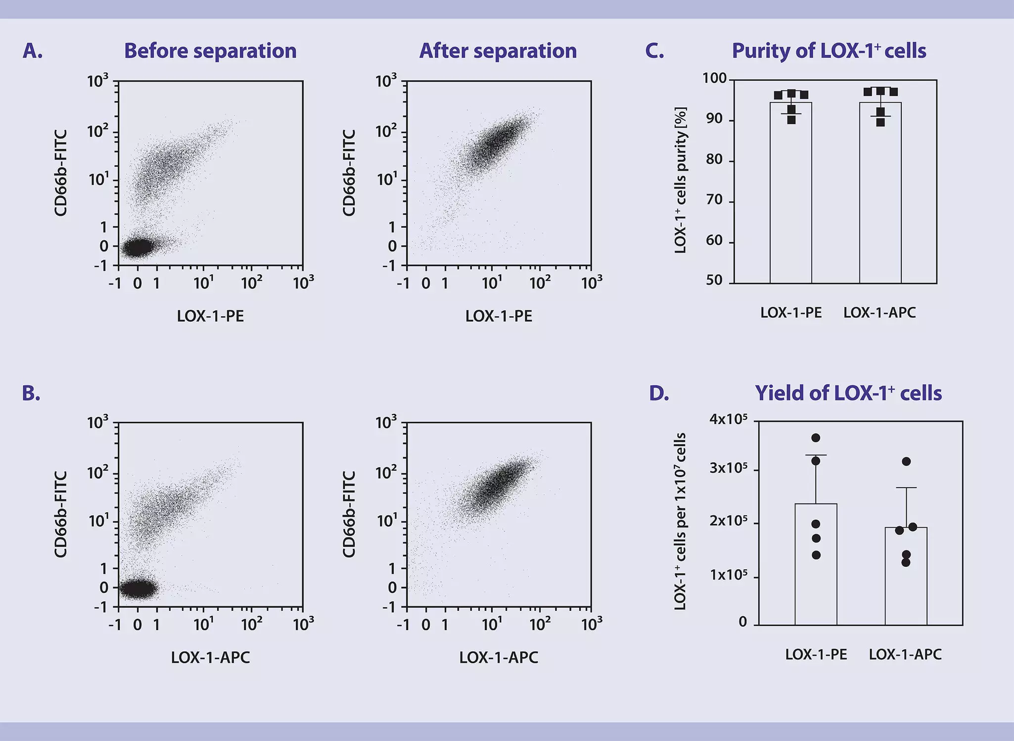
This application protocol describes an exemplary procedure, which can be used in order to isolate LOX-1+ cells. Here, LOX-1 cells were isolated from cord blood mononuclear cell suspension from one individual.
When working with anticoagulated cord blood, cord blood mononuclear cells should be isolated by density gradient centrifugation, e.g., using Ficoll-Paque™. For details regarding the preparation of mononuclear cells refer to the Resources section of the Diamond CD34 Isolation Kit, human.
▲Note: To remove platelets after density gradient separation, resuspend cell pellet in buffer and centrifuge at 200×g for 10−15 minutes at 20 °C. Carefully aspirate supernatant. Repeat washing step.
▲Note: It is strongly recommended to screen biological samples for the presence of LOX-1+ cells before proceeding with the separation protocol.
1. Magnetic labeling
- Work fast, keep cells cold, and use pre-cooled solutions. This will prevent capping of antibodies on the cell surface and non-specific cell labeling.
- Volumes for magnetic labeling given below are for up to 107 total cells. When working with fewer than 107 cells, use the same volumes as indicated. When working with higher cell numbers, scale up all reagent volumes and total volumes accordingly (e.g. for 2×10⁷ total cells, use twice the volume of all indicated reagent volumes and total volumes).
- For optimal performance it is recommended to obtain a single cell suspension before magnetic labeling. Pass cells through 30 μm nylon mesh (e.g. Pre-Separation Filters (30 μm) (# 130-041-407)) to remove cell clumps which may clog the column. Moisten filter with buffer before use.
- The recommended incubation temperature is 2–8 °C. Higher temperatures and/or longer incubation times may lead to non-specific cell labeling. Working on ice may require increased incubation times.
1.1 Labeling of LOX-1+ cells using LOX-1 Antibody, anti-human, PE, REAfinity™
- Determine cell number.
- Centrifuge cell suspension at 300×g for 10 minutes. Aspirate supernatant completely.
- Resuspend cell pellet in 96 μL of buffer per 107 total cells.
- Add 4 μL of LOX-1 Antibody, anti-human, PE, REAfinity per 107 total cells.
- Mix well and incubate for 15 minutes in the refrigerator (2−8 °C).
- Wash cells by adding 1–2 mL of buffer per 107 cells and centrifuge at 300×g for 10 minutes. Aspirate supernatant completely.
- Resuspend cell pellet by adding 80 μL of buffer per 107 total cells.
- Add 20 μL of Anti-PE MicroBeads per 107 total cells.
- Mix well and incubate for 10 minutes in the refrigerator (2−8 °C).
- Wash cells by adding 1–2 mL of buffer per 10⁷ cells and centrifuge at 300×g for 10 minutes. Aspirate supernatant completely.
- Resuspend up to 108 cells in 500 μL of buffer.
- Proceed to magnetic separation.
1.2 Labeling of LOX-1+ cells using LOX-1 Antibody, anti-human, APC, REAfinity
- Determine cell number.
- Centrifuge cell suspension at 300×g for 10 minutes. Aspirate supernatant completely.
- Resuspend cell pellet in 96 μL of buffer per 107 total cells.
- Add 4 μL of LOX-1 Antibody, anti-human, APC, REAfinity per 107 total cells.
- Mix well and incubate for 15 minutes in the refrigerator (2−8 °C).
- Wash cells by adding 1–2 mL of buffer per 107 cells and centrifuge at 300×g for 10 minutes. Aspirate supernatant completely.
- Resuspend cell pellet by adding 80 μL of buffer per 10⁷ total cells.
- Add 20 μL of Anti-APC MicroBeads per 107 total cells.
- Mix well and incubate for 10 minutes in the refrigerator (2−8 °C).
- Wash cells by adding 1–2 mL of buffer per 10⁷ cells and centrifuge at 300×g for 10 minutes. Aspirate supernatant completely.
- Resuspend up to 108 cells in 500 μL of buffer.
- Proceed to magnetic separation.
2. Magnetic separation using MS or LS Columns
- Choose an appropriate MACS® Column and MACS Separator according to the number of total cells and the number of LOX-1+ cells.
- Always wait until the column reservoir is empty before proceeding to the next step.
- Place column in the magnetic field of a suitable MACS Separator. For details refer to the respective MACS Column data sheet.
- Prepare column by rinsing with the appropriate amount of buffer:
MS: 500 μL
LS: 3 mL - Apply cell suspension onto the column. Collect flow-through containing unlabeled cells.
- Wash column with the appropriate amount of buffer. Collect unlabeled cells that pass through and combine with the flow-through from step 3.
MS: 3×500 μL
LS: 3×3 mL
▲Note: Perform washing steps by adding buffer aliquots as soon as the column reservoir is empty. - Remove column from the separator and place it on a suitable collection tube.
- Pipette the appropriate amount of buffer onto the column. Immediately flush out the magnetically labeled LOX-1+ cells.
- (Optional) To increase the purity of LOX-1+ cells, the eluted fraction can be enriched over a second MS or LS Column. Repeat the magnetic separation procedure as described in steps 1 to 6 by using a new column.
- Condamine T, et al. (2016) Lectin-type oxidized LDL receptor-1 distinguishes population of human polymorphonuclear myeloid-derived suppressor cells in cancer patients. Sci. Immunol.;1(2):aaf8943. .
- Si Y, et al. (2019) Multidimensional imaging provides evidence for down-regulation of T cell effector function by MDSC in human cancer tissue. Sci. Immunol. 18;4(40):eaaw9159.