

Synonyms: TNF, DIF, TNFSF2, Tnfsf1a
EXTENDED VALIDATION
- Clone:
- MP6-XT22
- Type of antibody:
- Primary antibodies
- Isotype:
- rat IgG1κ
- Applications:
- ICFC, FA Glossary
- Select a conjugate:


Related products for TNF-α Antibody, anti-mouse


Intracellular flow cytometry applications
APC
FITC
PE
Recommended dilution:
1:10
Remark:
The antibody is suited for staining of formaldehyde-fixed cells.
Data source:
Miltenyi Biotec
Conjugate:
APC
Used products:
130-102-339
Description:
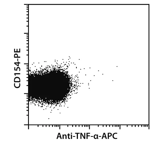
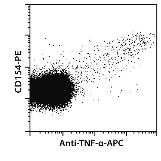
Mouse spleen cells were incubated with or without KLH for 6 hours. After 2 hours, brefeldin A was added. Cells were harvested, fixed, permeabilized, and intracellularly stained with Anti-TNF-α-PE and CD154-APC. Cells were analyzed by flow cytometry. Gating was performed according to CD4 expression and side scatter properties of the cells.
 view details
 view details
Functional assay applications
pure-functional grade
Data source:
Miltenyi Biotec
Type of functional assay:
neutralization of cytokine activity
Conjugate:
pure-functional grade
Used products:
130-095-725
Extended validation for TNF-α Antibody, anti-mouse
Sensitivity
Selected fluorochrome conjugated antibodies from Miltenyi Biotec were compared to commercially available hybridoma clones in flow cytometry analysis.
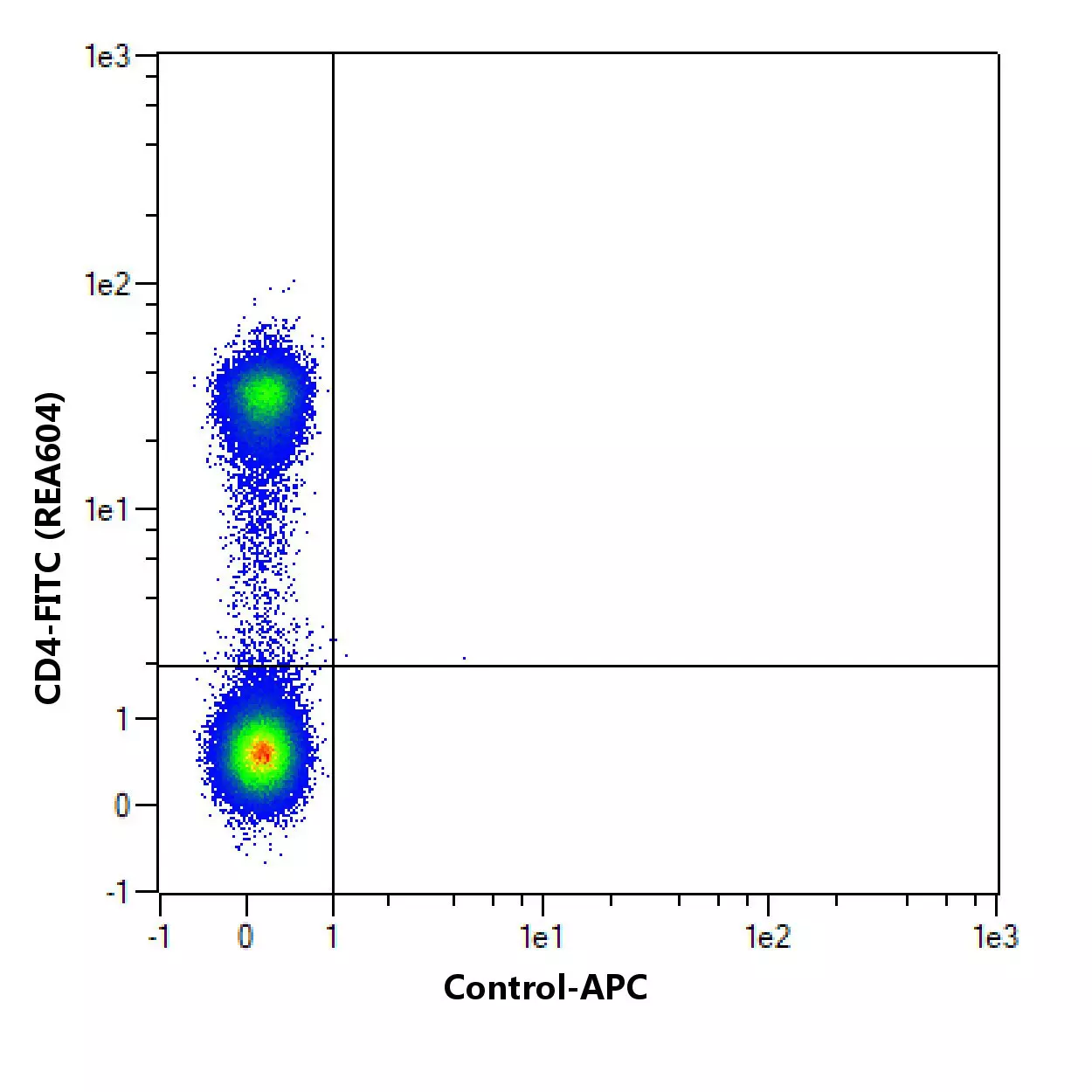
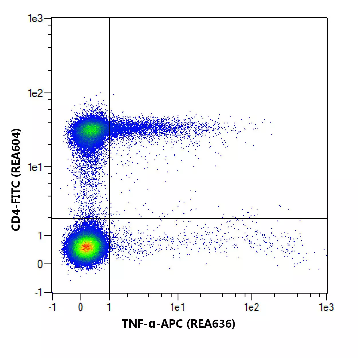
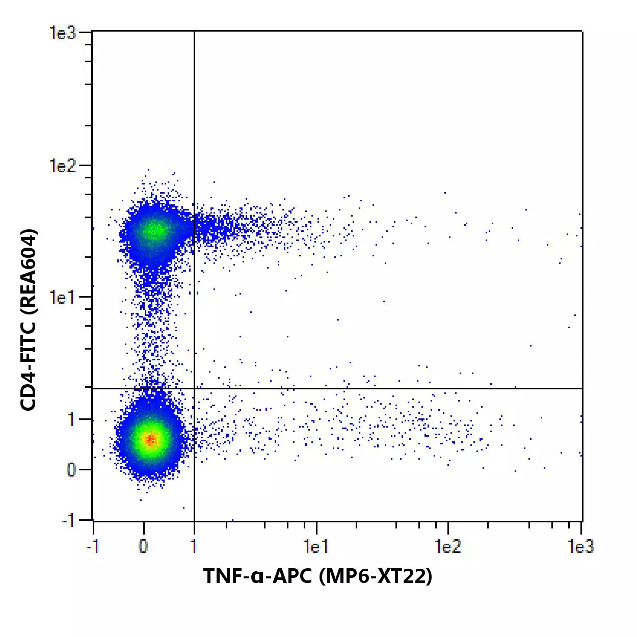
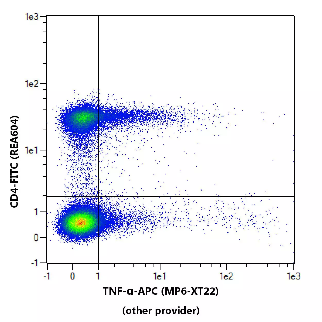
Flow cytometric comparison of different clones for TNF-α. Splenocytes from Balbc/c mice were stimulated with 50 ng/mL CD3 + 1 µg/mL CD28 for 2h, followed by an incubation with 1µg/mL brefeldin A for 4 hours. Cells were first stained with Viobility™ Fixable Dye followed by a suitable counterstaining. Cells were then fixed and permeabilized using the Inside Stain Set followed by intracellular staining with TNF-αantibodies. As a control, TNF-α antibody staining was omitted and cells were measured in the same channels. Flow cytometry was performed with the MACSQuant® Analyzer. Cell debris, dead cells, and cell doublets were excluded from the analysis based on scatter signals and Viobility 405/520 Fixable Dye fluorescence. No FcR Blocking Reagent was used. The recommended titers of respective antibodies from different suppliers were used.
Specifications for TNF-α Antibody, anti-mouse
Overview
Mouse tumor necrosis factor α (TNF-α) is secreted by macrophages, monocytes, neutrophils, CD4+ T cells, and NK cells. It has cytolytic activity against a range of tumor cells and is important in immune regulation. TNF-α binding to surface receptors elicits a wide array of biologic activities, including cytolysis and cytostasis of many tumor cell lines in vitro, hemorraghic necrosis of tumors in vivo, increased fibroblast proliferation, and enhanced chemotaxis and phagocytosis in neutrophils.
TNF, DIF, TNFSF2, Tnfsf1a
Detailed product information
Technical specifications
Clone
MP6-XT22
Clonality
monoclonal
Isotype
rat IgG1κ
Isotype control
Isotype Control Antibody, rat IgG1
Host
rat
Type of antibody
Primary antibodies
Species reactivity
mouse
Antigen
TNF-α
Molecular mass of antigen [kDa]
26
Entrez Gene ID
RRID
AB_2661142, AB_2661141, AB_10828341
Resources for TNF-α Antibody, anti-mouse
Certificates
To access a Certificate of Analysis (CoA) follow the link below and enter the specific lot of your product.
Should you wish to request additional documentation, such as a Safety Data Sheet (SDS) in other languages than English or Swedish, an SDS-replacement document, or a Certificate of Origin (CoO), please direct your inquiry to your local technical support team.
 Powered by Bioz