StraightFrom® Whole Blood CD56 MicroBeads were developed for the rapid positive selection of CD56+ cells directly from whole blood or bone marrow, minimizing hands-on time and maximizing yield of target cells. No sample preparation is required, including density gradient centrifugation or erythrocyte lysis.
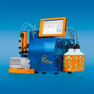
StraightFrom Whole Blood MicroBeads in combination with the autoMACS® Pro Separator allow standardization of the cell separation procedure and safe handling of hazardous samples.


Related products for StraightFrom® Whole Blood CD56 MicroBeads, human

StraightFrom® Whole Blood CD56 MicroBeads were developed for the rapid positive selection of CD56+ cells directly from whole blood or bone marrow, minimizing hands-on time and maximizing yield of target cells. No sample preparation is required, including density gradient centrifugation or erythrocyte lysis.
StraightFrom Whole Blood MicroBeads in combination with the autoMACS® Pro Separator allow standardization of the cell separation procedure and safe handling of hazardous samples.
The CD56 antigen is expressed by most NK cells and a small subset of T cells. Upon activation of NK cells, the surface expression of CD56 is increased.
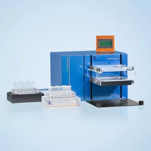
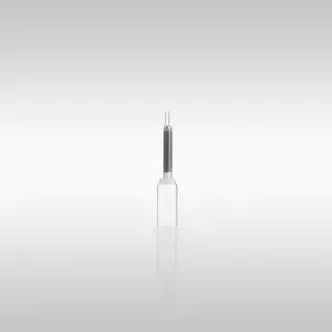
Anticoagulated cell samples of 0.25–15 mL are labeled with StraightFrom® Whole Blood CD56 MicroBeads to be subsequently separated with the autoMACS® Pro Separator, the MultiMACS™ Cell24 Separator Plus, or using the Whole Blood Column Kit.
CD56+ cells isolated using StraightFrom® Whole Blood CD56 MicroBeads are ideally suited for further flow cytometric, functional, or molecular analysis including lineage-specific chimerism analysis after allogeneic stem cell transplantation1,2.
autoMACS® Columns or Whole Blood Columns.
Separation of a whole blood sample using the StraightFrom® Whole Blood CD56 MicroBeads and the MultiMACS™ Cell24 Separator Plus with the Single-Column Adapter and Whole Blood Columns without washing step after labeling. Cells were fluorescently stained with CD56-PE, CD16‑APC, as well as CD45-VioBlue® and analyzed by flow cytometry using the MACSQuant® Analyzer. Cells were triggered via CD45-VioBlue, cell debris and dead cells were excluded from the analysis based on scatter signals and propidium iodide fluorescence.
- Willasch, A. et al. (2010) Enrichment of cell subpopulations applying automated MACS technique: purity, recovery and applicability for PCR-based chimerism analysis. Bone Marrow Transplant. 45: 181-189
- Meyer, R. G. et al. (2007) Prophylactic transfer of CD8-depleted donor lymphocytes after T-cell-depleted reduced-intensity transplantation. Blood 109: 374-382
- Koehl, U. et al. (2003) Quantitative analysis of chimerism after allogeneic stem cell transplantation by PCR amplification of microsatellite markers and capillary electrophoresis with fluorescence detection: the Frankfurt experience. Leukemia 17: 232-236