The StraightFrom® Buffy Coat CD56 MicroBeads, human have been developed for the rapid positive selection of CD56+ cells directly from buffy coat using the MultiMACS™ Cell24 Separator Plus for semi-automated separation, an autoMACS® NEO for fully automated separation or a MACS® Separator for manual separation. To enable direct processing of a full buffy coat, please consider the datasheet of the MicroBeads or the ‘Materials’ tab of the respective protocol to ensure the required columns and buffers are included on the kit or need to be purchased separately. </


Related products for StraightFrom® Buffy Coat CD56 MicroBeads, human
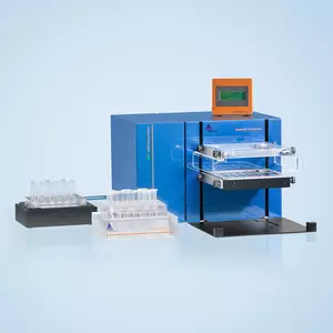


The StraightFrom® Buffy Coat CD56 MicroBeads, human have been developed for the rapid positive selection of CD56+ cells directly from buffy coat using the MultiMACS™ Cell24 Separator Plus for semi-automated separation, an autoMACS® NEO for fully automated separation or a MACS® Separator for manual separation. To enable direct processing of a full buffy coat, please consider the datasheet of the MicroBeads or the ‘Materials’ tab of the respective protocol to ensure the required columns and buffers are included on the kit or need to be purchased separately.
The CD56 antigen is expressed by most NK cells and a small subset of T cells. Upon activation of NK cells, the surface expression of CD56 is increased.
Buffy coats are labeled with StraightFrom® Buffy Coat CD56 MicroBeads to be subsequently separated with the autoMACS® NEO Separator, the MultiMACS™ Cell24 Separator Plus, or manually with a MACS® Separator.
The purified CD56+ cells are well suited for further flow cytometric, functional, or molecular analysis.
Separation of a buffy coat sample using the StraightFrom® Buffy Coat CD56 MicroBead Kit and the MultiMACS™ Cell24 Separator Plus with the Single-Column Adapter and Whole Blood Columns. Cells were fluorescently stained with CD56-PE, CD3‑APC, as well as CD45-VioBlue® and analyzed by flow cytometry using the MACSQuant® Analyzer. Cells were triggered via CD45-VioBlue, cell debris and dead cells were excluded from the analysis based on scatter signals and propidium iodide fluorescence.