The Neonatal Cardiomyocyte Isolation Kit, mouse has been developed for the untouched isolation of vital murine cardiomyocytes (P0-P3). Enriched cardiomyocytes are fully functional and can be used for downstream applications.


Related products for Neonatal Cardiomyocyte Isolation Kit, mouse

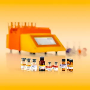
The Neonatal Cardiomyocyte Isolation Kit, mouse has been developed for the untouched isolation of vital murine cardiomyocytes (P0-P3). Enriched cardiomyocytes are fully functional and can be used for downstream applications.
One of the most used experimental model in cardiac research is the isolation of vital cardiac cells from neonatal hearts. Especially the isolation and subsequent culture of cardiomyocytes for biochemical, physiological, pharmacological and morphological studies has an high impact in the field of cardiovascular research. The Neonatal Cardiomyocyte Isolation Kit, mouse has been designed for the enrichment of untouched neonatal cardiomyocytes from dissociated neonatal mouse hearts. Non-cardiomyocytes are directly magnetically labeled by utilizing a cocktail of MicroBead-conjugated antibodies specific for non-cardiomyocytes. Isolation of pure unlabeled cardiomyocytes is achieved by depletion of magnetically labeled cells. The isolation of cardiomyocytes has been tested utilizing neonates aged from postnatal day 0 (P0) to postnatal day 3 (P3). For opimimal results, the Neonatal Heart Dissociation Kit has to be used prior cardiomyocyte isolation using this kit.
- Enrichment of untouched cardiomyocytes from neonatal (P0- P3) mouse hearts.
- Culture of enriched cardiomyocytes for biochemical, physiological, pharmacological, and morphological studies.
MS or autoMACS® Columns.
Neonatal cardiomyocytes were isolated from P2 CD-1® hearts using the Neonatal Heart Dissociation Kit, mouse and rat, the Neonatal Cardiomyocyte Isolation Kit, mouse, an MS Column, and a MiniMACS™ Separator. Subsequently, cells were fixed and stained using the Inside Stain Kit (# 130-090-477) and a FITC-conjugated antibody specific for cardiomyocytes (anti-α-actinin). Cells were analyzed by flow cytometry using the MACSQuant® Analyzer. Cell debris were excluded from the analysis based on scatter signals.
- Lakhal-Littleton, S. et al. (2015) Cardiac ferroportin regulates cellular iron homeostasis and is important for cardiac function. Proc. Natl. Acad. Sci. U.S.A. 112 (10): 3164-3169
- Trantidou, T. et al. (2015) Biorealistic cardiac cell culture platforms with integrated monitoring of extracellular action potentials. Sci Rep : 1-13
- O' Meara, C. C. et al. (2015) Transcriptional reversion of cardiac myocyte fate during mammalian cardiac regeneration. Circ. Res. 116 (5): 804-815