The MSC Phenotyping Cocktail Kit, anti-human, REAfinity enables the fast and standardized characterization and quantification of cultured human MSCs by flow cytometry based on the defined ISCT standards. The kit includes a set of ISCT proposed positive and negative markers as a ready-to-use phenotyping cocktail and also a corresponding isotype control cocktail. To ensure superior lot-to-lot consistency, low background, and high staining specificity the kit applies recombinantly engineered REAfinity antibodies.
For this Kit, an Express Mode add-on module for the MACSQuantify™ software


Related products for MSC Phenotyping Cocktail Kit, anti-human, REAfinity®

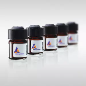
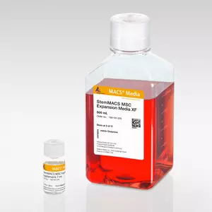

The MSC Phenotyping Cocktail Kit, anti-human, REAfinity enables the fast and standardized characterization and quantification of cultured human MSCs by flow cytometry based on the defined ISCT standards. The kit includes a set of ISCT proposed positive and negative markers as a ready-to-use phenotyping cocktail and also a corresponding isotype control cocktail. To ensure superior lot-to-lot consistency, low background, and high staining specificity the kit applies recombinantly engineered REAfinity antibodies.
For this Kit, an Express Mode add-on module for the MACSQuantify™ software is offered, designed to simplify and standardize the identification of mesenchymal stem cells (MSCs) on the MACSQuant® Analyzer by automated gating and data analysis: MSC Phenotyping Kit Express Mode
MSC Phenotyping Cocktail, anti-human, REAfinity containing:
- CD14 Antibody, anti-human, PE, REAfinity (clone: REA599)
- CD19 Antibody, anti-human, PE, REAfinity (clone: REA675)
- CD34 Antibody, anti-human, PE, REAfinity (clone: REA1164)
- CD45 Antibody, anti-human, PE, REAfinity (clone: REA747)
- CD73 Antibody, anti-human, APC, REAfinity (clone: REA804)
- CD90 Antibody, anti-human, FITC, REAfinity (clone: REA897)
- CD105 Antibody, anti-human, Vio® Bright V423, REAfinity (clone: REA794)
- HLA-DR Antibody, anti-human, VioGreen™, REAfinity (clone: REA805)
MSC Control Cocktail, anti-human, REAfinity containing:
- REA Control Antibody (S), human IgG1, FITC, REAfinity (clone: REA293)
- REA Control Antibody (S), human IgG1, PE, REAfinity (clone: REA293)
- REA Control Antibody (S), human IgG1, APC, REAfinity (clone: REA293)
- REA Control Antibody (S), human IgG1, Vio® Bright V423, REAfinity (clone: REA293)
- REA Control Antibody (S), human IgG1, VioGreen, REAfinity (clone: REA293)
CD73 Antibody, anti-human, APC, REAfinity (clone: REA804)
CD73 Antibody, anti-human, PE, REAfinity (clone: REA804)
CD90 Antibody, anti-human, FITC, REAfinity (clone: REA897)
CD105 Antibody, anti-human, Vio® Bright V423, REAfinity (clone: REA794)
CD73 Antibody, anti-human, Biotin, REAfinity (clone: REA804)
Biotin Antibody, anti-human, VioGreen, REAfinity (clone: REA746)
Instrument details
Flow cytometer equipped with a red (640 nm), a blue (488 nm), and a violet (405 nm) laser.
Cultured bone marrow derived MSCs were stained with the MSC Phenotyping Cocktail Kit, anti-human, REAfinity and Propidium iodide (PI) for dead cell exclusion. Cells were analyzed by flow cytometry using the MACSQuant® Analyzer 10. As a preliminary step for exclusion of debris a gate in forward scatter (FSC) versus side scatter (SSC) was set (A). A gate for viable cells (B) as well as for exclusion of doublets in forward scatter height (FSC-H) versus forward scatter area (FSC-A) (C) was set. Cells were analyzed for expression of CD73 (D), CD90 (E), CD105 (F), non-MSC markers (CD14, CD19, CD34, CD45) (G), and HLA-DR (H). Each histogram was overlaid with the corresponding isotype control (grey curves) to identify positively stained cells.
Aliquot 1 of cultured MSCs was stained with the MSC Phenotyping Cocktail and aliquot 2 with the Isotype Control Cocktail. The fractions were analyzed by flow cytometry using the MACSQuant® Analyzer 10. Each histogram was overlaid with the corresponding isotype control to identify positively stained cells. In order to distinguish between activated MSC and non-MSCs, the HLA-DR antibody (conjugated to different fluorochromes) has been used in combination with other MSC-negative markers.
To access a Certificate of Analysis (CoA) follow the link below and enter the specific lot of your product.
Should you wish to request additional documentation, such as a Safety Data Sheet (SDS) in other languages than English or Swedish, an SDS-replacement document, or a Certificate of Origin (CoO), please direct your inquiry to your local technical support team.
- Kolle, S.-F. A. et al. (2020) Ex vivo‐expanded autologous adipose tissue‐derived stromal cells ensure enhanced fat graft retention in breast augmentation: A randomized controlled clinical trial. Stem Cells Transl Med 9 (11): 1277-1286
- Egger, D. et al. (2019) From 3D to 3D: isolation of mesenchymal stem/stromal cells into a three-dimensional human platelet lysate matrix. Stem Cell Res Ther. 10: 248
- Dominici, M. et al. (2006) Minimal criteria for defining multipotent mesenchymal stromal cells. The International Society for Cellular Therapy position statement. Cytotherapy 8: 315-317
- Damerau, A. et al. (2021) A Human Osteochondral Tissue Model Mimicking Cytokine-Induced Key Features of Arthritis In Vitro. Int J Mol Sci 22 (1): 128
- Rahman, M. S. et al. (2020) The FGF, TGFβ and WNT axis Modulate Self-renewal of Human SIX2+ Urine Derived Renal Progenitor Cells. Sci Rep 10: 739
- Jungbluth, P. et al. (2019) Human iPSC-derived iMSCs improve bone regeneration in mini-pigs. Bone Res. 7: 32
- Sekine, K. et al. (2020) Generation of human induced pluripotent stem cell-derived liver buds with chemically defined and animal origin-free media. Sci Rep 10: 17937
- Gaber, T. et al. (2020) Synaptotagmin-13 Is a Neuroendocrine Marker in Brain, Intestine and Pancreas. Int J Mol Sci 21 (3): 865
- Widholz, B. et al. (2019) Pooling of Patient-Derived Mesenchymal Stromal Cells Reduces Inter-Individual Confounder-Associated Variation without Negative Impact on Cell Viability, Proliferation and Osteogenic Differentiation. Cells 8 (6): 633
- Ling, J. et al. (2020) Enhancing the Efficacy of Stem Cell Therapy with Glycosaminoglycans. Stem Cell Reports 14 (1): 105-121
- Ho, Y. K. et al. (2020) A highly efficient non-viral process for programming mesenchymal stem cells for gene directed enzyme prodrug cancer therapy. Sci Rep 10: 14257
- Chan, C. et al. (2021) Co-localisation of intra-nuclear membrane type-1 matrix metalloproteinase and hypoxia inducible factor-2α in osteosarcoma and prostate carcinoma cells. Oncol Lett 21 (2): 158
- Gutermuth, A. et al. (2019) Descemet's Membrane Biomimetic Microtopography Differentiates Human Mesenchymal Stem Cells Into Corneal Endothelial-Like Cells. Cornea 38 (1): 110
- Hernández, J. J. et al. (2021) Dodging COVID-19 infection: low expression and localization of ACE2 and TMPRSS2 in multiple donor-derived lines of human umbilical cord-derived mesenchymal stem cells. J. Transl. Med. 19: 149
- Le Naour, A. et al. (2020) Tumor cells educate mesenchymal stromal cells to release chemoprotective and immunomodulatory factors. J. Mol. Cell Biol. 12 (3): 202
- Sekine, K. et al. (2020) Robust detection of undifferentiated iPSC among differentiated cells. Sci Rep 10: 10293
- Eremichev, R. et al. (2021) Scar-Free Healing of Endometrium: Tissue-Specific Program of Stromal Cells and Its Induction by Soluble Factors Produced After Damage. Front Cell Dev Biol. (9)