
Synonyms: Itga2, gpIa, VLA-2α, VLA-2alpha, DX5
RECOMBINANT
- Clone:
- REAL133
- Type of antibody:
- Releasable antibodies, Primary antibodies, Recombinant antibodies
- Applications:
- FC Glossary
- Select a conjugate:
- Select a size:


Other sizes
Related products for CD49b Antibody, anti-mouse, REAlease®



Flow cytometry applications
APC
FITC
PE
Recommended dilution:
1:50
Data source:
Miltenyi Biotec
Conjugate:
APC
Used products:
130-122-702, 130-112-104
Description:
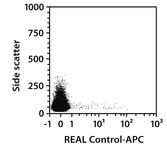
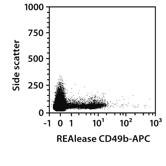
Splenocytes from C57BL/6 mice were stained with REAlease CD49b Complexes or with the corresponding REAL Control (left images) and analyzed by flow cytometry using the MACSQuant® Analyzer. Cell debris and dead cells were excluded from the analysis based on scatter signals and propidium iodide (PI) fluorescence.
 view details
 view details
Specifications for CD49b Antibody, anti-mouse, REAlease®
Overview
Clone REAL133 is an antibody fragment derived from the full CD49b antibody molecule. It displays no binding to Fc receptors. The recombinantly engineered antibody fragments are multimerized to form the REAlease Complex to bind markers with high avidity.
Clone REAL133 recognizes the mouse CD49b antigen, a single-pass type I membrane protein also known as integrin alpha-2 (ITGA2). CD49b together with CD29 (ITGB1) forms the adhesion molecule very late antigen-2 (VLA-2), which binds mainly to collagens I, II, and XI. VLA-2 is responsible for adhesion of platelets and other cells to collagens, modulation of collagen and collagenase gene expression, force generation, and organization of newly synthesized extracellular matrix. It is also a receptor for laminins, collagen C-propeptides, and E-cadherin. Mice homozygous for a null mutation in the CD49b gene die very early in embryogenesis. CD49b is expressed on the vast majority of mouse natural killer (NK) cells and on a subset of NKT cells. It is not as mouse strain-restricted as other NK cell markers, for example, NK1.1, and is expressed by most common inbred mouse strains. Bone marrow (BM) resident memory T helper (Tʜ) cells also express CD49b and the homing of adoptively transferred bone marrow (BM) memory Tʜ cells to BM can be efficiently blocked by antibodies to CD49b. This points to a decisive role of CD49b in the trafficking of memory Th cell precursors to the BM niches for memory Tʜ cells.
The REAlease Kits consist of the respective fluorochrome-conjugated REAlease Complexes and the REAlease Support Kit for removal of the REAlease Complexes and optional relabeling with different fluorochrome-conjugated REAlease Complexes.
Itga2, gpIa, VLA-2α, VLA-2alpha, DX5
Detailed product information
Technical specifications
Clone
REAL133
Clonality
monoclonal
Isotype control
Control Antibody
Host
cell line
Type of antibody
Releasable antibodies, Primary antibodies, Recombinant antibodies
Species reactivity
mouse
Antigen
CD49b
Distribution of antigen
NK cells, NKT cells
RRID
AB_2751448, AB_2751075, AB_2784232, AB_2751084, AB_2784231, AB_2784233
Resources for CD49b Antibody, anti-mouse, REAlease®
Certificates
To access a Certificate of Analysis (CoA) follow the link below and enter the specific lot of your product.
Should you wish to request additional documentation, such as a Safety Data Sheet (SDS) in other languages than English or Swedish, an SDS-replacement document, or a Certificate of Origin (CoO), please direct your inquiry to your local technical support team.
Citations for CD49b Antibody, anti-mouse, REAlease®
Publications
- Hanazawa, A. et al. (2013) CD49b/CD69-dependent generation of resting T helper cell memory. Front Immunol 4: 183
- Edelman, J. M. et al. (1994) The mouse VLA-2 homologue supports collagen and laminin adhesion but not virus binding. Cell Adhes. Commun. 2 (2): 131-143
- Gagliani, N. et al. (2013) Coexpression of CD49b and LAG-3 identifies human and mouse T regulatory type 1 cells. Nat Med 19 (6): 739-746