
Synonyms: Spn, B-cell differentiation antigen LP-3, GALGP, Ly-48, Sialophorin, CD43
RECOMBINANT EXTENDED VALIDATION
- Clone:
- REA364 | 1B11
- Type of antibody:
- Primary antibodies, Recombinant antibodies
- Isotype:
- recombinant human IgG1
- Applications:
- FC, MICS, IF, IHC Glossary
- Select a conjugate:


Related products for CD43 Glycoform Antibody, anti-mouse, REAfinity™


Flow cytometry applications
APC
APC-Vio 770
PE
PE-Vio 770
PerCP-Vio 700
Species tested:
mouse
Recommended dilution:
1:50
Remark:
The antibody is suited for staining of formaldehyde-fixed cells.
Data source:
Miltenyi Biotec
Conjugate:
APC
Used products:
130-132-428
Description:
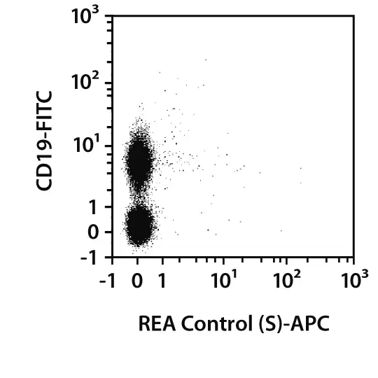
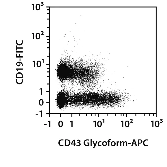
Splenocytes from C57BL/6 mice were stained with CD43 Glycoform antibodies or with the corresponding REA control antibodies (left image), as well as with CD19 antibodies. Flow cytometry was performed with the MACSQuant® Analyzer. Cell debris and dead cells were excluded from the analysis based on scatter signals and propidium iodide fluorescence or 4',6-diamidino-2-phenylindole (DAPI) fluorescence, as in the case of tandem conjugates.
 view details
 view details
MICS (MACSima Imaging Cyclic Staining) applications
APC
PE
Fixation:
frozen – Acetone
Species tested:
mouse
Recommended dilution:
1:50
Data source:
Miltenyi Biotec
Conjugate:
APC
Used products:
130-132-428
Testing grade:
Fixation:
frozen – PFA
Species tested:
mouse
Recommended dilution:
1:50
Data source:
Miltenyi Biotec
Conjugate:
APC
Used products:
130-132-428
Testing grade:
Extended validation for CD43 Glycoform Antibody, anti-mouse, REAfinity™
Specificity
In order to compare the epitope specificity of an antibody, the clone being used is compared with other known clones recognizing the same antigen in a competition assay.
Other clones
Overlap in epitope recognition with REA364
EBioR2/60
low
1B11
high
S11
low
S7
low
Cells were incubated with an excess of purified unconjugated CD43 Glycoform (REA364) antibody followed by staining with fluorochrome-conjugated antibodies from other known clones targeting the same antigen. Fluorescence intensity was then used to assess epitope overlap: a minimal or absent signal indicated high epitope overlap, an intermediate signal suggested partial overlap, and a strong signal reflected low overlap.
Sensitivity
Selected fluorochrome conjugated antibodies from Miltenyi Biotec were compared to commercially available hybridoma clones in flow cytometry analysis.
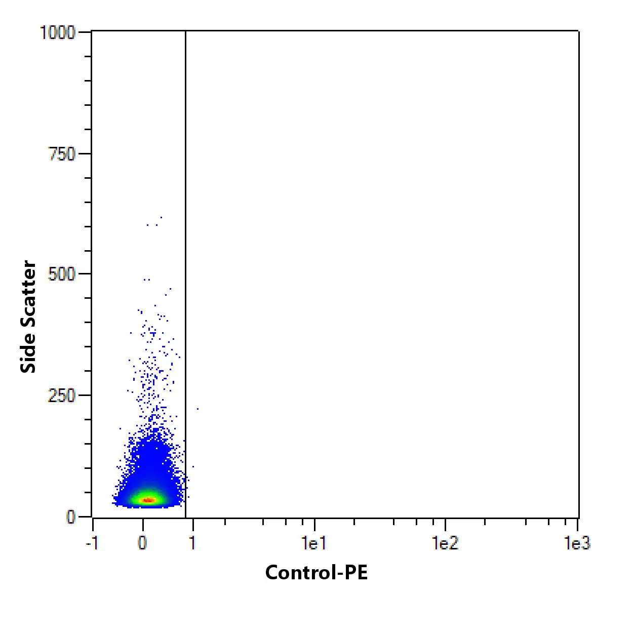
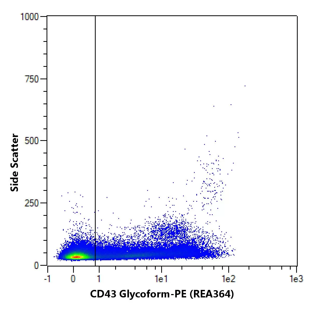
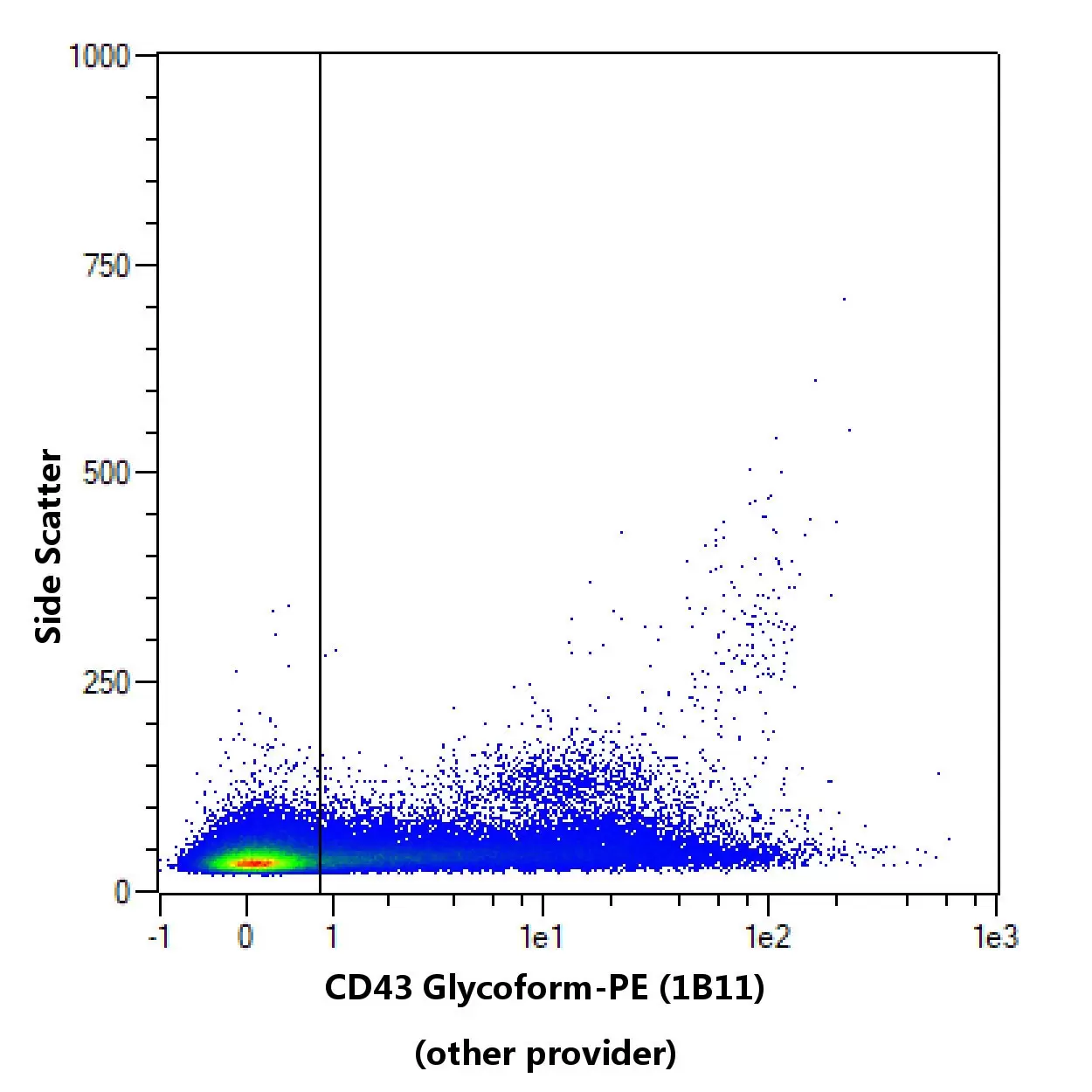
Flow cytometric comparison of different clones for ( CD43 Glycoform specificity from PIM). C57BL/6 Mouse splenocytes were stained with CD43 Glycoform antibodies and with a suitable counterstaining. As a control, CD43 Glycoform antibody staining was omitted and cells were measured in the same channels. Flow cytometry was performed with the MACSQuant® Analyzer. Cell debris, dead cells, and cell doublets were excluded from the analysis based on scatter signals and 4',6-diamidino-2-phenylindole (DAPI) fluorescence. No FcR Blocking Reagent was used. The recommended titers of respective antibodies from different suppliers were used.
To provide an indication on how an antibody performs after fixation of cells, in-house data on staining results before and after fixation with 3.7% formaldehyde using Miltenyi Biotec antibodies are provided. Different experimental settings may lead to different results.
No fixation
Post fixation

Comparison of staining pattern on non-fixed and fixed cells using CD43 Glycoform (REA364). The performance of the antibody after fixation was tested by comparing the staining pattern on fresh (no fixation) versus fixed (post fixation) cells.
Specifications for CD43 Glycoform Antibody, anti-mouse, REAfinity™
Overview
Clone REA364 recognizes the activation-associated glycoform (130-kD glycoform) of the murine CD43 antigen, a cell-surface glycoprotein which is also known as sialophorin or leukosialin. CD43 is a heavily O-glycosylated protein which is expressed in a variety of hematopoietic lineage cells, including T lymphocytes. It has been reported to have two glycoforms, one with a molecular mass of 115 kD that is expressed on all T cells and one with a mass of 130 kD that is expressed only on activated T cells. Biochemical analysis showed that CD43 on resting T cells carries tetrasaccharides (115-kD glycoform), whereas activated T cells carry branched hexasaccharide core two O-glycans (130-kD glycoform). Expression of the 130-kD glycoform of CD43 on all T cells in transgenic mice shows a reduction in primary T-cell immune responses that is probably due to the antiadhesive function of this glycoform.
Additional information: Clone REA364 displays negligible binding to Fc receptors.
Spn, B-cell differentiation antigen LP-3, GALGP, Ly-48, Sialophorin, CD43
Detailed product information
Technical specifications
Clone
REA364
Clonality
monoclonal
Isotype
recombinant human IgG1
Isotype control
REA Control Antibody, human IgG1
Host
human cell line
Type of antibody
Primary antibodies, Recombinant antibodies
Species reactivity
mouse
Antigen
CD43 Glycoform
Molecular mass of antigen [kDa]
38
Distribution of antigen
B cells, T cells, lymphocytes
Entrez Gene ID
RRID
AB_2905011, AB_2658145, AB_2658149, AB_2658144, AB_2658148
Recombinant antibodies at Miltenyi Biotec
As an innovation-powered company with a commitment to quality and excellence, Miltenyi Biotec applies cutting-edge methods to discover and develop newer and better antibodies to satisfy both market demand and regulatory requirements.
The Miltenyi way of generating best-in-class antibodies and antibody conjugates
At Miltenyi Biotec, we opt for recombinant antibodies, which enable sequence verification and optimization for lot-to-lot consistency, making them superior to hybridoma-based antibodies.
Infusing our unique expertise
Recombinants are the result of combining genetic material from different immunized animals, and using display technologies to screen in-house antibody libraries for the best antibodies. Additional analyses on existing hybridoma-based antibodies can also give rise to sequences contributing to novel recombinants.
Maximizing value for your research
By further re-engineering these antibody sequences, we can precisely control their properties to achieve the following benefits:
reduce undesirable binding to Fc gamma receptors,
- eliminate destabilizing amino acid motifs,
- increase specific bonding affinity for stronger, more accurate targeting, and
- optimize fluorochrome conjugation for brighter and clearer signals.
In a final step, by synthesizing and cloning the resulting gene sequence, we achieve an expression vector that develops a cell line, which in turn produces a special recombinant antibody.
Better science with quality antibodies
Already in 2015, 100 of the world’s leading scientists collectively called for better standards and reproducibility in antibody production, proposing recombinant antibodies to circumvent variability (Baker, M. Reproducibility crisis: Blame it on the antibodies. Nature 521, 274–276 (2015). https://doi.org/10.1038/521274a). Moreover, by applying reverse engineering, which is encouraged by the EU Directive 2016/943 r, Miltenyi Biotec acts “[i]n the interest of innovation and fostering competition…” (§16).
Resources for CD43 Glycoform Antibody, anti-mouse, REAfinity™
Certificates
To access a Certificate of Analysis (CoA) follow the link below and enter the specific lot of your product.
Should you wish to request additional documentation, such as a Safety Data Sheet (SDS) in other languages than English or Swedish, an SDS-replacement document, or a Certificate of Origin (CoO), please direct your inquiry to your local technical support team.
Citations for CD43 Glycoform Antibody, anti-mouse, REAfinity™
Publications
- Alcaide, P. et al. (2007) The 130-kDa glycoform of CD43 functions as an E-selectin ligand for activated Tʜ1 cells in vitro and in delayed-type hypersensitivity reactions in vivo. J. Invest. Dermatol. 127 (8): 1964-1972
- Carlow, D. A. et al. (1999) A novel CD8 T cell-restricted CD45RB epitope shared by CD43 is differentially affected by glycosylation. J Immunol 163 (3): 1441-1448
- van den Berg, T. K. et al. (2001) Cutting Edge: CD43 functions as a T cell counterreceptor for the macrophage adhesion receptor Sialoadhesin (Siglec-1). J Immunol 166: 3637- 3640