This kit is suitable for the identification and enumeration of dead cells, such as apoptotic or necrotic cells, by flow cytometry.


Related products for Annexin V-FITC Kit
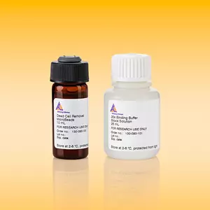
This kit is suitable for the identification and enumeration of dead cells, such as apoptotic or necrotic cells, by flow cytometry.
The Annexin V-FITC Kit contains reagents for the simultaneous fluorescent detection of apoptotic, necrotic, and dead cells through the specific binding of FITC-labeled Annexin V to phosphotidylserine (PS). The kit consists of Annexin V-FITC, Annexin V Binding Buffer, and Propidium Iodide Solution.
In most normal, viable eukaryotic cells, the negatively charged phospholipid PS is located in the cytosolic leaflet of the plasma membrane lipid bilayer.1 PS redistribution from the inner to the outer leaflet is an early and widespread event during apoptosis.1,2 However, in necrosis, PS becomes accessible due to the disruption of membrane integrity.2 Apart from necrosis and apoptosis, PS also becomes accessible in activated platelets3, in certain cell anomalies such as sickle cell anemia4, in erythrocyte senescence5, upon degranulation of mast cells6, and in certain stages of B cell differentiation7. PS exposure by apoptotic cells serves as a trigger for the recognition and removal by macrophages.8,9
Annexin V is a 35 kDa phospholipid-binding protein and has a high affinity for PS in the presence of physiological concentrations of calcium (Ca2+).10
Jurkat cells cultured with staurosporine (50 nM) for 15 h, were stained with Annexin V-FITC and PI and analyzed by flow cytometry.
Staining principle of PS-exposing cells with Annexin V-FITC.
To access a Certificate of Analysis (CoA) follow the link below and enter the specific lot of your product.
Should you wish to request additional documentation, such as a Safety Data Sheet (SDS) in other languages than English or Swedish, an SDS-replacement document, or a Certificate of Origin (CoO), please direct your inquiry to your local technical support team.
- Kuypers, F. A. et al. (1996) Detection of altered membrane phospholipid asymmetry in subpopulations of human red blood cells using fluorescently labeled annexin V. Blood 87: 1179-1187
- Fadok, V. A. et al. (2000) A receptor for phosphatidylserine-specific clearance of apoptotic cells. Nature 405: 85-90
- Demo, S. D. et al. (1999) Quantitative measurement of mast cell degranulation using a novel flow cytometric annexin-V binding assay. Cytometry 36: 340-348
- Moss, S. E. et al. (1991) Diversity in the Annexin family. In: Novel Calcium Binding Proteins, Springer Verlag : 535-566
- Fadok, V. A. et al. (1992) Exposure of phosphatidylserine on the surface of apoptotic lymphocytes triggers specific recognition and removal by macrophages. J Immunol 148: 2207-2216
- Martin, S. J. et al. (1995) Early redistribution of plasma membrane phosphatidylserine is a general feature of apoptosis regardless of the initiating stimulus: inhibition by overexpression of Bcl-2 and Abl. J. Exp. Med. 182: 1545-1556
- Thiagarajan, P. et al. (1990) Binding of annexin V/placental anticoagulant protein I to platelets. Evidence for phosphatidylserine exposure in the procoagulant response of activated platelets. J. Biol. Chem. 265: 17420-17423
- Koopman, G. et al. (1994) Annexin V for flow cytometric detection of phosphatidylserine expression on B cells undergoing apoptosis. Blood 84: 1415-1420
- Schroit, A. J. et al. (1991) Transbilayer movement of phospholipids in red cell and platelet membranes. Biochim. Biophys. Acta 1071: 313-329
- Dillon, S. R. et al. (2001) Annexin V binds to positively selected B cells. J Immunol 166: 58-71