Isolation and cultivation of O4-positive oligodendrocytes from neonatal mouse brain
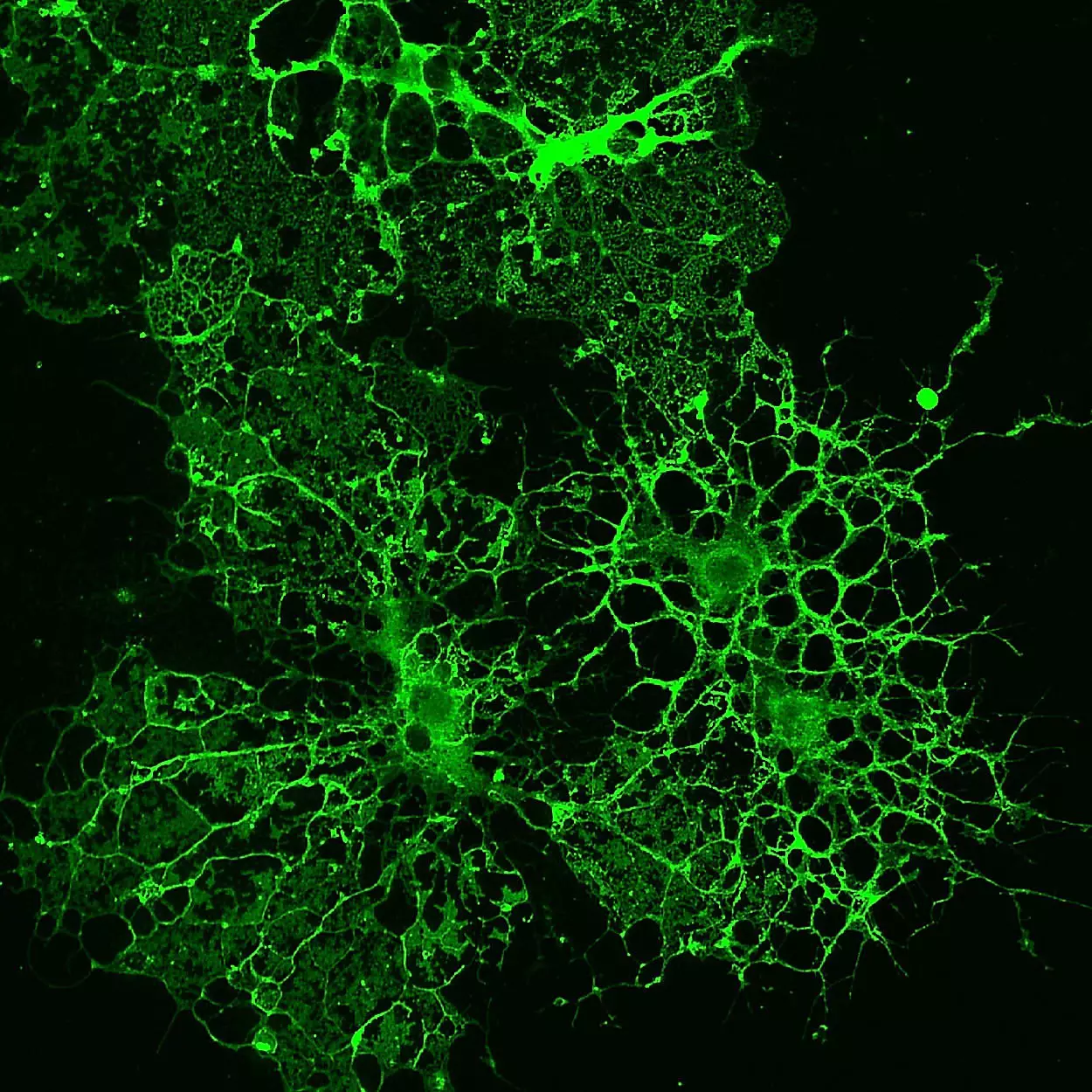
This application protocol describes the isolation of highly purified and viable oligodendrocytes from neonatal mouse brain tissue. Brain tissue from mice younger than P8 is dissociated into a single-cell suspension and Anti-O4 MicroBeads are used to isolate oligodendrocytes. The O4 antigen, a sulfatide of the glycosphingolipid class, is a marker for oligodendrocytes. During oligodendrocyte development, O4 expression begins on late oligodendrocyte progenitors that are A2B5-positive. While A2B5 expression disappears, O4 continues to be expressed. O4 expression is also found in Schwann cells. The isolation of O4+ cells leads to highest purities if P3–P7 (postnatal day 3–7) rodents are used. Mouse brain tissue derived from P3–P7 CD-1® mice contains approximately 5–10% O4+ cells.
Protocol
Isolation and cultivation of O4-positive oligodendrocytes from neonatal mouse brain
45 min
Dissociation of brain tissue
50 min
Isolation of O4-positive oligodendrocytes
30 min
Flow cytometric analysis
5 d
Cell culture
1 h
Immunocytochemical staining of cultured cells
Protocol

This protocol includes detailed instructions for preparing necessary reagents and materials and then carrying out each step.
 View details