Isolation and cultivation of neurons from neonatal mouse brain
This application protocol describes the generation of highly purified and viable neurons from neonatal mouse brain tissue. Brain tissue from mice younger than P8 is dissociated into a single-cell suspension and neurons are then isolated using an indirect magnetic labeling system that depletes non-neuronal cells like astrocytes, oligodendrocytes, microglia, endothelial cells, and fibroblasts. The cell number and composition of the resulting highly pure neuronal cell fraction varies according to mouse age and brain region. This isolation protocol has been tested with CD-1® mice aged from embryonic day 18 (E18) to adult.
Protocol
Isolation and cultivation of neurons from neonatal mouse brain
45 min
Dissociation of brain tissue
50 min
Isolation of neurons
30 min
Flow cytometric analysis
7 d
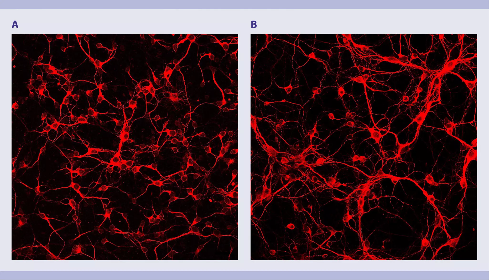
Cell culture
1–24 h
Immunocytochemical staining of cultured cells
Adjust cell culture time to meet experimental objectives.
This protocol includes detailed instructions for preparing necessary reagents and materials and then carrying out each step.
D-PBS/BSA buffer: Prepare a solution containing Dulbecco’s phosphate-buffered saline (D-PBS) with Ca2+ and Mg2+ and 0.5% bovine serum albumin (BSA) by diluting MACS® BSA Stock Solution 1:20 with DPBS. Keep buffer cold (2–8 °C). Prepare fresh on the day of use and degas the buffer, as air bubbles could block the column.
Preparation of cell culture plates
Coat a 24-well culture dish with 0.01% poly-L-lysine overnight at 37 °C. Wash three times with ddH2O the following day.
Preparation of medium for cell culture
For cell culture, prepare MACS Neuro Medium containing 2% MACS NeuroBrew®-21, 1% penicillin/streptomycin, and 0.5 mM L-glutamine.
Dissociate mouse neonatal brain using the Neural Tissue Dissociation Kit – Postnatal Neurons. Follow the protocol of the kit data sheet.
Download data sheet
Isolate neurons from the single-cell suspension using the Neuron Isolation Kit, mouse. Follow the protocol of the kit data sheet.
Download data sheet
- The recommended antibody dilution for labeling of cells is 1:10 for up to 1×10⁶ cells/50 μL of DPBS/BSA buffer.
- Volumes given below are for up to 1×10⁶ nucleated cells. When working with fewer than 1×10⁶ cells, use the same volumes as indicated. When working with higher cell numbers, scale up all reagent volumes and total volumes accordingly (e.g. for 2×10⁶ nucleated cells, use twice the volume of all indicated reagent volumes and total volumes).
- Analyze 100 μL of the positive and 100 µL of the negative fractions. Optionally, also analyze 20 μL of the original fraction.
- Centrifuge at 300×g for 5 minutes. Aspirate supernatant completely.
- Resuspend up to 1×10⁶ nucleated cells per 45 μL of D-PBS/BSA buffer (see section "Things to prepare in advance of tissue dissociation and cell culture").
- Add 5 μL of Biotin Antibody, PE.
- Mix well and incubate for 10 minutes in the dark in the refrigerator (2−8 °C).
▲ Note: Higher temperatures and/or longer incubation times may lead to non-specific cell labeling. Working on ice requires increased incubation times. - Wash cells by adding 1 mL of DPBS/BSA buffer and centrifuge at 300×g for 10 minutes. Aspirate supernatant completely.
- Resuspend cell pellet in a suitable amount of D-PBS/BSA buffer for analysis by flow cytometry, e.g., using the MACSQuant® Analyzer 10.
- Plate 1×10⁵ cells in 50 μL of prepared medium as a drop in the middle of each well of a coated 24-well plate (see section "Things to prepare in advance of tissue dissociation and cell culture").
- Let cells settle for 30 minutes at 37 °C in the incubator.
- Carefully add 450 μL of prepared medium to each well.
- Take off the medium to remove non-attached and dead cells.
- Add 500 μL of prepared medium.
- Maintain the culture by replacing 50% of medium every other day.
Things to prepare in advance
Staining buffer: Prepare a solution containing autoMACS® Running Buffer with FcR Blocking Reagent, mouse (1:10).
Immunocytochemical staining of cultured neurons
- Wash cells 3× with PBS.
- Fix cells with 2% PFA for 10 minutes at room temperature.
- Wash cells 3× with PBS.
▲Note: When working with CD68 antibodies, add 0.2% TRITON X-100 in PBS, incubate for 10 minutes at room temperature, and wash cells 3× with autoMACS® Running Buffer. Do not treat cells with TRITON X-100 before staining with ACSA-2, O4, or PSA-NCAM antibodies.
▲Note: Fixed cells can be stored in azide-containing buffer at 2–8 °C for up to one week. - Add staining buffer (see section "Things to prepare in advance") and incubate for 10 minutes at room temperature.
- Discard staining buffer.
- Add pure antibody of choice in staining buffer to the cells and incubate in the dark. For incubation temperature and time as well as recommended antibody concentration, refer to the table below.
- Wash cells 3× with autoMACS Running Buffer.
- Add a corresponding secondary antibody in staining buffer to the cells and incubate in the dark. For incubation temperature and time as well as recommended antibody concentration, refer to the table below.
- Wash cells 3× with autoMACS Running Buffer.
▲Note: For co-staining with additional antibodies repeat step 6–9. - Store cells in autoMACS Running Buffer.
- Cells are now ready for immunofluorescence microscopy.
▲Note: Samples can be stored at 2–8 °C in the dark for up to one week.
▲Note: When working with cells cultured on coverslips, the coverslips need to be mounted onto slides before imaging.
Table 1: Recommended antibody panel and staining conditions.
| Primary antibody | Secondary antibody | |||||
| Antibody | Incubation temperature | Incubation time | Recommended antibody concentration | Secondary antibody | Incubation temperature | Incubation time |
| ACSA-2 | Room temperature | 10 minutes | 1–5 µg/mL | IgG2b Antibody, anti-rat | Room temperature | 10 minutes |
| GLAST (ACSA-1) | Room temperature | 10 minutes | 1–5 µg/mL | IgG2a Antibody, anti-mouse | Room temperature | 10 minutes |
| O4 | Room temperature | 10 minutes | 5–10 µg/mL | IgM Antibody, anti-mouse | Room temperature | 10 minutes |
| PSA-NCAM | Room temperature | 10 minutes | 5–10 µg/mL | IgM Antibody, anti-mouse | Room temperature | 10 minutes |
| CD11b | Room temperature | 10 minutes | 5–10 µg/mL | IgG2bκ Antibody, anti-rat | Room temperature | 10 minutes |
| CD68 | 2–8 °C | Overnight | 5–10 µg/mL | IgG2a Antibody, anti-rat | Room temperature | 1 hour |
| CD171 (L1CAM) | 2–8 °C | Overnight | 5–10 µg/mL | IgG2a Antibody, anti-rat | Room temperature | 1 hour |