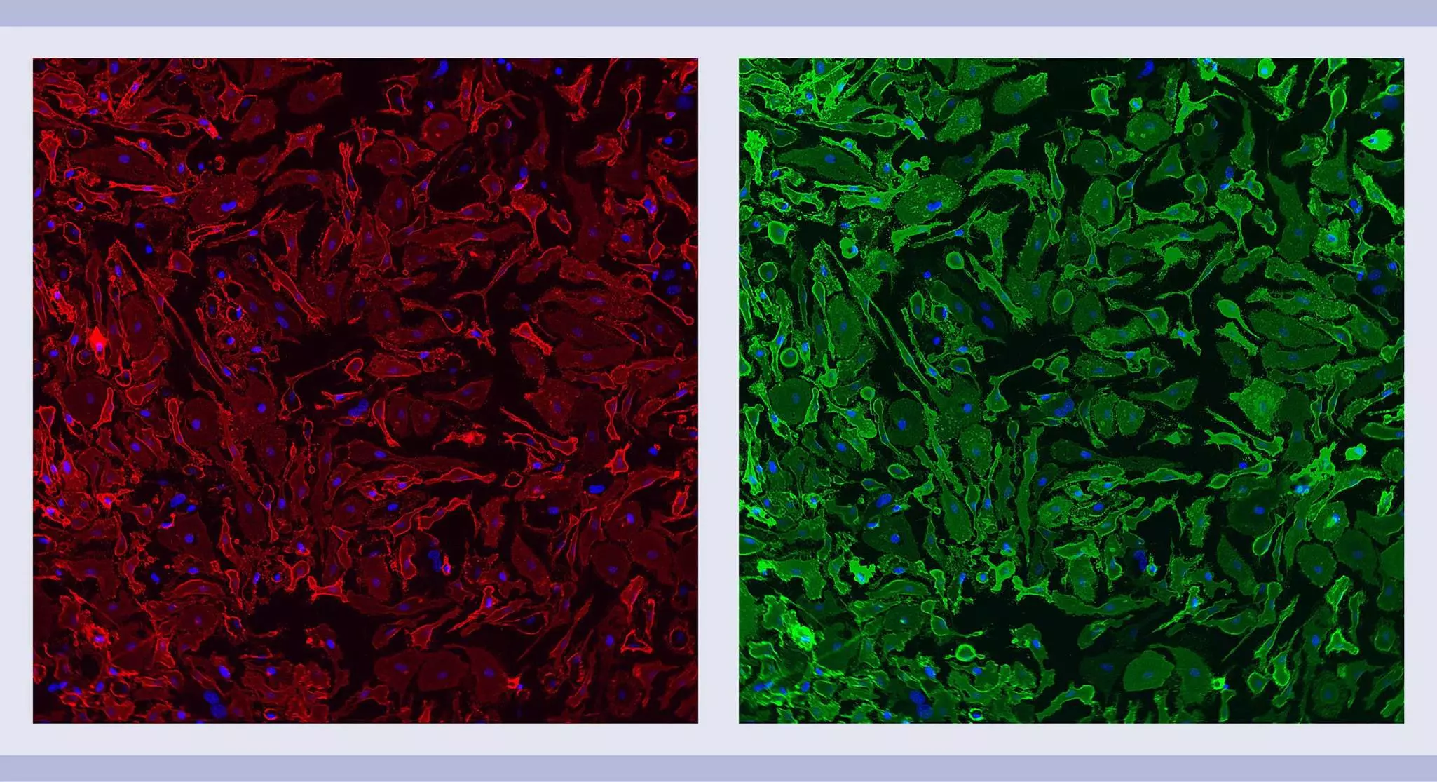
Isolation and cultivation of microglia from neonatal mouse or rat brain
This application protocol describes the isolation of highly purified and viable microglia from neonatal brain tissue. Brain tissue from mice or rats younger than P8 is dissociated into single-cell suspensions via enzymatic digestion of the extracellular matrix and gentle mechanical dissociation. CD11b (Microglia) MicroBeads, mouse or CD11b/c (Microglia) MicroBeads, rat are used to isolate microglia from the single-cell suspension. CD11b (Microglia) MicroBeads isolate CD11b+ cells, whereas the CD11b/c (Microglia) antibody appears to recognize a common epitope shared between CD11b and CD11c. CD11b, also known as integrin alpha M (ITGAM) or Mac-1, is a component of complement factor 3 (CR3). CD11c, also called integrin alpha X (ITGAX), is a component of complement receptor 4 (CR4). CD11b and CD11c are expressed on microglia, macrophages, monocytes, granulocytes, NK cells, and dendritic cells.
Protocol
Isolation and cultivation of microglia from neonatal mouse or rat brain
45 min
Dissociation of brain tissue
1 h
Isolation of microglia
30 min
Flow cytometric analysis
7 d
Cell culture
1–24 h
Immunocytochemical staining of cultured cells
Adjust cell culture time to meet experimental objectives.
This protocol includes detailed instructions for preparing necessary reagents and materials and then carrying out each step.
PB buffer: Prepare a solution containing phosphate-buffered saline (PBS), pH 7.2, and 0.5% bovine serum albumin (BSA) by diluting MACS® BSA Stock Solution 1:20 with PBS. Keep buffer cold (2−8 °C). Prepare fresh on the day of use and degas the buffer, as air bubbles could block the column.
▲ Note: BSA can be replaced by other proteins such as mouse serum albumin, mouse serum, or fetal bovine serum (FBS).
Preparation of cell culture plates
Coat a 24-well culture dish with 0.01% poly-L-lysine overnight at 37 °C. Wash three times with ddH2O the following day.
Preparation of medium for cell culture
- Prepare the following cell culture medium: DMEM containing 10% FBS, 1% penicillin/streptomycin, and 2 mM L-glutamine.
- (Optional) Add 20 ng/mL M-CSF (# 130-094-129) to enhance microglia growth and survival.
Use the Neural Tissue Dissociation Kit (T) or the Neural Tissue Dissociation Kit (P) to dissociate brain tissue and prepare a single-cell suspension. Follow the protocol of the kit data sheet.
Download data sheet
Good to know
The data sheet for the Neural Tissue Dissociation Kits includes a set of tips and hints to improve the quality and yield of the dissociation procedure. Refer to the data sheet appendix if, for example, the yield of viable cells is too low or a cell pellet will not form.
Isolate microglia from the single-cell suspension using either the CD11b (Microglia) MicroBeads, human and mouse or the CD11b/c (Microglia) MicroBeads, rat. Follow the protocol of the corresponding kit data sheet.
Download data sheet
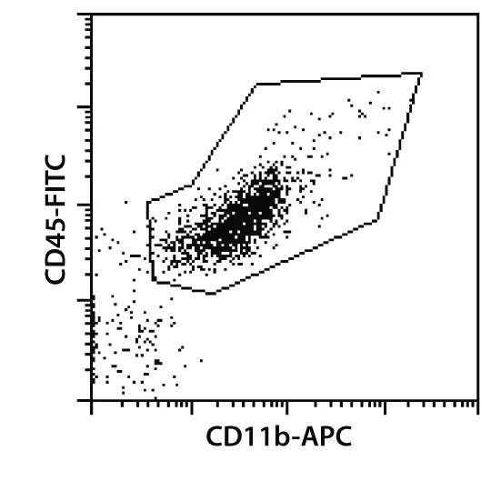
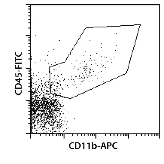
Figure 1: CD11b+ microglia isolated from neonatal mouse brain tissue. CD11b+ cells were isolated from mouse neural cell suspension using CD11b (Microglia) MicroBeads, an MS Column, and a MiniMACS™ Separator. Cells were fluorescently stained with CD11b-APC and CD45-FITC. Cell debris and dead cells were excluded from the analysis based on scatter signals and propidium iodide fluorescence.
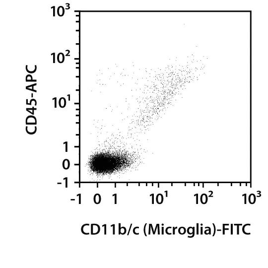
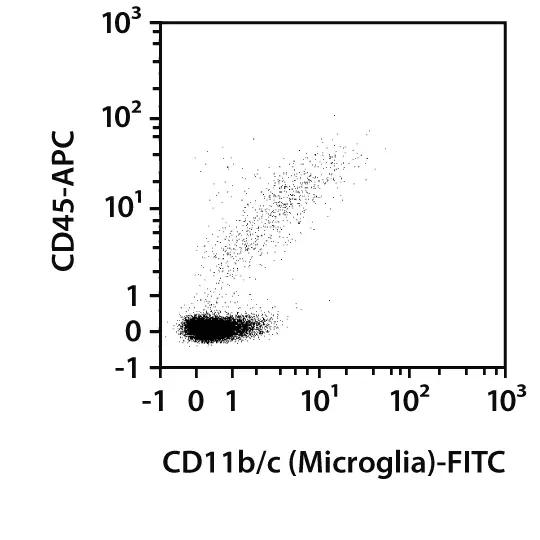
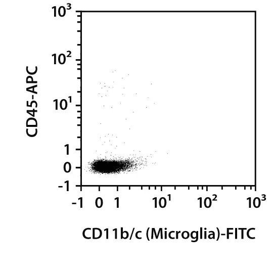
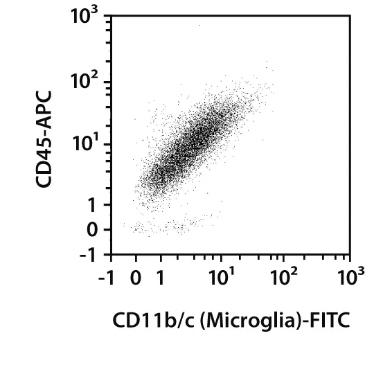
Figure 2: CD11b/c+ microglia isolated from neonatal rat brain tissue. CD11b/c+ cells were isolated from P3 rat brain tissue using the Neural Tissue Dissociation Kit (P), the gentleMACS™ Dissociator, CD11b/c (Microglia) MicroBeads, a MiniMACS™ Separator, and two MS Columns. Cells were fluorecently stained with CD11b/c-FITC in combination with CD45-APC and analyzed by flow cytometry using the MACSQuant® Analyzer. Cell debris and dead cells were excluded from the analysis based on scatter signals and propidium iodide fluorescence.
Analysis of mouse microglia
- The recommended antibody dilution for labeling cells is 1:10 for up to 1×10⁷ cells/100 μL of PB buffer.
- Volumes given below are for up to 1×10⁷ nucleated cells. When working with fewer than 1×10⁷ cells, use the same volumes as indicated. When working with higher cell numbers, scale up all reagent volumes and total volumes accordingly (e.g. for 2×10⁷ nucleated cells, use twice the volume of all indicated reagent volumes and total volumes).
- Use 100 μL of the CD11b+ fraction for analysis. Optionally, also analyze 100 μL of the negative fraction and 20 μL of the original fraction.
- Resuspend up to 1×10⁷ nucleated cells per 100 μL of PB buffer (see section "Things to prepare in advance of cell isolation and cell culture").
▲ Note: Always use freshly prepared buffer. - Add 2 µL of CD11b Antibody, anti-mouse, APC and CD45 Antibody, anti-mouse, FITC.
- Mix well and incubate for 10 minutes in the refrigerator (2−8 °C) in the dark.
▲ Note: Higher temperatures and/or longer incubation times may lead to non-specific cell labeling. Working on ice requires increased incubation times. - Wash cells by adding 1 mL of PB buffer and centrifuge at 300×g for 10 minutes. Aspirate supernatant completely.
- Resuspend cell pellet in a suitable amount of PB buffer for analysis by flow cytometry, e.g., using the MACSQuant® Analyzer 10.
Analysis of rat microglia
- The recommended antibody dilution for labeling cells is 1:10 for up to 1×10⁶ cells/50 μL of buffer.
- Volumes given below are for up to 1×10⁶ nucleated cells. When working with fewer than 1×10⁶ cells, use the same volumes as indicated. When working with higher cell numbers, scale up all reagent volumes and total volumes accordingly (e.g. for 2×10⁶ nucleated cells, use twice the volume of all indicated reagent volumes and total volumes).
- Use 100 μL of the CD11b/c+ fraction for analysis. Optionally, also analyze 100 μl of the negative fraction and 20 μL of the original fraction.
- Resuspend up to 1×10⁶ nucleated cells per 45 μL of buffer.
▲ Note: Always use freshly prepared buffer. - Add 5 µl CD11b/c Antibody, anti-rat, FITC and CD45 Antibody, anti-rat, APC.
- Mix well and incubate for 10 minutes in the refrigerator (2−8 °C) in the dark.
▲ Note: Higher temperatures and/or longer incubation times may lead to non-specific cell labeling. Working on ice requires increased incubation times. - Wash cells by adding 1 mL of buffer and centrifuge at 300×g for 10 minutes. Aspirate supernatant completely.
- Resuspend cell pellet in a suitable amount of buffer for analysis by flow cytometry, e.g., using the MACSQuant Analyzer 10.
- Plate 1×10⁵ cells in 50 μL of pre-warmed prepared medium as a drop in the middle of each well of a coated 24-well plate (see section "Things to prepare in advance of cell isolation and cell culture").
- Let cells settle down for 30 minutes at 37 °C in the incubator.
- Carefully add 450 μL of prepared medium to each well.
- Maintain the culture by replacing 50% of medium every other day.
Things to prepare in advance
Staining buffer: Prepare a solution containing autoMACS® Running Buffer with FcR Blocking Reagent, mouse in a ratio of 1:10, e.g., add 1 mL FcR Blocking Reagent to 9 mL autoMACS Running Buffer.
- Wash cells 3× with PBS.
- Fix cells with 2% PFA for 10 minutes at room temperature.
- Wash cells 3× with PBS.
▲ Note: When working with CD68 antibodies, add 0.2% TRITON™ X-100 in PBS, incubate for 10 minutes at room temperature, and wash cells 3× with autoMACS Running Buffer.
▲ Note: Fixed cells can be stored in azide-containing buffer at 2–8 °C for up to one week. - Add staining buffer (see section "Things to prepare in advance") and incubate for 10 minutes at room temperature.
- Discard staining buffer.
- Add primary antibody in staining buffer to the cells with a final concentration of 5–10 μg/mL and incubate at room temperature in the dark for 10 minutes.
When working with CD68 antibodies incubate at 2–8 °C overnight in the dark - Wash cells 3× with autoMACS Running Buffer.
- Add a corresponding secondary antibody in staining buffer to the cells.
When working with CD11b antibodies incubate at room temperature in the dark for 10 minutes.
When working with CD68 antibodies incubate at room temperature in the dark for 1 hour. - Wash cells 3× with autoMACS Running Buffer.
▲ Note: For co-staining with additional antibodies repeat steps 6–9. - Store cells in autoMACS Running Buffer.
- Cells are now ready for immunofluorescence microscopy.
▲ Note: Samples can be stored at 2–8 °C in the dark for up to one week.
▲ Note: When working with cells cultured on coverslips, the coverslips need to be mounted onto slides before imaging.