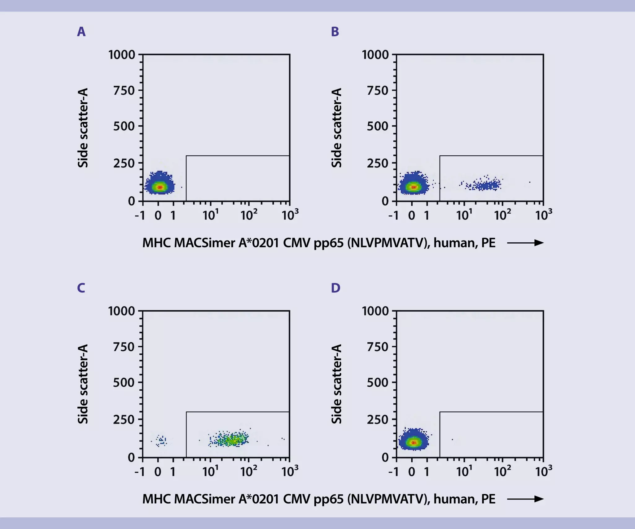
- Work fast, keep the cells cold, and use pre-cooled solutions. This will prevent capping of antibodies on the cell surface and a non-specific cell labeling.
- Volumes for magnetic labeling given below are for up to 5×10⁷ cells per LS Column. When working with fewer than 5×10⁷ cells, use the same volumes as indicated. When working with higher cell numbers, scale up the number of LS Columns used (e.g., for 1.5×10⁸ cells use 3 LS Column with 5×10⁷ cells per LS Column). Cells can be labeled using the autoMACS® NEO Separator.
- The recommended incubation temperature is +2 to +8 °C. Higher temperatures and/or longer incubation times may lead to non-specific cell labeling. Working on ice may require increased incubation times.
- The centrifugal force (RFC) and centrifugation time mentioned below are recommendations. The optimal relative RCF and centrifugation time may be different depending on the cell sample.
- Use a MHC MACSimer FleX Kit for peptide loading with the peptide of interest or use pre-loaded MACSimers.
- Determine cell number.
▲ Note: Use freshly isolated PBMCs from human blood. - Transfer 5×10⁷ PBMCs into a 15 mL conical tube through a Pre-Separation Filter (70 µm) .
- Fill up to 15 mL with separation buffer through the same Pre-Separation Filter (70 µm).
- Centrifuge cell suspension at 300×g for 10 minutes. Aspirate supernatant completely.
- Resuspend the cells thoroughly in 500 µL of separation buffer.
- Transfer 10 µL of the cell suspension into one well of a round-bottom 96-well plate. Add 40 µL of separation buffer to the well. Store the plate on ice until antibody co-staining. The sample is used as a control for the antibody co-staining (MACSimer-unstained sample).
- Carefully add the appropriate amount MHC MACSimer A*0201 CMV pp65 (NLVPMVATV), human, PE to the conical tube and mix well.
▲ Note: Depending on sample composition (e.g. whole blood, isolated PBMCs, enriched T cells) and the individual application field, the optimum amount of peptide-loaded MHC MACSimers for T cell staining can greatly vary. It is recommended to perform an application-specific titration assay prior to final experiment setup. A titer of 1:11 is recommended as a starting point. - Incubate for 20 minutes in the dark in the refrigerator (+2 to +8 °C).
- Wash cells by adding 5 mL of separation buffer per tube and centrifuge at 300×g for 10 minutes. Aspirate supernatant completely.
- Resuspend cell pellet in 400 µL of separation buffer per tube and add 100 µL of Anti-PE MicroBeads UltraPure per tube. Mix well.
- Incubate for 15 minutes in the refrigerator (+2 to +8 °C).
- Wash cells by adding 5 mL of separation buffer per tube and centrifuge at 300×g for 10 minutes. Aspirate supernatant completely.
- Resuspend cells in 500 µL of separation buffer per tube.
- Transfer 10 µL in another well of the round-bottom 96-well plate. Add 40 µL of separation buffer to the well and place plate on ice. The sample is used as a control for the separation efficiency (MACSimer-stained pre-isolation sample).
- Proceed to magnetic separation.
 View details