Human hematopoietic stem cells


1 Introduction to human hematopoietic stem cells 2 Human HSCs in peripheral blood and bone marrow 2.1 Cell subsets, frequencies, and marker expression 2.2 Miltenyi Biotec application protocols for human HSCs from peripheral blood and bone marrow 2.3 Sample preparation of peripheral blood and bone marrow 2.4 Magnetic separation of human HSCs from peripheral blood and bone marrow 2.5 Characterization of human HSCs by flow cytometry 2.6 Cell culture of human HSCs
1 Introduction to human hematopoietic stem cells
Human hematopoietic stem cells (HSCs) are tissue-specific adult stem cells capable of differentiating into all blood cell types to ensure the homeostasis of blood throughout life. The first signs of hematopoiesis (blood cell formation) occur during embryonic development in blood islands within the yolk sack and in the aorta-gonad-mesonephros region. These early HSCs migrate in later developmental stages into the fetal liver, which serves as the main compartment of prenatal hematopoiesis. Late in fetal development, HSCs migrate to bone marrow where adult hematopoiesis takes place throughout life (PMID: 29358215).
Human HSCs were the first somatic stem cells to be discovered. Research on human HSCs date back to their discovery in the 1960s (PMID: 18948588), and has led to the development of fundamental concepts in stem cell biology, such as the stem cell niche, stem cell self-renewal, and asymmetric cell division. HSCs are used as a model to understand hematopoiesis regulation in humans and to elucidate mechanisms driving stem cell self-renewal or the directed differentiation towards specific blood cell types. Clinically, HSCs or HSC-enriched fractions are used to treat blood-related diseases like leukemia, sickle cell, β-thalassemia, or severe combined immunodeficiency (SCID) (PMID: 30670463, 30858502, 27106799, 22278184).
2 Human HSCs in peripheral blood and bone marrow
Besides human bone marrow, HSCs can be found and isolated from blood sources such as apheresis products or umbilical cord blood. Depending on the starting material, HSC content within the source varies from 0.1 to 2.5%.
2.1 Cell subsets, frequencies, and marker expression
At a glance: Human HSCs in peripheral blood, blood products, and bone marrow
Human CD34 was the first differentiation marker recognized on hematopoietic stem and progenitor cells (HSPCs) and is the classical marker used to obtain enriched populations of HSCs for research and clinical applications. It is a well-established marker for primitive HSCs and is additionally expressed on further differentiated human progenitor cells (PMID: 8547630). The presence or absence of additional markers can be used to further define the target cell population (e.g., CD38–; CD133+, CD90+, CD45RA–).
CD133 was first detected on HSCs using a monoclonal antibody that was raised against CD34+ blood cells (PMID: 9389720). As CD34+ cells can be generated in vitro from CD133+CD34– cells (PMID: 15342935), CD133+ cells appear to be ancestral to CD34+ cells. Gene expression profiles and repopulation assays provided further evidence for a more primitive phenotype of CD133+ cells compared to CD34+ cells (PMID: 17253947, 9831864). CD117 can also be used for the separation of human HSPCs from peripheral blood, umbilical cord blood, or bone marrow. Human HSCs show a CD34+ CD117 low phenotype in contrast to mouse HSCs possessing a CD34–CD117+ phenotype (PMID: 9831864). We recommend the comprehensive overview of Rieger and Schroeder for detailed information about the different human HSC markers for HSC isolation (PMID: 23209149).

 View details
2.2 Miltenyi Biotec application protocols for human HSCs from peripheral blood and bone marrow
Miltenyi Biotec has created dedicated application protocols to work with and analyze human HSCs.
2.3 Sample preparation of peripheral blood and bone marrow
Samples containing human HSCs can be obtained from various sources, including bone marrow, umbilical cord blood, or mobilized apheresis. To study human HSCs, it may be necessary to prepare samples upfront to, for example, isolate mononuclear cells. For detailed information, see the MACS Handbook chapter Blood (human).
 MACS Handbook:
2.4 Magnetic separation of human HSCs from peripheral blood and bone marrow
Miltenyi Biotec has developed numerous products for straightforward magnetic separation of human HSCs. Positive selection enables direct labeling and separation of human HSCs, whereas depletion strategies effectively remove non-HSCs to isolate untouched HSCs. For details on MACS® Technology, see the MACS Handbook chapter Magnetic cell separation.
MACS Handbook:
2.4.1 Isolation of human HSCs
At a glance: Kits and reagents for the separation of human HSCs from blood, blood products, and bone marrow
| Starting material | Isolation strategy | Comments | Automation | Product |
| Peripheral blood, umbilical cord blood, apheresis harvest, differentiated ES and iPS cells, bone marrow | Positive selection of target cells | Use for debris-rich starting materials or samples with low HSC content | Yes | CD34 MicroBead Kit UltraPure, human |
| Peripheral blood, umbilical cord blood, apheresis harvest, differentiated ES and iPS cells, bone marrow | Positive selection of target cells | Yes | CD34 MicroBead Kit, human | |
| Umbilical cord blood, bone marrow, apheresis product | Two-step positive selection of target cells | Use to isolate primitive CD38– subset of CD34+ for further phenotypic or functional characterization | Yes | CD34+CD38– Cell Isolation Kit, human |
| Umbilical cord blood, bone marrow, apheresis product | Two-step positive selection of target cells | Use to isolate highly pure Lin– CD34+ HSCs | Yes | Diamond CD34 Isolation Kit, human |
| Umbilical cord blood, bone marrow, apheresis product | Positive selection of target cells | For positive selection or depletion of CD133+ cells of hematopoietic or endothelial origin | Yes | CD133 MicroBead Kit Hematopoietic Tissue, human |
| Umbilical cord blood, bone marrow, apheresis product | Depletion of non-target cells | Depletes cells expressing lineage markers | Yes | Lineage Cell Depletion Kit, human |
CD34 MicroBead Kit UltraPure, human
Human HSC-containing samples are routinely processed over a density gradient centrifugation to preenrich mononuclear cells as starting material for subsequent HSC isolation.
CD34 MicroBeads and related isolation kits allow a positive selection of human HSCs and progenitor cells from various starting materials. CD34 is a well-established marker for these cells, and is also expressed on hemangioblasts, endothelial progenitor cells, and mature endothelial cells (PMID: 19067770). The CD34 MicroBead Kit UltraPure, human, is especially formulated to handle debris-rich samples. Similarly, the CD133 MicroBead Kit – Hematopoietic Tissue, human, enables positive selection of human HSCs and progenitor cells, optimized for hematopoietic tissues.
 View details
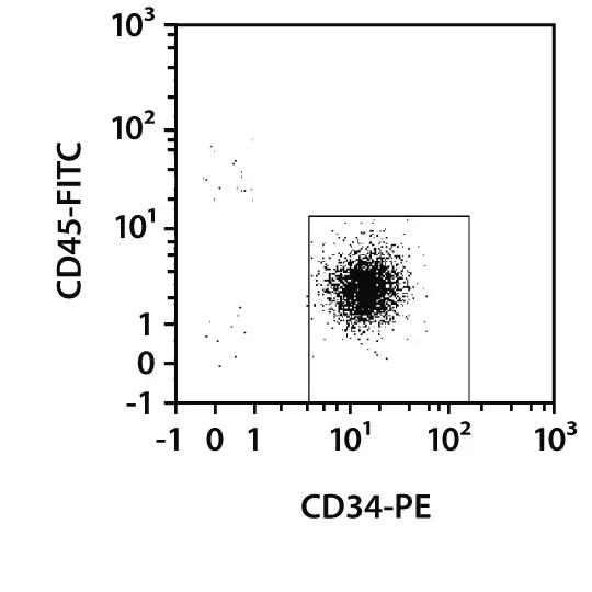
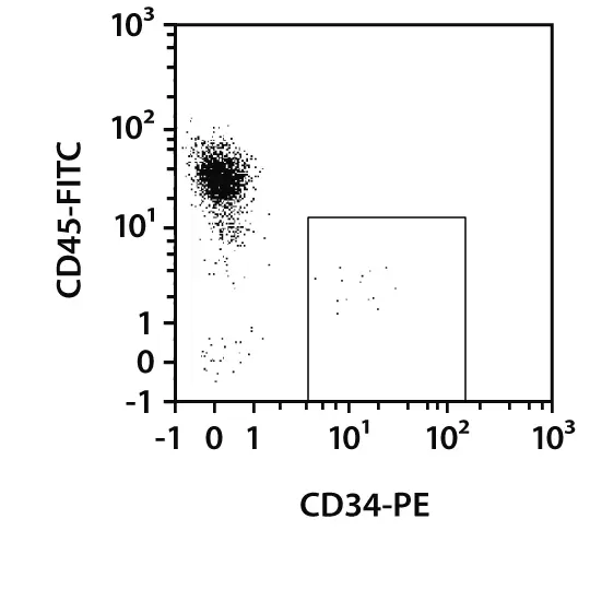
CD34+ cells isolated from a debris-rich PBMC sample. The CD34 MicroBead Kit UltraPure, human, was used to separate human HSCs from a sample of PBMCs. Cells were stained with CD34-PE, CD45-FITC, and propidium iodide solution. Flow cytometry analysis shows high purity after enrichment.
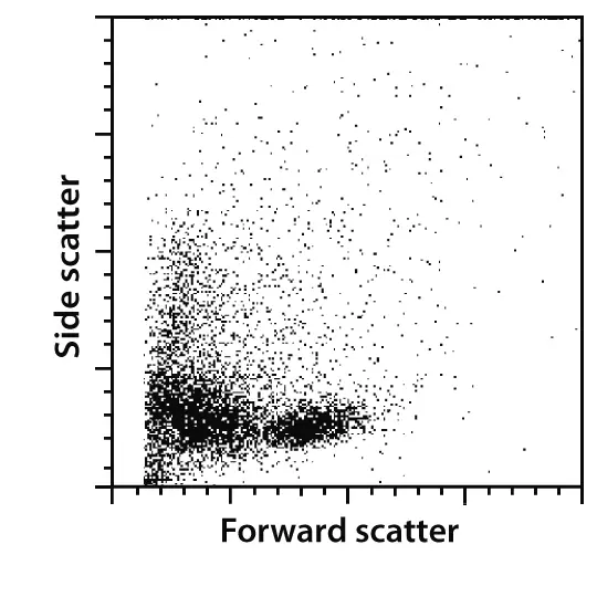
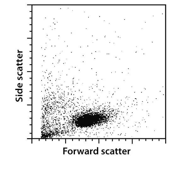
A comparison of the ungated forward scatter versus side scatter profile of the same sample after separation with the CD34 MicroBead Kit, human, or the CD34 MicroBead Kit UltraPure, human, demonstrates reduced debris carry-over with the CD34 MicroBead Kit UltraPure, human.
CD34+CD38– Cell Isolation Kit, human
CD38 is absent on most primitive CD34+ cells. The CD34+CD38– Cell Isolation Kit, human, is designed to isolate the more primitive CD38– subpopulation of CD34+ cells. The two-step isolation of CD34 enrichment followed by CD38 depletion only takes 2 hours.
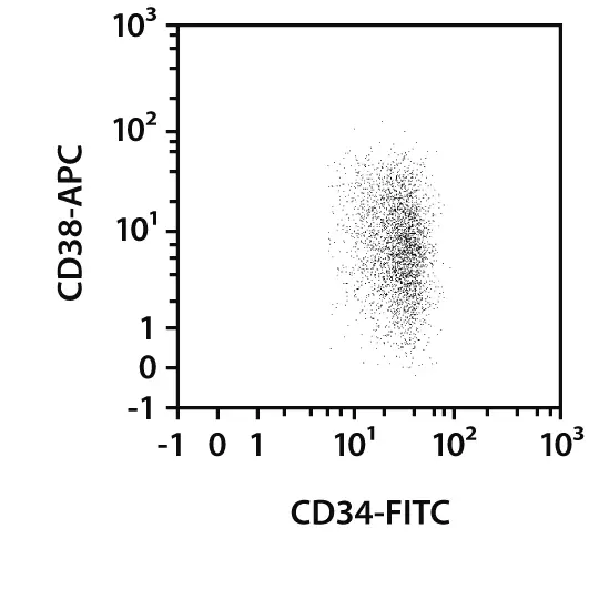
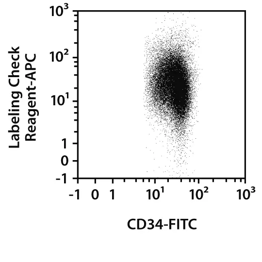
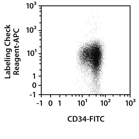
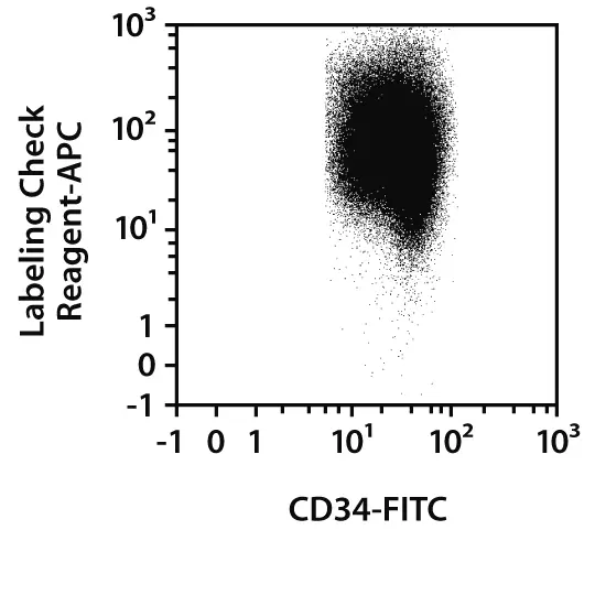
CD34+CD38– cells isolated from human cord blood mononuclear cells. CD34+ CD38– cells were isolated by the two-step isolation process (1. CD34 enrichment, 2. CD38 depletion) of the CD34+ CD38– Cell Isolation Kit, human. Cells were fluorescently stained with CD34-FITC, CD38-APC, CD45-VioBlue, Labeling Check Reagent‑APC, and Propidium Iodide Solution and analyzed by flow cytometry using the MACSQuant® Analyzer. Cell debris and dead cells were excluded from the analysis based on scatter signals and propidium iodide fluorescence. All shown plots were gated on CD34+ cells.
Lineage Cell Depletion Kit, human
The Lineage Cell Depletion Kit, human, is designed to magnetically label mature cells (T cells, B cells, NK cells, dendritic cells, monocytes, granulocytes, and erythroid cells) and their differentiation-committed precursors. Depletion of the labeled cells results in the enrichment of untouched human HSCs and progenitor cells.
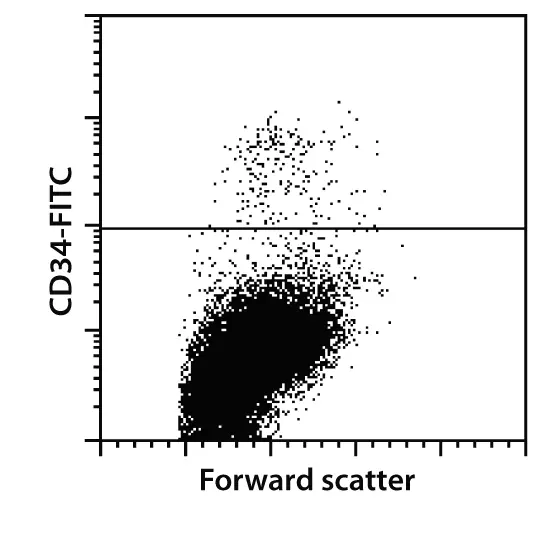

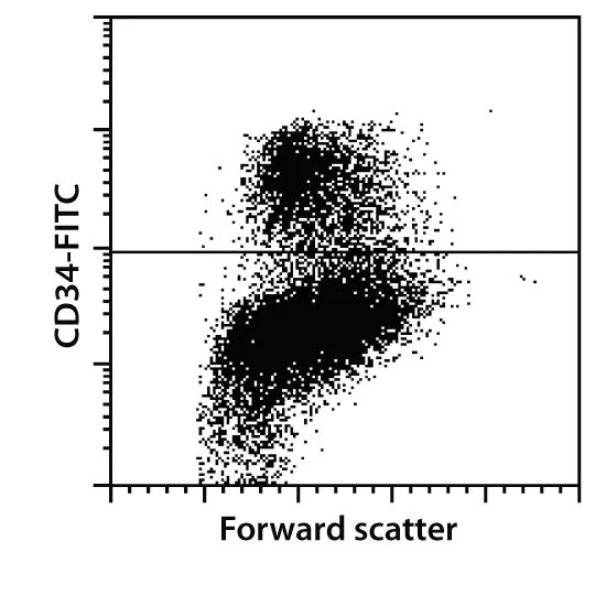
Analysis of a bone marrow mononuclear cell (BM-MNC) population before and after lineage cell depletion. Lineage-negative cells were isolated from BM-MNCs using the Lineage Cell Depletion Kit, human, and an LS Column. Cells were fluorescently stained with CD34-FITC and all fractions were analyzed by flow cytometry.
2.5 Characterization of human HSCs by flow cytometry
At a glance: Dedicated antibody cocktails for flow cytometry analysis of human HSCs
| Cocktail markers | Isolation kit or reagent used | Product |
| CD133/2-PE, CD34-APC, CD45-FITC | CD133 MicroBead Kit – Hematopoietic Tissue, human | CD34/CD133 Stem Cell Analysis Cocktail Kit, anti-human |
| CD34-PE, CD45-FITC | CD34 MicroBead Kit, human, CD34 MicroBead Kit UltraPure, human, or Indirect CD34 MicroBead Kit, human | CD34 Stem Cell Analysis Cocktail, anti-human |
Human HSCs are routinely analyzed by flow cytometry based on various extracellular and intracellular markers. The MC Antibody Cocktails simplify the flow cytometry analysis of cells separated by MACS Technology. All antibodies are optimally titrated to be simply added to an aliquot of a cell fraction before analysis.
Miltenyi Biotec also offers a wide range of unique and standard monoclonal antibodies for research of human HSCs and progenitor cells, including CD34, CD38, CD133, CD90, CD117, and Anti-Sca-1. Fluorochrome-conjugated MACS Antibodies are perfectly suited for the identification, enumeration, and characterization of human HSCs, which can be combined into panels tailored to specific research needs.

 View details
An online tool to quickly construct the right multicolor flow cytometry panel for each research project can be accessed via the related documents panel to the right. For details about Miltenyi Biotec antibodies and dyes for cell analysis, see MACS Antibodies and kits and support reagents.
Related content:
 Related products:
2.6 Cell culture of human HSCs
2.6.1 Expansion of human HSCs
At a glance: Expansion and supplement products
| Use | Comments | Product |
| Expansion | StemMACS HSC Expansion Media XF, human | |
| Supplement | Cytokine cocktail out of recombinant human Flt3-Ligand (Flt3-L), stem cell factor (SCF), thrombopoietin (TPO) | StemMACS HSC Expansion Cocktail, human |
The number of HSCs available in human samples is often insufficient for downstream applications. Human HSCs can be expanded in culture with HSC-relevant growth factors to obtain higher cell numbers or to elucidate mechanisms and signaling pathways driving self-renewal or differentiation. Early-acting cytokines – human stem cell factor (SCF), Flt3-Ligand (Flt3-L), thrombopoietin (TPO), and optional interleukin-3 (IL-3) – expand primitive HSCs, although current HSC culture systems allow only limited expansion and maintenance of primitive HSCs that are able to engraft and completely restore the entire blood system (PMID: 28801069)
StemMACS™ HSC Expansion Media XF is specifically formulated for the robust expansion of human HSCs from umbilical cord blood, bone marrow, or peripheral blood. The StemMACS HSC Expansion Cocktail, human, an optimized set of cytokines (human SCF, Flt3-Ligand and TPO), can be added to the medium to create a xeno-free culture environment that preserves high levels of primitive CD34+CD133+ and CD34+ CD45RA– cells.

MACS Handbook:
 Related PDFs:
2.6.2 Colony-forming unit (CFU) analysis of human HSCs
At a glance: Kits and reagents for the functional analysis of human HSCs by HSC-CFU assays
| Use | Comments | Product |
| CFU analysis by light microscopy (conventional HSC-CFU assay) | Available in different formulations containing methylcellulose, with or without erythropoietin (Epo) | StemMACS HSC-CFU Media, human |
| CFU analysis by flow cytometric readout (user-independent HSC-CFU assay kit) | Highly standardized by combining methylcellulose-free cell culture with a flow cytometric readout | StemMACS HSC-CFU Assay Kit, human |
HSC-CFU assays are used to examine the proliferation and differentiation potential of HSCs in vitro. The cells are cultured in medium supplemented with growth factors that drive differentiation into the myeloid lineage, resulting in outgrowth of a distinct colony from a single cell. A human HSC can give rise to one of six colony types which in turn are the precursors of cells that constitute the human blood, see figure below. The HSC-CFU assays provide results on the total colony count and types of a sample, based on which conclusions about the hematopoietic system of the sample´s donor can be made.

The StemMACS HSC-CFU Media, human, for conventional HSC-CFU assays and the StemMACS HSC-CFU Assay Kit, human, for user-independent HSC-CFU assays, are produced under tightly controlled manufacturing conditions and use highly qualified raw materials to provide a consistent and optimally performing CFU assay. They are designed to maximize growth and differentiation of progenitor cells and allow the clonal progeny of a single cell to grow into a distinct colony.
To perform the conventional CFU assay, Miltenyi Biotec offers different formulations of StemMACS HSC-CFU Media, human, that are standardized, semi-solid media based on methylcellulose in IMDM, and supplemented with FBS, BSA, and different growth factors. The cultivation of the cells takes place in 6-well plates or 35 mm dishes over 14 days. During this time cells gradually differentiate into specific colonies that can be identified microscopically based on their distinct morphological characteristics. The microscopic readout and classification of each colony can be challenging, especially for inexperienced users, and requires excessive training in order to obtain accurate results. An online tutorial describing how to set up and conduct the conventional HSC-CFU assay, plus pitfalls to avoid, can be accessed in the related documents panel to the right.
At a glance: A comparison of StemMACS HSC-CFU Media formulations and uses
| HSC-CFU complete with Epo | HSC-CFU complete w/o Epo | HSC-CFU lite with Epo | |
| The formulation supports growth of: | |||
| CFU-G, CFU-M, CFU-GM | Yes | Yes | Yes |
| BFU-E, CFU-E, CFU-GEMM | Yes | No | Yes |
| Components: | |||
| Methylcellulose in IMDM | 1% | 1% | 1% |
| Fetal bovine serum (FBS) | 30% | 30% | 30% |
| Bovine serum albumin (BSA) | 1% | 1% | 1% |
| L-glutamine | 2 mM | 2 mM | 2 mM |
| 2-mercaptoethanol | 0.1 nM | 0.1 nM | 0.1 nM |
| Stem cell factor (SCF) | 50 ng/mL | 50 ng/mL | 50 ng/mL |
| GM-CSF | 20 ng/mL | 20 ng/mL | 10 ng/mL |
| G-CSF | 20 ng/mL | 20 ng/mL | – |
| IL-3 | 20 ng/mL | 20 ng/mL | 10 ng/mL |
| IL-6 | 20 ng/mL | 20 ng/mL | – |
| Erythropoietin (Epo) | 3 U/mL | – | 3 U/mL |
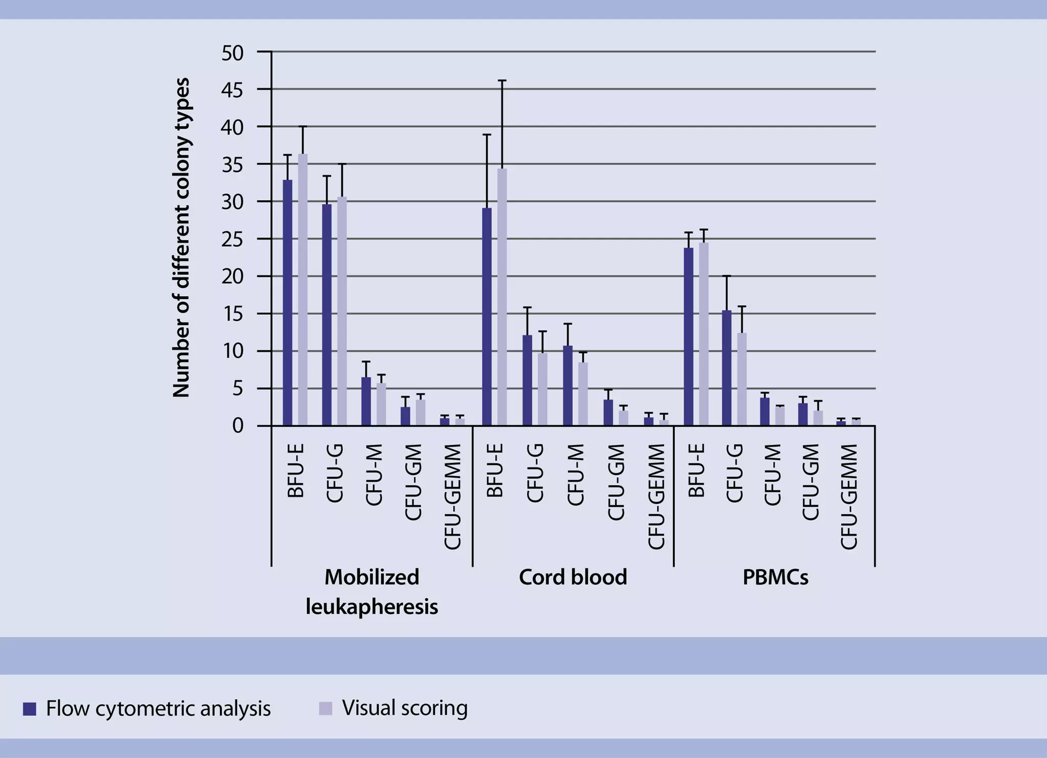
In addition to the conventional HSC-CFU assay, Miltenyi Biotec provides an advanced version that was developed to standardize and automate the scoring of colonies, the StemMACS HSC-CFU Assay Kit, human. This kit includes a methylcellulose-free culture medium for the cultivation of colonies in 96-well plates, as well as a fluorochrome conjugated antibody cocktail to detect all colony types by a flow cytometric readout. Based on the individual surface marker combinations (CD15, CD14, CD235a) the colonies can be easily identified, thus eliminating the need for user-dependent visual scoring under a microscope. The StemMACS HSC-CFU Assay Kit, human, provides user-independent and clear results on the total count of colonies, as well as the individual types that can be found in a sample. Furthermore, for an automated and high throughput acquisition of the plates, the MACSQuant® Analyzer X can be utilized. For colony identification via flow cytometric readout when using the MACSQuant Analyzer, we provide an analysis excel sheet and gating template. These analysis tools are optimized for the MACSQuantify Software version 2.11 and upwards. To receive these analysis tools, please contact your local technical support.

Related content:
2.6.3 Viral transduction of human HSCs
At a glance: Kits and reagents for viral transduction of human HSCs
| Use | Comments | Product |
| Viral transduction | Does not have to be pre-coated on cell culture surfaces | Vectofusin-1 |
| Determination of vector copy numbers (VCN) | The primers included in the kit are designed to amplify an amplicon of the lentiviral gag gene and the human reference gene PTBP2. These primers are only compatible with the lentiviral vectors provided by Lentigen and Miltenyi Biotec. | MACS COPYcheck Kit, human |
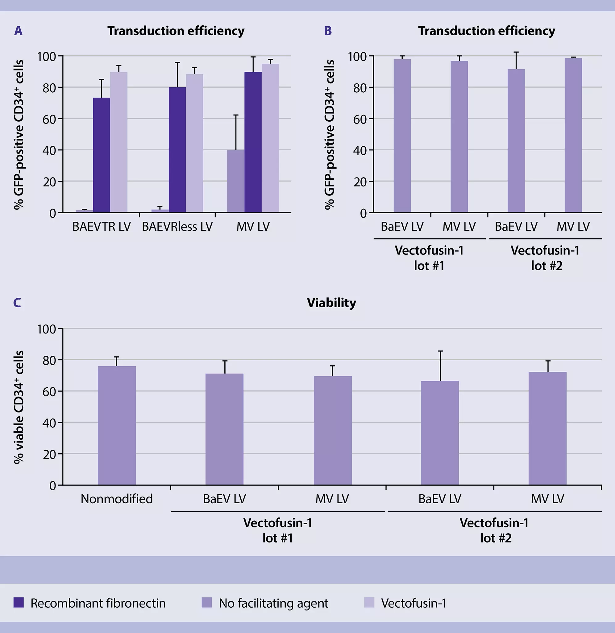
Viral transduction is a fast and efficient method to study gene function or modulate gene expression. It is also a potential approach for gene modification in the context of gene therapies. The modification of human HSCs with retroviral and lentiviral vectors often requires the presence of a transduction-enhancing reagent (PMID: 30788387). Polycationic reagents induce aggregation of vector particles and facilitate binding of the vectors to target cells via electrostatic interaction. Bridging molecules, such as recombinant fibronectin, increase the co-localization of virus and target cell.
Miltenyi Biotec offers the novel transduction enhancer Vectofusin-1®, a fully synthetic non-toxic cationic amphipathic peptide that supports high transduction levels with small amounts of retroviral vector. When added to culture medium, Vectofusin-1 promotes the entry of several retroviral pseudo-types into target cells (PMID: 31578886). Vectofusin-1 is a registered trademark of Genethon.
You have previously chosen International as your location.
What do you want to do?