A complete workflow solution for large-scale 3D light sheet microscopy


A complete workflow solution for large-scale 3D light sheet microscopy
See the big picture and dive deep into the details
Vasculatures and neuronal networks are complex structures, spanning a spectrum from micrometers to centimeters in a 3D space. Imagine analyzing them as a whole, without the need for tissue sectioning. Revolutionize your research with our state-of-the-art, high-resolution, automated 3D light sheet microscopy solution, the UltraMicroscope Platform, designed for samples of various sizes. Examine your intact samples entirely, delving into intricacies at the single-cell level.
Best of all, it's simpler than you think! Miltenyi Biotec provides an all-encompassing workflow solution, saving you time and resources. This integrated approach is suitable for all who want to streamline their research, whether you’re a seasoned microscopy expert or just starting your 3D light sheet imaging journey!
3D light sheet imaging workflow
Content
- Staining
- Tissue clearing
- Imaging
- Image processing
Acquiring high-quality 3D light sheet microscopy data from large samples, such as organoids, whole rodent organs, or even human organs, involves four key steps:
- Staining: The sample is labeled with fluorescent antibodies that specifically target desired cell types or molecules. Thorough epitope labeling throughout the entire 3D structure and achieving high signal-to-noise ratios are crucial for reliable analysis.
- Tissue clearing: Opaque tissue samples must be rendered transparent to enable the excitation of fluorochromes deep within the sample.
- Imaging: The labeled and cleared sample is then imaged using a light sheet microscope designed for large samples. Optics with long working distances are employed for both overview and high-resolution images.
- Image processing: The acquired image data is processed using specialized software to optimize the results.
Miltenyi Biotec provides comprehensive solutions for all four steps.
Step 1: Staining
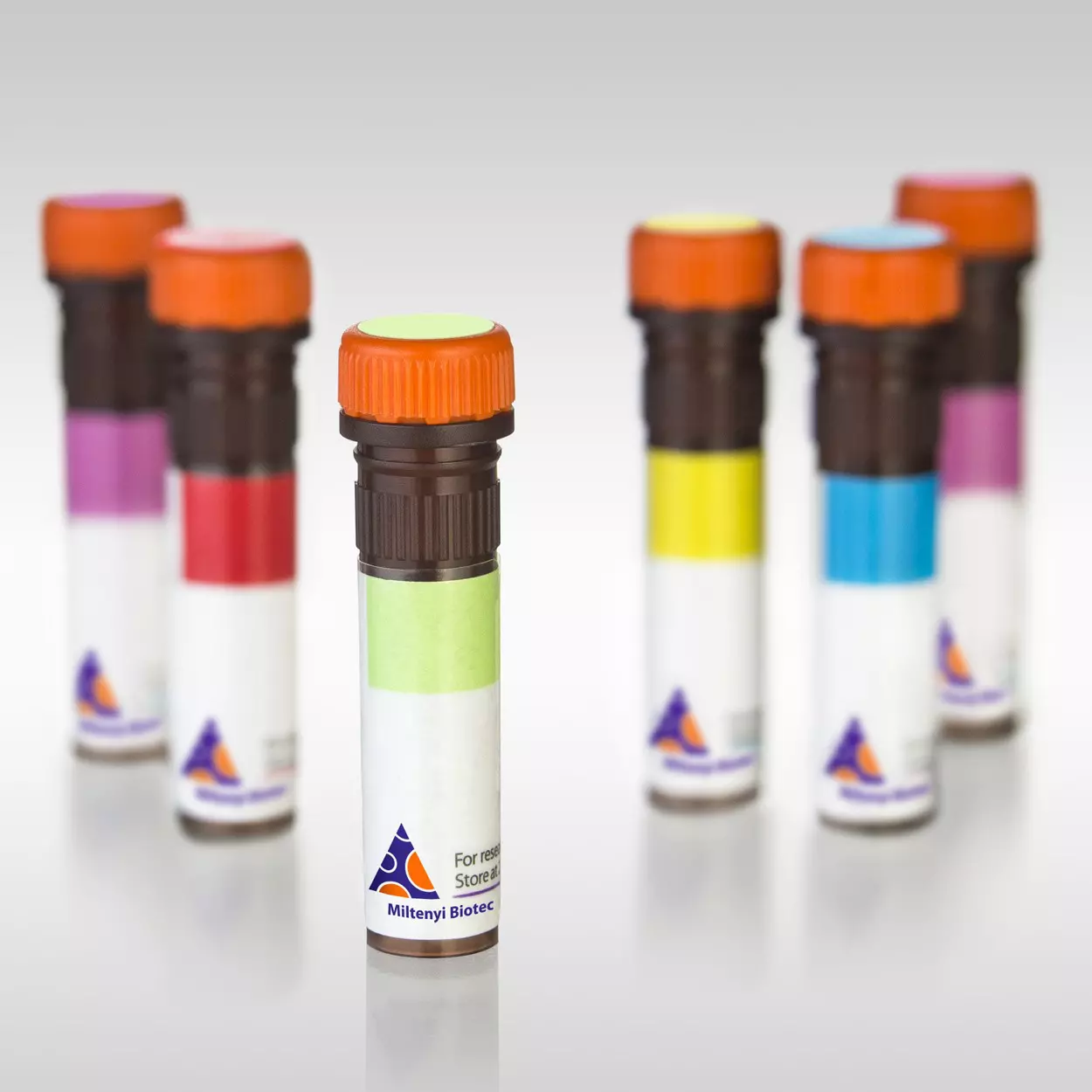
In light sheet microscopy, samples are usually stained with fluorescent dyes or antibodies that bind to specific molecules of interest, allowing users to detect and distinguish complex structures in the tissue.
Staining large tissue samples with antibodies can be expensive and time-consuming. Our 3D-immunofluorescence (IF) antibodies are specifically designed and validated for whole-mount staining of large cleared samples, including mouse brain, human tumor samples, and cerebral brain organoids. Conjugated to bright and photostable Vio® Dyes, our REAfinity® Recombinant Antibodies expedite and simplify your research with shorter experiment times and optimal signal-to-noise ratios. For maximum reliability and conclusive results, all our 3D-IF antibodies are functionally validated with the MACS® Deep Clearing and MACS Clearing Kits, and have been extensively tested and reported as compatible with various tissue clearing methods.
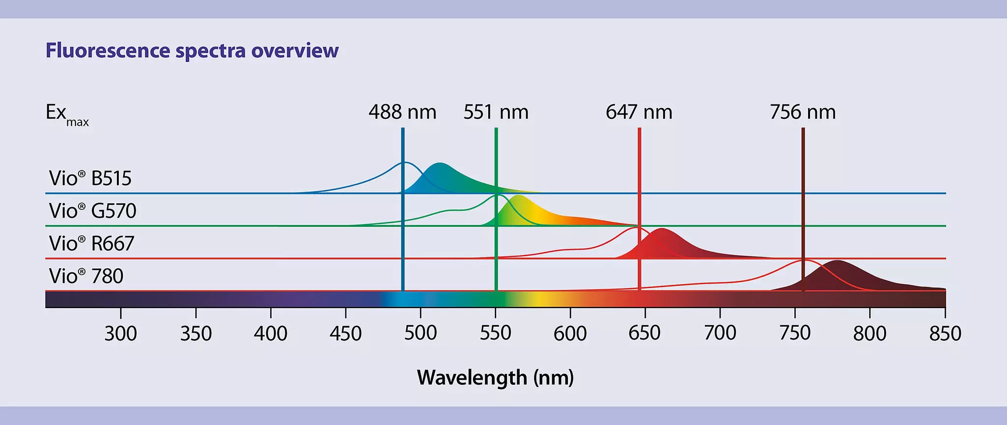
Our 3D-IF–validated antibodies are conjugated to Vio Dyes, which are known for their exceptional brightness, high photostability, and excellent signal-to-background ratios. When performing four-channel staining, you benefit from fluorophores that closely resemble commonly used Alexa Fluor® dyes and are optimally matched to the laser and filter combinations widely used in 3D imaging.
Validated 3D-IF antibodies provide conclusive 3D imaging data
The video shows a human cerebral organoid, whole-mount stained with Ki-67 Antibody, anti-human/mouse, Vio R667, REA183 (yellow); SOX2 Antibody, anti-human, Vio B515, REA320 (magenta); and β-Tubulin 3 Antibody, anti-human/mouse, Vio G570, REA1152 (cyan). The sample was cleared using the MACS Clearing Kit and imaged with the UltraMicroscope Platform.
3D-IF antibodies are compatible with other tissue clearing methods like iDISCO+
This video gives you an intriguing insight into the complexity of the mouse brain. It showcases outstanding data obtained by Gubra, a CRO and Biotech Company from Denmark, using Miltenyi Biotec's Neurofilament Antibody (anti-human/mouse, Vio R667, REAfinity) to stain neurofilaments. The sample was cleared using the iDISCO+ method and imaged with the UltraMicroscope Platform.
It is by far the best TH antibody we have ever tested.

Step 2: Tissue clearing

After staining, it's crucial to render opaque tissue samples transparent – a process known as tissue clearing. This is of most importance to excite fluorescent molecules deep inside the sample and achieve distinct images of fine details within large tissues.
Creating a new clearing protocol from scratch can be time-consuming and labor-intensive. Our ready-to-use, non-toxic MACS Deep Clearing Kit and MACS Clearing Kit efficiently render your samples transparent for optimal 3D imaging results.
Good to know: Our non-toxic MACS Imaging Solution (refractive index = 1.556–1.562) is ideally suited for imaging your stained and cleared samples with the UltraMicrosope Platform.
Organic solvent–based tissue clearing at its best
The MACS Clearing Kits offer optimized solutions for immunolabeling and clearing of tissue samples of various sizes:
- MACS Deep Clearing Kit – ideal for immunolabeling and clearing tissue samples larger than 5 × 5 × 5 mm³, up to the size of a whole adult mouse brain. It enables thorough antibody penetration and outstanding clearing throughout the entire sample.
- MACS Clearing Kit – designed for smaller tissues up to 5 × 5 × 5 mm³, offering fast, efficient, and reliable clearing performance.
Video
Fast, efficient, and non-toxic tissue clearing of a whole mouse brain using the MACS Deep Clearing Kit
This video demonstrates an adult mouse brain that has undergone tissue clearing with the MACS Deep Clearing Kit. It showcases the final stage of the protocol, in which the sample is rendered transparent for volumetric fluorescence imaging using light sheet microscopy. In this step, the sample’s refractive index is adjusted to match that of the MACS Imaging Solution (1.556–1.562) within 24 hours.
I appreciate the simplicity and effectiveness of the MACS® Clearing Kit. Its easy-to-use design eliminates the need for laborious reagent preparation, with no compromise on the quality of our results. Plus, it utilizes a non-toxic clearing solution, which is a huge bonus!

Step 3: Imaging
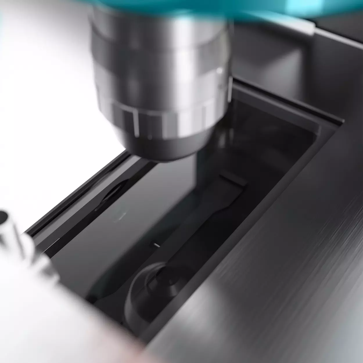
The heart of the 3D imaging workflow lies in acquiring high-quality images using a light sheet microscope. The UltraMicroscope Platform – featuring the UltraMicroscope Blaze™ and UltraMicroscope Choros™ – is equipped with cutting-edge technology and state-of-the-art light sheet optics, delivering exceptional image quality across a wide range of imaging scales, from macroscopic to subcellular levels. You can visualize samples as large as a whole cleared mouse or even entire human organs, encompassing everything from the overview to the finest details.
The UltraMicroscope´s automated functions, combined with LightSpeed Mode, revolutionize 3D imaging by enabling efficient, high-throughput data acquisition. Mount multiple samples in the microscope and process them in batches, eliminating the need for manual intervention or monitoring. This significantly reduces imaging time and enhances productivity.
Good to know: To further streamline 3D imaging, Miltenyi Biotec offers a selection of application-specific sample holders: the MACS UltraMounts. When combined with predefined imaging settings, batch acquisition, and automated processing pipelines, they unlock a high-throughput imaging experience with minimal hands-on intervention. The result is maximum efficiency, consistency, and scalability in your 3D imaging workflows.
Video
Automated 3D light sheet imaging
The UltraMicroscope Platform lets you easily create detailed 3D images of complex biological systems such as an entire brain – without sectioning and with minimal training and hands-on time.
UltraMicroscope technology allows us to see every single cancer metastasis in the whole bodies of transparent mice and we can also see if drugs are targeting all those tiny micro-metastases. UltraMicroscope technology will be a powerful tool for drug development in oncology.

Step 4: Image processing
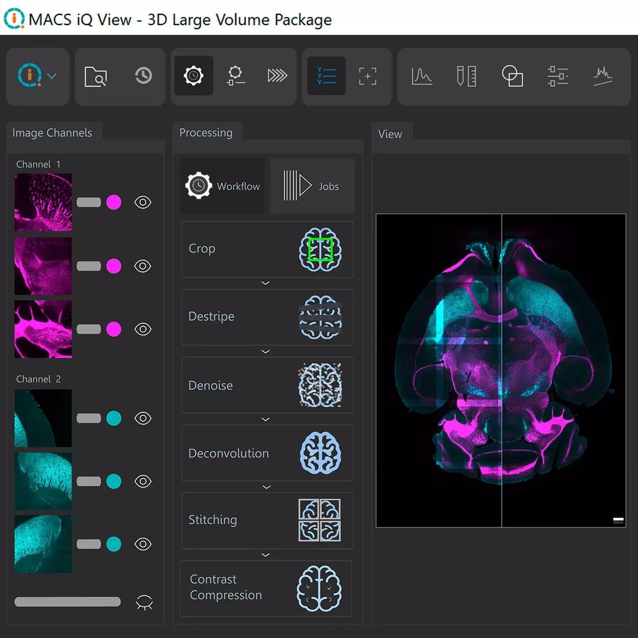
Image processing is an important step in light sheet microscopy, as it allows researchers to enhance the quality and interpretability of the acquired data.
MACS iQ View – 3D Large Volume is a software package specifically designed for processing light sheet images acquired with the UltraMicroscope Blaze and UltraMicroscope Choros.
The software package includes a comprehensive suite of image processing modules that can be used to perform a variety of tasks, including 3D cropping, destriping, denoising, deconvolution, stitching, and contrast compression. Additionally, the software supports batch processing, enabling users to queue and initiate various workflows across multiple datasets. This feature proves especially valuable when dealing with extensive datasets and imaging multiple samples using the UltraMicroscope Platform, particularly in the LightSpeed Mode, ensuring a smooth processing workflow.
MACS iQ View – 3D Large Volume interfaces with the UltraMicroscope Platform’s acquisition software, ImSpector. Once connected, you can schedule individual processing steps or custom workflows and assign them directly to measurements while setting up your imaging experiment in ImSpector. After acquisition, processing starts automatically in MACS iQ View – 3D Large Volume, eliminating the need to return to your computer.
Video
Image processing with MACS iQ View – 3D Large Volume
This video shows an XY-plane of data from dopaminergic neurons in the whole mouse brain (cyan, dopaminergic neurons labeled with Tyrosine Hydroxylase Antibody, anti-human/mouse/rat, Vio® G570, REAfinity™) before (left) and after (right) processing with MACS iQ View – 3D Large Volume. The image was processed with Destripe (standard setting and 20-pixel stripe size) to remove striping artifacts, Deconvolution (subtle setting) to enhance resolution, and Contrast Compression to visualize both dim and bright structures at the same time (Structure mode).
MACS® iQ View – 3D Large Volume is a great new tool that enables us to quickly transition from raw images to 3D reconstructions. Its processing functions, particularly those specific for light-sheet images like stripe artifact removal, simplify the segmentation of images for quantitative analysis. Additionally, stitching works very well and very fast, even when handling large files.




You have previously chosen International as your location.
What do you want to do?