San Diego Convention Center
San Diego, California, USA
Miltenyi Biotec booth #4627
April 17–22, 2026 | San Diego, USA


In San Diego, we are moving beyond the limits of standard workflows. Whether you are interested in spatial multiomics, high-quality antibodies, or tumor dissociation, discover practical approaches to investigate the tumor microenvironment and advance your immuno-oncology research.
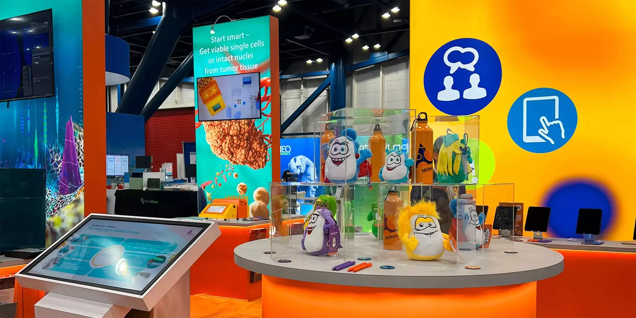
Visit our booth #4627 and step into an experience designed to elevate your data
and provide fresh perspectives you can bring back to the lab.*
San Diego Convention Center
San Diego, California, USA
Miltenyi Biotec booth #4627
April 19–22, 2026 | Exhibit opening hours
Sunday, April 19 | 12:00–05:00 p.m.
Monday, April 20 | 09:00 a.m.–05:00 p.m.
Tuesday, April 21 | 09:00 a.m.–05:00 p.m.
Wednesday, April 22 | 09:00 a.m.–12:00 p.m.
*Not affiliated with or endorsed by AACR.
Elevate your booth experience: Take a photo in our hot air balloon, share it with the community, and receive a limited-edition MACS Antibodies T-shirt.
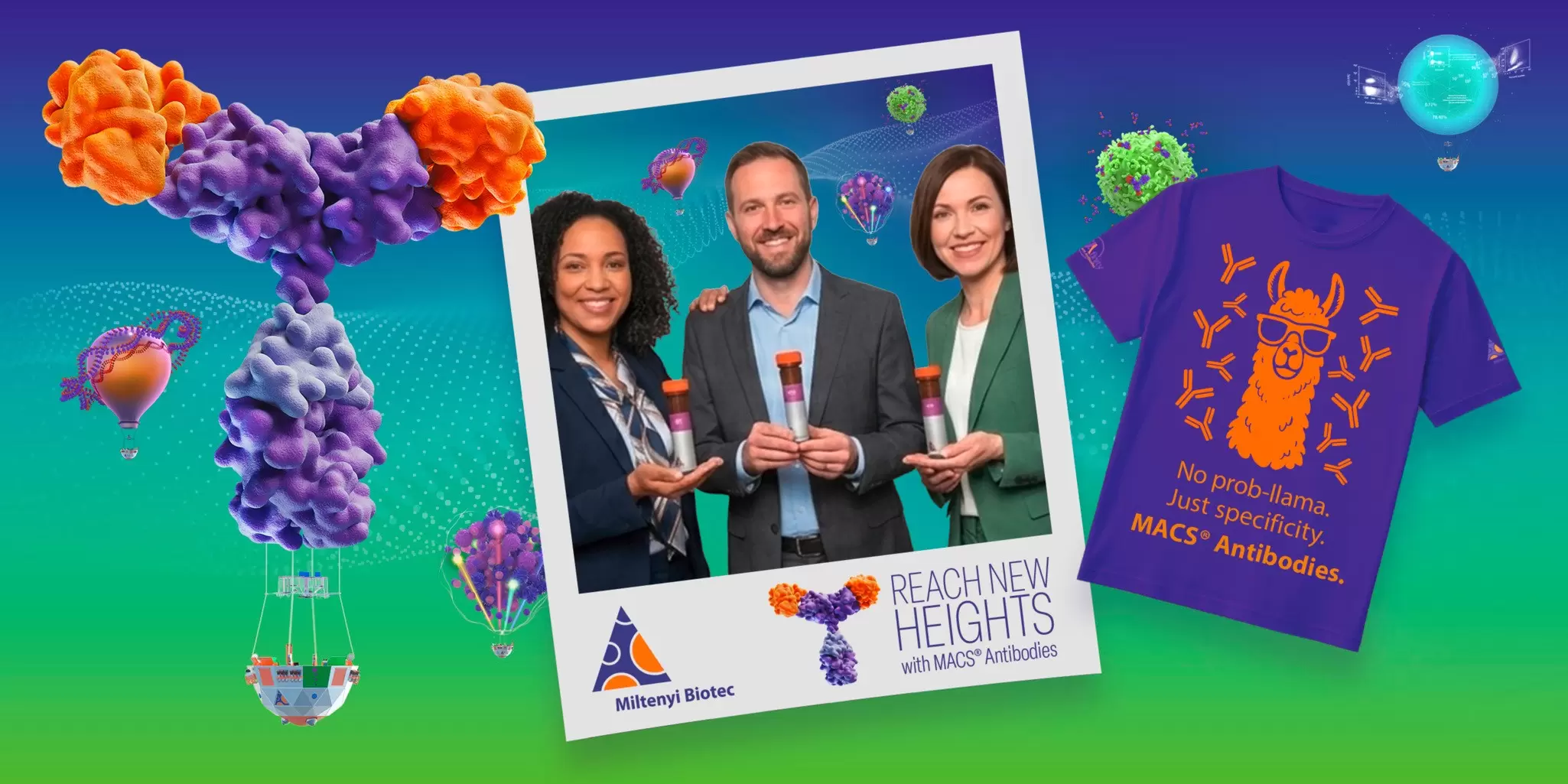


Navigate the workflow discovery trail, learn more about our automated and standardized solutions, and get a chance to win a prize.
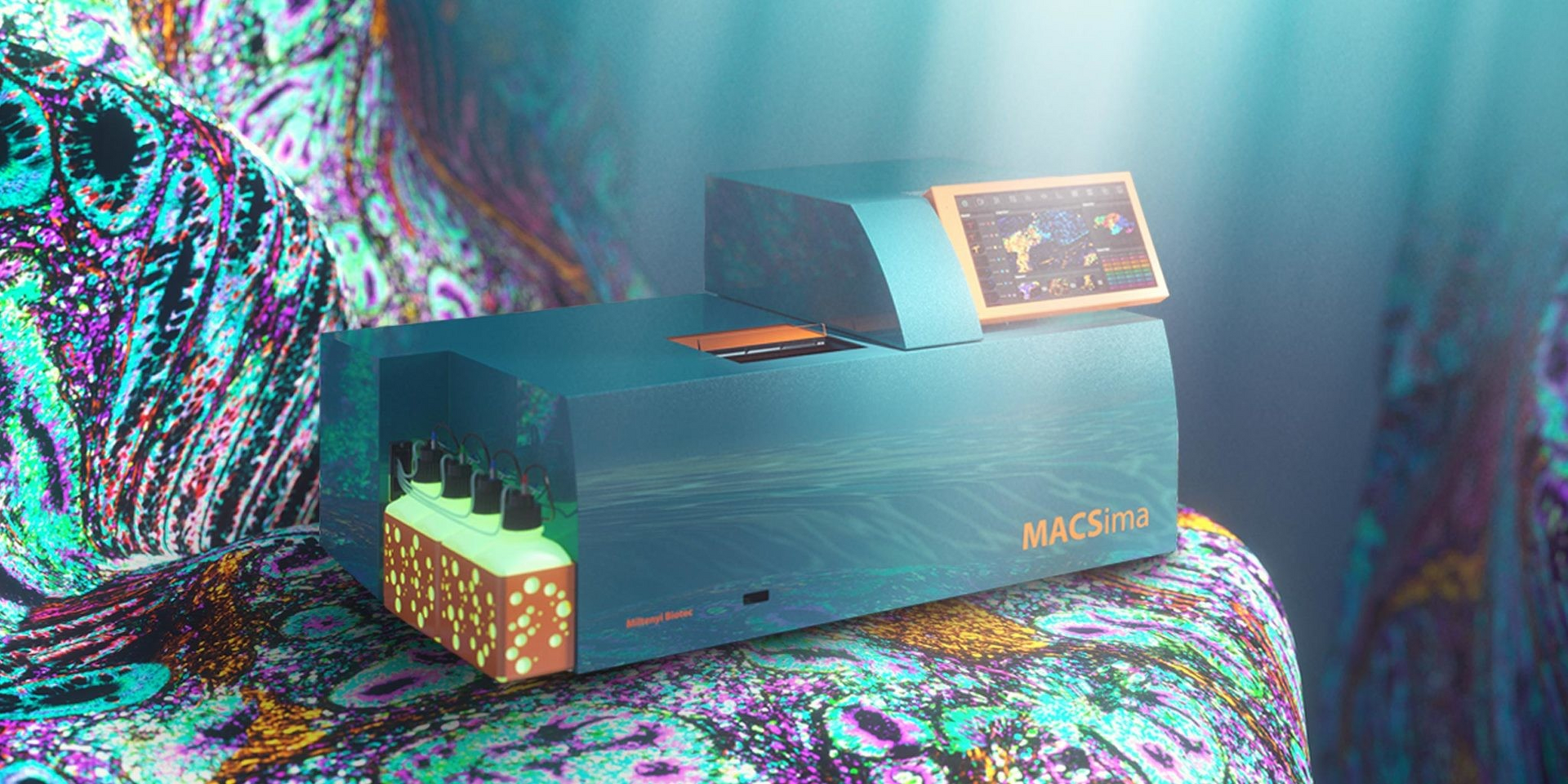
Spatial biology enables detailed analysis of molecular processes within native tissue architecture. Using MACSima Imaging Cycling Staining (MICS), hundreds of proteins and dozens of RNA markers can be detected on the same section, generating high‑dimensional datasets that enhance cellular characterization and improve therapeutic target selection.
At the Spotlight Theater, we will demonstrate the application of the MACSima Platform in two preclinical settings. First, establishing an integrated immunophenotyping and precision oncology workflow for pediatric tumors. Second, developing a multiomics monitoring approach that maps the localization and activity of transgenic TCR T cells in situ.
Join us for this exclusive session featuring guest speakers from world-renowned cancer centers.*
Tuesday, April 21 | 12:30–01:30 p.m.
(Lunch included)
Spotlight Theater D

Christian Seitz, MD
Head of the Stem Cell Transplantation and Cell and Gene Therapy Program, Hopp Children’s Cancer Center Heidelberg (KiTZ), Germany
Abstract
Precision oncology programs based on bulk RNA sequencing, whole-genome sequencing, and methylome profiling have transformed cancer care through molecular classification and identification of actionable genomic alterations.
However, bulk omics technologies have inherent limitations, particularly in the context of precision immunotherapy. Current workflows lack spatially resolved information on tumor-immune interactions, functional immune states, and protein-level heterogeneity of therapeutic targets.
As a result, key determinants of treatment response, including target antigen distribution, MHC expression, immune checkpoint protein localization, and immunosuppressive microenvironmental niches, are not adequately captured for clinical decision-making.
Here, we report the establishment and validation of an integrated immune phenotyping and precision oncology workflow for pediatric oncology, based on Miltenyi Biotec’s MACSima Spatial Biology Platform.
We developed and rigorously validated a >60-marker panel for fresh-frozen tumor sections, enabling comprehensive profiling of tumor, immune, and stromal cell populations, extracellular matrix components, immune checkpoint molecules, as well as cell surface and intracellular target antigens.
This spatial immunotherapy panel and integrated analysis workflow enable identification of actionable immune targets across pediatric cancers, support patient stratification into biomarker-driven basket trials, and guide rational combination immunotherapy strategies.
A feasibility study integrating this workflow into the INFORM (INdividualized therapy FOr high-Risk childhood Malignancies) precision oncology program is currently underway.
Speaker information
Christian Seitz, MD, is the Head of Stem Cell Transplantation and Cell and Gene Therapy Program at KiTZ, Hopp Children’s Cancer Center in Heidelberg, Germany, since 2024. Before that, he worked as a pediatric hematologist–oncologist at the University Children’s Hospital Tübingen, Germany.
He studied medicine (2007–2014) at universities in Ulm and Frankfurt, Germany, as well as at the National Cancer Institute in Bethesda, USA. He received his doctorate, Dr. med., from Goethe University Frankfurt.
In 2014, he started his clinical training in pediatrics under Rupert Handgretinger at the University Children’s Hospital Tübingen, focusing on stem cell transplantation and cellular therapies, and was board certified in 2020.
His scientific work focuses on the development and clinical translation of modular chimeric antigen receptor (CAR) technologies and the interaction of CAR T cells in the tumor microenvironment.

Sophia Scheuermann, PhD
Postdoctoral Researcher & Lab Head at Seitz Lab, and PerVision Scientific Coordinator at the University Children's Hospital of Tübingen, Germany
Abstract
Single-cell analysis of complex tumor tissues is essential for understanding mechanisms of therapeutic response and resistance. High-plex spatial multiomics enables deep phenotypic characterization of tissues, revealing spatial organization and cellular interactions that are critical for evaluating immunotherapies in solid tumors.
While new immunotherapies like transgenic T cell receptor (TCR)-T cell therapies represent promising treatment modalities, their efficacy is often limited by challenges such as an immunosuppressive tumor microenvironment (TME), insufficient T cell persistence, and immune evasion by HLA downregulation. Comprehensive and spatially resolved monitoring of these therapies is therefore crucial for advancing future clinical trials.
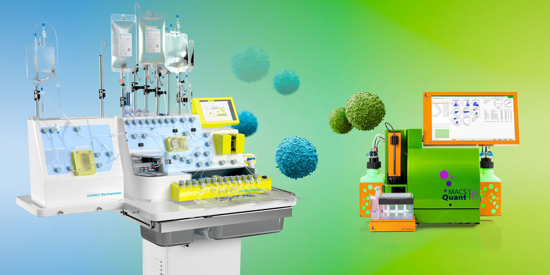
In this study, we established a multimodal monitoring workflow to investigate localization and activity of transgenic TCR-T cells in situ. Sequential FFPE tissue samples were collected from a patient treated with transgenic TCR-T cells generated using the CliniMACS Prodigy® Platform. Samples were analyzed using classical immunohistochemistry (IHC), bulk TCR sequencing, and high-plex spatial proteomics on the MACSima Platform. Furthermore, we employed a novel in situ sequencing-by-synthesis approach by Miltenyi Biotec to directly detect therapeutic TCR-T cells combined with multiplexed protein profiling of 60 markers on the same tissue section.
This integrative spatial multiomics approach enabled detailed characterization of immune cell composition, immune signatures, and cellular crosstalk within the TME. Importantly, we achieved specific detection of therapeutic TCR-T cells, allowing unprecedented analysis of their spatial context and cellular neighborhoods. Our results highlight that merging conventional protein staining with spatial in situ sequencing offers a powerful new lens into tumor ecosystems, paving the way to advancement of precision immunotherapy.
Speaker information
Dr. Sophia Scheuermann is an immunologist and spatial biology researcher specializing in immuno‑oncology and advanced tissue imaging. She defended her PhD thesis “Spatial Immuno-Oncological Profiling of Cancer Using Multiplexed Tissue Imaging” in 2025 in the Advanced Cellular Therapies Lab of Dr. Christian Seitz at the University Children’s Hospital Tübingen, Germany.
Since 2023, she coordinates the clinical trial Personalized Vaccination in Fusion + Sarcoma Patients (PerVision) in collaboration with the German Cancer Consortium (DKTK) and the German Cancer Research Center (DKFZ), focusing on translational approaches to individualized cancer immunotherapy.
She serves as a postdoctoral researcher and head of the AG Seitz – Translational Immunotherapy group at the Hopp Children’s Cancer Center Heidelberg and also expanded her expertise in immunology during a research stay at the University of British Columbia, Vancouver, Canada, in 2019.
Dr. Scheuermann’s work bridges advanced imaging technologies, immunotherapy development, and translational oncology, with a strong commitment to improving therapeutic strategies for young patients with cancer.
*This Exhibitor Spotlight Theater is a promotional activity and is not approved for continuing education credit. The content of this Exhibitor Spotlight Theater are the opinions of the presenter and do not represent the position or the opinion of the American Association for Cancer Research® (AACR) or its members.
The scientific posters from our R&D specialists are now available for download and offer
fresh insights into the future of cancer research.
#742 Unlocking 3D: TumorMACS™ Media as a dual-purpose solution for primary 2D and 3D tumor organoid cultures
Presenter: Benjamin Theek
#817 Spatial characterization of T cell receptors at subcellular resolution using in situ sequencing-by-synthesis and spatial protein detection on the same tissue section
Presenter: Hansueli Meyer
#1225 3D spatial multiomics characterization of ovarian cancer tissue samples
Presenter: Lena Nolte
#3718 CAR detection reagents – Pioneering advances in CAR T analytic
Presenter: Stephan Wilmes
#4281 Armoring ROR1 CAR T cells with IL18 improves CAR T cell function and cytotoxicity against pancreatic cancer cell lines in vitro and in vivo
Presenter: Bal Krishna Chand Thakuri
The video shows the 3D rendered visualization of a pancreatic carcinoma. GFP-expressing tumor cells are shown in green, infiltrating CAR T cells in purple, and the vasculature in orange. Imaging was performed using UltraMicroscope technology.
Do you want to get in contact with us? Send your inquiry to:
Jonathan Druge, Global Marketing
You have previously chosen International as your location.
What do you want to do?