The Tumor Cell Isolation Kit, human enables the enrichment of human tumor cells from primary specimens using MACS® Cell Separation technology.
While depleting tumor associated cells such as lymphocyte subpopulations, fibroblasts, and endothelial cells in a fast and easy protocol, the tumor cells remain untouched, improving any downstream analysis.


Related products for Tumor Cell Isolation Kit, human
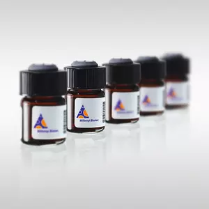


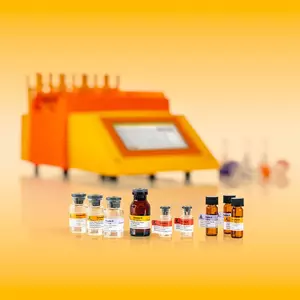


The Tumor Cell Isolation Kit, human enables the enrichment of human tumor cells from primary specimens using MACS® Cell Separation technology.
While depleting tumor associated cells such as lymphocyte subpopulations, fibroblasts, and endothelial cells in a fast and easy protocol, the tumor cells remain untouched, improving any downstream analysis.
The Tumor Cell Isolation Kit, human has been designed for the enrichment of untouched human tumor cells from primary specimens. During the growth phase in vivo, tumor tissue is vascularized and infiltrated by cells of non-tumor origin, including heterogeneous lymphocyte subpopulations, fibroblasts, and endothelial cells.
The level of infiltration is highly dependent on multiple factors like tumor subtype, growth rate, and affected organ site. However, even when these factors are constant, the amount and composition of infiltrating cells are highly variable, which makes accurate molecular downstream analyses difficult.
The contaminating non-tumor cells lead to hybridization of non-tumor cell derived mRNA molecules to probes on microarrays and a significant reduction of sensitivity caused by measurement of irrelevant signals during next-generation sequencing or proteome analysis. In addition, the culture of human tumor cells is frequently hampered by fibroblasts overgrowing the target cells. For optimal results, the Tumor Cell Isolation Kit, human should be used in combination with the Tumor Dissociation Kit, human (# 130-095-929) and gentleMACS™ Dissociators.
LS or autoMACS® Columns.
EpCAM+ human breast carcinoma cells were isolated from a heterogeneous sample of human cells using the Tumor Cell Isolation Kit, human, an LS Column, and a QuadroMACS™ Separator after the tissue was dissociated using the Tumor Dissociation Kit, human and the gentleMACS™ Octo Dissociator. The cells were fluorescently stained with CD326 (EpCAM)‑VioBlue® and CD31‑PE and analyzed by flow cytometry using the MACSQuant® Analyzer. Cell debris and dead cells were excluded from the analysis based on scatter signals and propidium iodide fluorescence.
EpCAM positive human breast carcinoma cells were isolated from a heterogeneous sample of human cells using the Tumor Cell Isolation Kit, human, an LS Column, and a QuadroMACS™ Separator. Subsequently, the unseparated fraction and the negative fraction were cultured for seven days in expansion medium (90% RPMI-1640, 2mM L-Glutamine, 10% FBS, 100 U/mL penicillin, and 100 U/mL streptomycin). After fixation, cells were stained using antibodies specific for vimentin (red), labeling fibroblasts, and EpCAM (green), labeling tumor cells. Cell nuclei were stained with DAPI (blue).
- Schäfer, D. et al. (2021) Identification of CD318, TSPAN8 and CD66c as target candidates for CAR T cell based immunotherapy of pancreatic adenocarcinoma. Nat Commun 12: 1453
You have previously chosen International as your location.
What do you want to do?