NK cells


1 Introduction 2 NK cells from peripheral blood 3 NK cells from other tissues (lymphoid, non-lymphoid)
1 Introduction
Natural killer (NK) cells are part of the innate immune system and mediate responses against viruses, parasites, bacteria, and tumor cells. Additionally, NK cells contribute to the adaptive immune response by linking innate and the adaptive immunity through their receptor FcγRIIIA (CD16).
2 NK cells from peripheral blood
Two distinct subsets of human NK cells have been identified based on CD56 expression: CD56dim and CD56bright NK cells (PMID: 3086432). These NK cell subsets lack CD3 expression and are phenotypically and functionally distinct. Natural killer T (NKT) cells are also included in this chapter as they also express the CD56 marker on their surface. However, NKT cells do express CD3. Additional NK cell subsets can be identified according to their tissue distribution. These have a different repertoire and effector functions compared to peripheral blood NK cells.
 Related PDFs:
2.1 Peripheral blood NK cell subsets
At a glance: NK cell subsets in peripheral blood
| Cell subset | Frequency | Markers | Function |
| CD16brightCD56dim/+ NK cells | ≥90% of peripheral blood NK cells | CD16+, CD56+, KIRs+, CCR7–, L-selectin– | High number of cytotoxic and cytolytic granules Antibody-dependent cell-mediated cytotoxicity functionLymphokine-activated killer cell activityNatural cytotoxicityMigrate to site of acute inflammation (early arrival) |
| CD16dim/–CD56bright NK cells | ≤10% of peripheral blood NK cells | CD56+, KIRs–/low, CCR7+, L-selectin+ | Lymphokine-activated killer cell activity Cytokine production Immunoregulation Migrate to secondary lymphoid organs |
The majority of human peripheral blood NK cells are CD56dim (90%) and express high levels of CD16; a minority (10%) are CD56bright and CD16dim/neg.
CD56bright NK cells are known for their capacity to produce and secrete cytokines, such as granulocyte–macrophage colony-stimulating factor (GM-CSF), IFN-γ, interleukin (IL)-10, IL-13, and TNF-β. Resting CD56bright and CD56dim NK cell subsets show differences in their NK cell receptor repertoires (PMID: 9469418). CD56bright NK cells express the high/intermediate affinity IL-2 receptor that confers the capacity to expand in vitro and in vivo in response to low doses of IL-2 (PMID: 7678599, 1692080). CD56bright NK cells do not express CD16 and express low levels of CD69, killer-cell immunoglobulin-like receptors (KIRs), and intracellular perforin (PMID: 15536127). Resting CD56bright cells exhibit low cytotoxicity, but after activation with IL-2 or IL-12, CD56bright cells exhibit similar or enhanced cytotoxicity against targets compared to CD56dim cells.
CD56dim NK cells are considered terminally differentiated and mature NK cells that are enriched in bone marrow, blood, and spleen (PMID: 12480696, 15536127, 16606675). CD56dim NK cells are cytolytic and efficient effectors of natural and antibody-dependent target cell lysis. In contrast to CD56bright NK cells, resting CD56dim NK cells express only the intermediate-affinity IL-2 receptor and proliferate weakly in response to high doses of IL-2 in vitro (PMID: 1370410). An important feature of CD56dim NK cells is the expression of KIRs and CD16 and the absence of CCR7 and L-selectin. CD56dim cells also express perforin and granzymes, and are more cytotoxic against NK cell–sensitive targets (K562 and COLO205 cell lines) than CD56bright NK cells (PMID: 2530273).
2.2 Miltenyi Biotec application protocols for peripheral blood NK cells
Miltenyi Biotec has created dedicated application protocols to isolate and analyze NK cells.
2.3 Sample preparation of peripheral blood
Dedicated solutions from Miltenyi Biotec allow the isolation of NK cells directly from whole blood. In addition, NK cells can also be isolated from PBMCs generated by density gradient centrifugation or using the MACSprep™ PBMC Isolation Kit, human. Other starting materials such as whole blood, buffy coat or cone, leukocyte reduction system chamber (LRSC), and leukapheresis products are also appropriate to isolate NK cells. For more detailed information, see the MACS Handbook chapter Blood (human).
 MACS Handbook:
2.4 Magnetic separation of peripheral blood NK cells
Miltenyi Biotec has developed numerous products for the straightforward magnetic separation of CD56+ cells. These cells can be isolated either straight from whole blood, buffy coat, or LRSC without density gradient centrifugation and erythrocyte lysis, or from PBMCs after density gradient centrifugation.
For details on MACS® Cell Separation Technology, see the MACS Handbook chapter Magnetic cell separation.
MACS Handbook:
2.4.1 Isolation of NK and NKT cells straight from whole blood or blood products
At a glance: Kits and reagents for the separation of NK and NKT cells from peripheral blood
| Starting material | Isolation strategy | Comments | Automation | Product |
| Whole blood | Positive selection of target cells | CD56 is predominantly expressed on NK and NKT cells. | Yes* | StraightFrom Whole Blood CD56 MicroBead Kit, human |
| Buffy coat | Positive selection of target cells | CD56 is predominantly expressed on NK and NKT cells. | Yes** | StraightFrom Buffy Coat CD56 MicroBead Kit, human |
| LRSC | Positive selection of target cells | CD56 is predominantly expressed on NK and NKT cells. | Yes** | StraightFrom LRSC CD56 MicroBead Kit, human |
| Whole blood | Depletion of non-target cells | No | MACSxpress NK Cell Isolation Kit, human | |
| *Automation options range from fully automated benchtop solutions such as the autoMACS® NEO Separator to high-throughput platforms such as the MultiMACS™ Cell24 Separator Plus or MultiMACS X. **Semi- or fully automated high-throughput cell separation with the MultiMACS™ Cell24 Separator Plus or MultiMACS X. | ||||
The StraightFrom® CD56 MicroBead Kits were developed for the rapid isolation of CD56+ cells directly from whole blood, buffy coat, or LRSC. The kits require no prior sample preparation and are optimized for a specific starting material. Subsequent depletion of CD3+ cells using CD3 MicroBeads, human yields NK cells of high purity.
StraightFrom® Buffy Coat CD56 MicroBead Kit
 View details
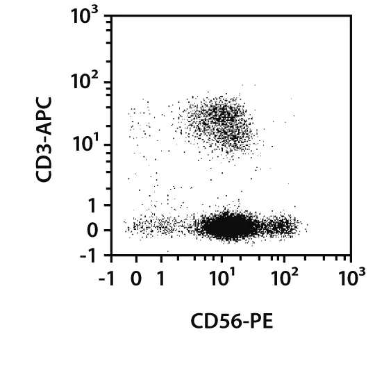
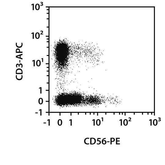
Fast isolation of CD56+ cells from buffy coat. Separation was performed with the StraightFrom Buffy Coat CD56 MicroBead Kit and the MultiMACS™ Cell24 Separator Plus with the Single-Column Adapter and Whole Blood Columns. Cells were fluorescently stained with CD56-PE, CD3-APC, and CD45-VioBlue® and analyzed by flow cytometry on the MACSQuant® Analyzer. Cells were triggered via CD45-VioBlue. Cell debris and dead cells were excluded from the analysis based on scatter signals and propidium iodide fluorescence.
The MACSxpress® NK Cell Isolation Kit isolates untouched NK cells directly from small (2 mL) or larger volumes (up to 30 mL) of whole blood . Non-target cells are removed by immunomagnetic depletion while erythrocytes are simultaneously sedimented to yield untouched target cells of high purity.
MACSxpress® NK Cell Isolation Kit, human
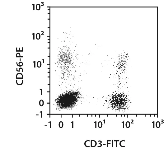
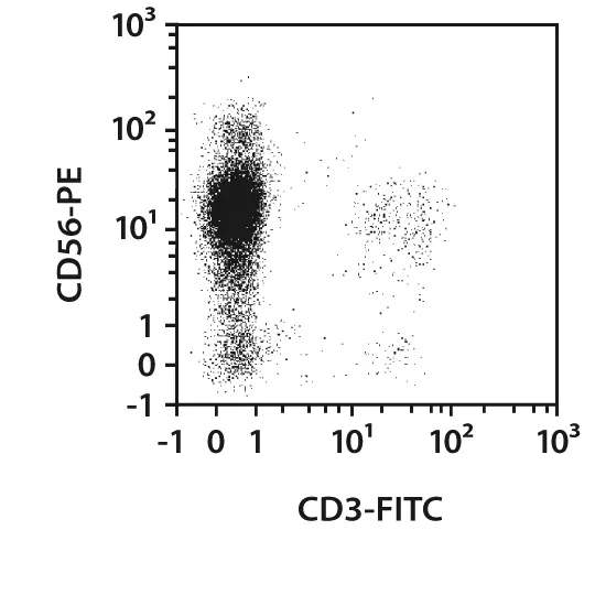
Untouched NK cells from whole blood. The MACSxpress NK Cell Isolation Kit, a MACSmix™ Tube Rotator, and a MACSxpress Separator were used to separate NK cells from 30 mL of human EDTA-anticoagulated whole blood. Isolated cells were fluorescently stained with CD45-VioBlue, CD3-FITC, and CD56-PE, and analyzed by flow cytometry using the MACSQuant Analyzer. Cell debris, non-leukocytes, and dead cells were excluded from the analysis based on CD45 expression, scatter signals, and propidium iodide fluorescence.
The StraightFrom™ MicroBead Kits were developed for the rapid positive selection of target cells directly from whole blood, buffy coat, LRSC, or Leukopak®. None of the kits require any prior sample preparation, like density centrifugation, erythrocyte lysis. or cell count. The appropriate kit is chosen based on the starting material.
With the StraightFrom Buffy Coat CD8 MicroBead Kit, human, CD8+ cells are separated from an entire buffy coat in less than 30 minutes. The kit can be used in combination with a QuadroMACS™ Separator for manual separation, but is best combined with the MultiMACS™ Cell24 Separator Plus for fast and convenient semi-automated separation.
2.4.2 Isolation of NK and NKT cells from PBMCs
At a glance: Kits and reagents for the separation of NK and NKT cells from PBMCs
| Starting material | Isolation strategy | Comments | Automation | Product |
| PBMCs | Positive selection of target cells | CD56 is predominantly expressed on NK and NKT cells. | Yes* | CD56 MicroBeads, human |
| PBMCs | Depletion of non-target cells | Yes* | NK Cell Isolation Kit, human | |
| PBMCs | Positive selection of target cells | CD56 is predominantly expressed on NK and NKT cells. Antibody/MicroBead labeling is removable. | Yes* | REAlease CD56 MicroBead Kit, human |
| *Automation options range from fully automated benchtop solutions such as the autoMACS® NEO Separator to high-throughput platforms such as the MultiMACS™ Cell24 Separator Plus or MultiMACS X. | ||||
Instead of working directly with whole blood or whole blood products, one can process the samples by density gradient centrifugation to pre-enrich peripheral blood mononuclear cells (PBMCs). PBMCs then serve as starting material for NK and NKT cell isolation by the following options.
CD56 MicroBeads, human enable positive selection or depletion of CD56+ cells by direct magnetic labeling. Positive selection by the REAlease® CD56 MicroBead Kit, human, followed by depletion of CD3+ cells with CD3 MicroBeads, human leads to pure and non-activated NK cells.

 View details
The NK Cell Isolation Kit, human enables fast isolation of untouched NK cells from human PBMCs. Non-NK cells (i.e. T cells, monocytes, neutrophils, eosinophils, B cells, dendritic cells, granulocytes, and erythroid cells) are labeled with a cocktail of biotin-conjugated antibodies. Subsequently, these non-target cells are magnetically labeled with the NK Cell MicroBead Cocktail and depleted to yield highly pure NK cells.

2.4.3 Isolation of NK cell subsets from PBMCs
At a glance: Kits and reagents for the separation of NK cell subsets from PBMCs
| Starting material | Isolation strategy | Comments | Automation | Product |
| PBMCs | Depletion of non-target cells and subsequent positive selection of target cells | Isolation of CD56+CD8+ cells based on the CD8 marker | Yes | CD56+CD8+/CD8– NK Cell Isolation Kit, human |
| PBMCs | Depletion of non-target cells and subsequent positive selection of target cells | Isolation of CD56+CD16+ cells based on the CD16 marker | Yes | CD56+CD16+ NK Cell Isolation Kit, human |
| PBMCs | Depletion of non-target cells and subsequent positive selection of target cells | Depletion of non-NK cells and CD56+CD16+ cells, followed by isolation of CD56+CD16– based on the CD56 marker | Yes | CD56+CD16– NK Cell Isolation Kit, human |
This table lists dedicated kits for different NK cell subsets. However, it is possible to isolate virtually any subset using the REAlease CD56 MicroBead Kit, human in combination with an appropriate additional cell separation reagent. The REAlease CD56 MicroBeads enable positive selection of CD56+ cells (NK and NKT cells). All labeling can then be easily removed for a second labeling and positive selection based on a desired marker.
2.5 Characterization of peripheral blood NK cells by flow cytometry
NK cell subsets and their differentiation status can be determined by flow cytometry based on their expression of cell surface markers, transcription factors, and their secretion of cytokines. Miltenyi Biotec offers a vast portfolio of conventional antibodies and recombinantly engineered REAfinity™ Antibodies for comprehensive analysis.
2.5.1 Flow cytometry panels
At a glance: Markers for the detection of NK cells by flow cytometry
| Basic NK lineage | Effector functions | Transcription factors | KIRs | Secreted factors | Other interesting markers |
| NKG2A | NKG2D | T-bet | pan-KIR2D | IFN-γ | PLZF |
| NKG2C | NKp44 | Eomes | KIR2DL1 | GM-CSF | FcεR1γ |
| CD56 | NKp46 | RORγ (t) | KIR2DL1/S1 | IL-12 (p35/p70) | Tim-3 |
| CD16 | NKp80 | GATA3 | KIR2DL3 | TNF-α | TIGIT |
| CD94 | DNAM-1 | TOX | KIR2DL2/L3 | CD279 (PD1) | |
| LILRB1 | CD25 | Helios | KIR2DS4 | PD-L2 | |
| CD57 | CD122 | KIR3DL1 | |||
| CD122 (IL-2Rβ) | Granzyme B | KIR3DL1/S1 | |||
| CD117 | TRAIL | KIR2DL4 | |||
| CD69 | KIR3DL1/DL2 | ||||
| 2B4/CD244 | |||||
| PLC–γ2 | |||||
| Perforin | |||||
| CD107a |
 Related products:
2.5.2 Analysis of cytokines and transcription factors
Miltenyi Biotec offers a range of solutions for the analysis of NK cell–associated cytokines.
- MACS® Antibodies can be used for intracellular staining of cytokines.
- MACS Cytokine Secretion Assays allow detection and enrichment of viable cytokine-secreting cells. The kits are available for a wide range of cytokines, including TNF-α, GM-CSF, IFN-α, IFN-γ, IL-2, IL-4, IL-5, IL-10, IL-12, IL-13, IL-17, and IL-22, and can be used for detailed characterization of NK cell subsets.
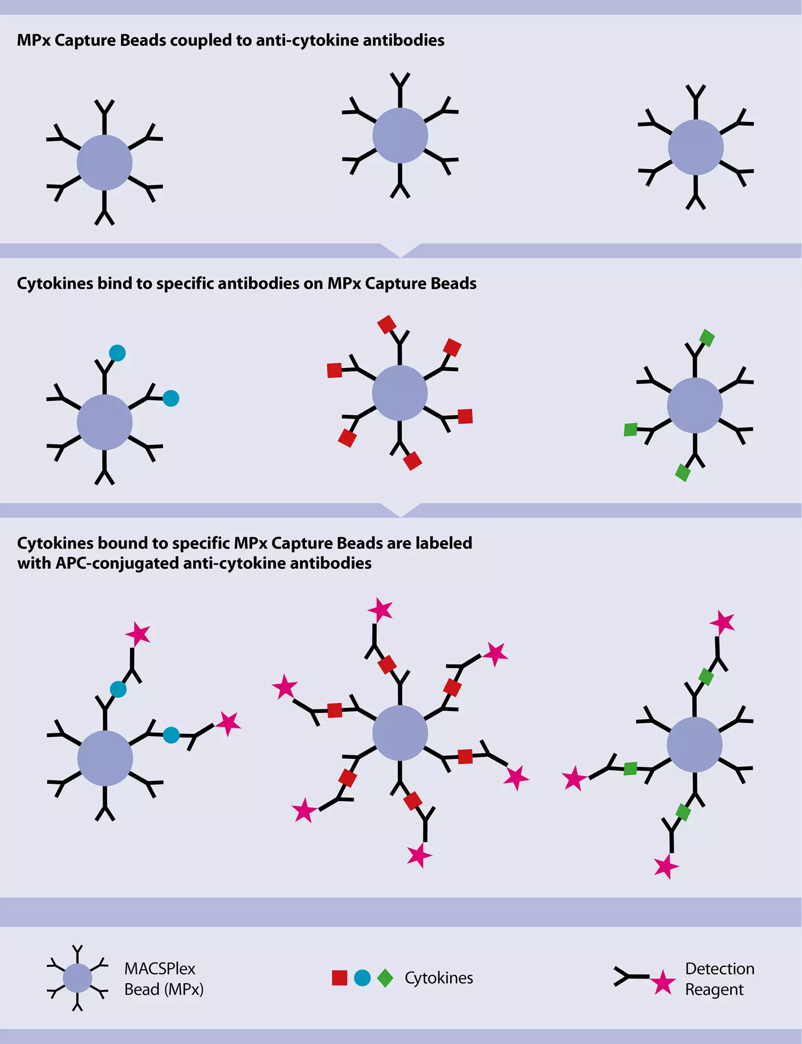
- MACSPlex Cytokine Kits are used for multiplex analysis of secreted cytokines in serum and cell culture supernatant, using a standard flow cytometer. Cytokines that can be analyzed in a single sample include: GM-CSF, IFN-α, IFN-γ, lL-2, IL-4, IL-5, IL-6, IL-9, IL-10, IL-12p70, IL-17A, and TNF-α.

2.6 Culture of NK cells
NK cells are cultured, for example, to assess their cytotoxic functions, for studies on how to enhance these functions, and to obtain larger numbers of cells for downstream applications.
MACS Handbook:
2.6.1 Cultivation and expansion of NK cells
| Use | Comments | Product |
| Culture medium | Designed to promote NK cell proliferation and activation, with minimal growth of unwanted cells (T cells, B cells, DCs, etc.) | NK MACS Medium |
| Supplement | Consistent, high-quality recombinant cytokines for successful cell culture. Available in premium, research, and GMP grades. | MACS Cytokines |
| Stimulation | Based on cell-sized particles loaded with activating antibodies. | NK Cell Activation/Expansion Kit, human |
NK MACS Medium, research grade is a cell culture medium developed specifically for NK cells. It has been used in a variety of applications and, in combination with MACS Cytokines, is an ideal starting point for reliable cultivation conditions.

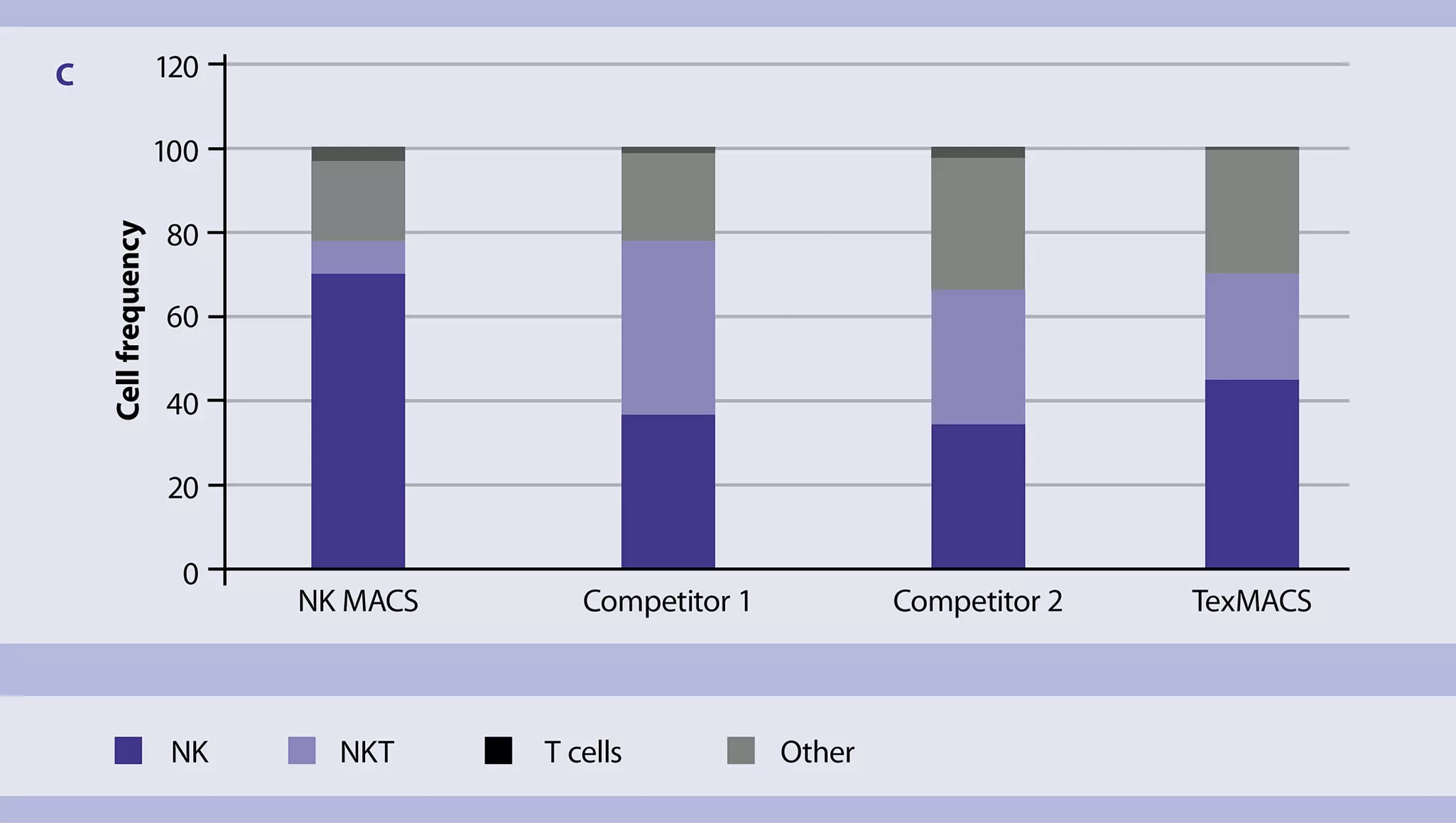
For detailed information about Miltenyi Biotec media optimized for NK cells, see the MACS Handbook chapter Cell culture.
NK cell activation is essential for a variety of downstream applications. Miltenyi Biotec offers polyclonal stimulation reagents that have been carefully designed to ensure optimal stimulation conditions.
The NK Cell Activation/Expansion Kit, human is based on large cell-sized particles loaded with biotinylated antibodies against CD2 and CD335 (NKp46) to activate and expand primary cells. These particles mimic antigen-presenting cells and, when applied at a specific bead-to-cell ratio, lead to efficient NK cell activation.
NK Cell Activation/Expansion Kit, human

NK cell expansion with the NK Cell Activation/Expansion Kit. Starting with NK cells isolated with the NK Cell Isolation Kit, human (n = 2), cells were stimulated at day 1 using the NK Cell Activation/Expansion Kit, human or left unstimulated by culturing in medium with IL‑2 alone.
3 NK cells from other tissues (lymphoid, non-lymphoid)
Although CD56dim NK cells are the predominant subset in blood, CD56bright NK cells are far more abundant in the human body due to their enrichment in lymphoid and non-lymphoid tissues. However, their proportions vary greatly from only a small percentage of total lymphocytes in secondary lymphoid organs to up to 50% or more of all lymphocytes in some organs, such as the uterus (PMID: 27121652).
NK cells are also known to infiltrate tumor tissue to varying degrees, depending on the type of tumor. For more information about tumor-infiltrating lymphocytes, see the MACS Handbook chapters Tumor tissue (human) and Tumor cells (human).
MACS Handbook:
You have previously chosen International as your location.
What do you want to do?