Employees in clean room attire having a conversation in a lab
Logo

Custom applications for automated cell manufacturing on the CliniMACS Prodigy®

Advance your transition from research to preclinical and clinical studies with an automated, GMP-compliant workflow within a closed, integrated system.

Our Custom Application (CAP) Services team partners with you to translate your manual protocols into a streamlined, tailor-made procedure on the CliniMACS Prodigy that meets your specific requirements. Every step is developed based on decades of experience in cell manufacturing and supported by rigorous workflow design and controlled documentation.

Why partner with us?

Icon transparent tailer made applications

Tailor-made applications that perfectly fit your requirements

Icon transparent engineering expertise

Engineering expertise for optimized and reproducible processes

Icon transparent easy implementation

Smooth implementation with installation and hands-on user introduction

Icon transparent compliance

Compliance made simple with GMP-supportive documentation

Discover what´s possible

From sample preparation to final product formulation, every step of your workflow can be customized to support the cell therapy of your choice. Benefit from decades of engineering expertise, deep technological insight, as well as comprehensive guidance and documentation throughout your project.

Let’s talk about your goals. Whether you are scaling up, tackling specific challenges such as formulation, or integrating external devices, we are here to help you overcome technical limitations and bring your cell therapy closer to clinical manufacturing.

Good to know: Our MACSQuant® Analyzers are the perfect fit for in-process and quality control testing. With the Custom Express Modes software add-on, you can simplify and automate flow cytometric analysis. These preprogrammed applications include predefined experiment settings, automated acquisition, and data analysis to support reproducible results with minimal hands-on effort.

Infographis showing a mixture of different cells in a cell culture bag

Preparation

Choose a starting material (fresh or frozen):
  • Leukapheresis product (mobilized or non-mobilized)
  • Whole blood
  • Buffy coat
  • Bone marrow
  • Cord blood
  • Dissociated tissue
  • Cultured cells
Define processing steps:
  • Density gradient centrifugation
  • Red blood cell reduction
  • Cell wash, concentration, and/or cell labeling
Infographis showing a mixture of different cells in a cell culture bag

Preparation

Choose a starting material (fresh or frozen):
  • Leukapheresis product (mobilized or non-mobilized)
  • Whole blood
  • Buffy coat
  • Bone marrow
  • Cord blood
  • Dissociated tissue
  • Cultured cells
Define processing steps:
  • Density gradient centrifugation
  • Red blood cell reduction
  • Cell wash, concentration, and/or cell labeling
Infographic demonstrating the magnetic field for column based magnetic cell sepataion

Separation

Design your strategy:
  • Target cell enrichment
  • Non-target cell depletion
  • Combination strategies
Choose target cells:
  • T cells (CD4+, CD8+, CD62L+, T regs)
  • NK cells
  • HSCs
  • iPSCs
Optimize key parameters including:
  • Purity
  • Recovery
  • Process time
Infographic demonstrating the magnetic field for column based magnetic cell sepataion

Separation

Design your strategy:
  • Target cell enrichment
  • Non-target cell depletion
  • Combination strategies
Choose target cells:
  • T cells (CD4+, CD8+, CD62L+, T regs)
  • NK cells
  • HSCs
  • iPSCs
Optimize key parameters including:
  • Purity
  • Recovery
  • Process time
Infographic showing viral transduction of a cell in a process generating CAR T cells

Genetic modification

Select a method:
  • Viral transduction (e.g. lentiviral vectors)
  • Electroporation with flexible parameters
Infographic showing viral transduction of a cell in a process generating CAR T cells

Genetic modification

Select a method:
  • Viral transduction (e.g. lentiviral vectors)
  • Electroporation with flexible parameters
Infographic showing the expansion of CAR T cells

Cell culture (adherent or suspension)

Customize culture conditions:
  • Media exchange
  • Cell wash
  • Addition of feed, reagents, and cytokines
  • Cell activation and expansion
  • External culture vessel integration
  • In-process control sample collection
  • Microscopic imaging
Infographic showing the expansion of CAR T cells

Cell culture (adherent or suspension)

Customize culture conditions:
  • Media exchange
  • Cell wash
  • Addition of feed, reagents, and cytokines
  • Cell activation and expansion
  • External culture vessel integration
  • In-process control sample collection
  • Microscopic imaging
Infographic showing CAR T cells in a cell culture bag

Final product formulation

Set formulation and filling parameters:
  • Choice of medium (fresh or cryopreservation)
  • Number, size, and type of final product bags
  • Final cell concentration and volume
  • Cell seeding and harvesting (partial, complete, or transfer to external culture devices)
Infographic showing CAR T cells in a cell culture bag

Final product formulation

Set formulation and filling parameters:
  • Choice of medium (fresh or cryopreservation)
  • Number, size, and type of final product bags
  • Final cell concentration and volume
  • Cell seeding and harvesting (partial, complete, or transfer to external culture devices)
Scheme of flow cytometer MACSQuant 16 for multiparameter cell analysis

Need automated IPC/QC?

Customize Express Modes for quick, automated flow gating, data reporting, and export
Scheme of flow cytometer MACSQuant 16 for multiparameter cell analysis

Need automated IPC/QC?

Customize Express Modes for quick, automated flow gating, data reporting, and export
Infographis showing a mixture of different cells in a cell culture bag

Preparation

Choose a starting material (fresh or frozen):
  • Leukapheresis product (mobilized or non-mobilized)
  • Whole blood
  • Buffy coat
  • Bone marrow
  • Cord blood
  • Dissociated tissue
  • Cultured cells
Define processing steps:
  • Density gradient centrifugation
  • Red blood cell reduction
  • Cell wash, concentration, and/or cell labeling
Infographis showing a mixture of different cells in a cell culture bag

Preparation

Choose a starting material (fresh or frozen):
  • Leukapheresis product (mobilized or non-mobilized)
  • Whole blood
  • Buffy coat
  • Bone marrow
  • Cord blood
  • Dissociated tissue
  • Cultured cells
Define processing steps:
  • Density gradient centrifugation
  • Red blood cell reduction
  • Cell wash, concentration, and/or cell labeling
Infographic demonstrating the magnetic field for column based magnetic cell sepataion

Separation

Design your strategy:
  • Target cell enrichment
  • Non-target cell depletion
  • Combination strategies
Choose target cells:
  • T cells (CD4+, CD8+, CD62L+, T regs)
  • NK cells
  • HSCs
  • iPSCs
Optimize key parameters including:
  • Purity
  • Recovery
  • Process time
Infographic demonstrating the magnetic field for column based magnetic cell sepataion

Separation

Design your strategy:
  • Target cell enrichment
  • Non-target cell depletion
  • Combination strategies
Choose target cells:
  • T cells (CD4+, CD8+, CD62L+, T regs)
  • NK cells
  • HSCs
  • iPSCs
Optimize key parameters including:
  • Purity
  • Recovery
  • Process time
Infographic showing viral transduction of a cell in a process generating CAR T cells

Genetic modification

Select a method:
  • Viral transduction (e.g. lentiviral vectors)
  • Electroporation with flexible parameters
Infographic showing viral transduction of a cell in a process generating CAR T cells

Genetic modification

Select a method:
  • Viral transduction (e.g. lentiviral vectors)
  • Electroporation with flexible parameters
Infographic showing the expansion of CAR T cells

Cell culture (adherent or suspension)

Customize culture conditions:
  • Media exchange
  • Cell wash
  • Addition of feed, reagents, and cytokines
  • Cell activation and expansion
  • External culture vessel integration
  • In-process control sample collection
  • Microscopic imaging
Infographic showing the expansion of CAR T cells

Cell culture (adherent or suspension)

Customize culture conditions:
  • Media exchange
  • Cell wash
  • Addition of feed, reagents, and cytokines
  • Cell activation and expansion
  • External culture vessel integration
  • In-process control sample collection
  • Microscopic imaging
Infographic showing CAR T cells in a cell culture bag

Final product formulation

Set formulation and filling parameters:
  • Choice of medium (fresh or cryopreservation)
  • Number, size, and type of final product bags
  • Final cell concentration and volume
  • Cell seeding and harvesting (partial, complete, or transfer to external culture devices)
Infographic showing CAR T cells in a cell culture bag

Final product formulation

Set formulation and filling parameters:
  • Choice of medium (fresh or cryopreservation)
  • Number, size, and type of final product bags
  • Final cell concentration and volume
  • Cell seeding and harvesting (partial, complete, or transfer to external culture devices)
Scheme of flow cytometer MACSQuant 16 for multiparameter cell analysis

Need automated IPC/QC?

Customize Express Modes for quick, automated flow gating, data reporting, and export
Scheme of flow cytometer MACSQuant 16 for multiparameter cell analysis

Need automated IPC/QC?

Customize Express Modes for quick, automated flow gating, data reporting, and export

The Custom Application journey

At every stage, we work closely with you to understand your specific needs and transform your manual processes into an optimized, automated workflow. Our modular approach enables a smooth transition from preclinical research to commercial manufacturing, balancing flexibility, speed, and cost efficiency.

  • Preclinical research: Based on your requirements we develop a CAP to establish a robust process foundation.
  • Clinical trials: Conduct early testing with our optional Risk Assessment Module to support a safe and compliant path to clinical evaluation.
  • Clinical and commercial manufacturing: Transition to a GMP-supportive CAP for commercial production with fixed parameters, comprehensive GMP documentation, and no added license fees.
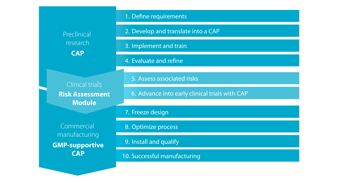
Image showing the workflow of our Custom Application (CAP) Service where we translate the customer manual protocol into a streamlined, tailor-made procedure on the CliniMACS Prodigy

Quality and safety you can rely on

Icons for Iso 9001 and Gamp

We uphold the highest international quality standards (ISO) to ensure consistency and reproducibility across every project. Regular audits confirm our compliance and commitment to quality excellence.

Our software development follows GAMP5 guidelines, ensuring risk-based validation and robust documentation of automated systems in pharmaceutical manufacturing.

With quality management practices aligned to GAMP5 principles, we deliver efficient, reliable, and fully documented software solutions you can trust.

Discover what’s possible

You have previously chosen International as your location.
What do you want to do?