Illustration of a dendritic cell
Logo

Efficient macrophage polarization and characterization: M1/M2 states, markers, and assays

Monocyte-derived macrophages are immune cells which derive from the differentiation of circulating monocytes and can adopt different phenotypes. M0 macrophages are characterized as naive and can polarize either into proinflammatory M1 or anti-inflammatory M2 macrophages. M1 macrophages have the ability to secrete proinflammatory cytokines and participate in inflammation, while M2 macrophages can phagocytose apoptotic cells and contribute to tissue remodeling.

Macrophages are a widely studied cell type. However, isolating, differentiating, and characterizing M1 and M2 macrophages in a consistent and reproducible manner remains a challenge.

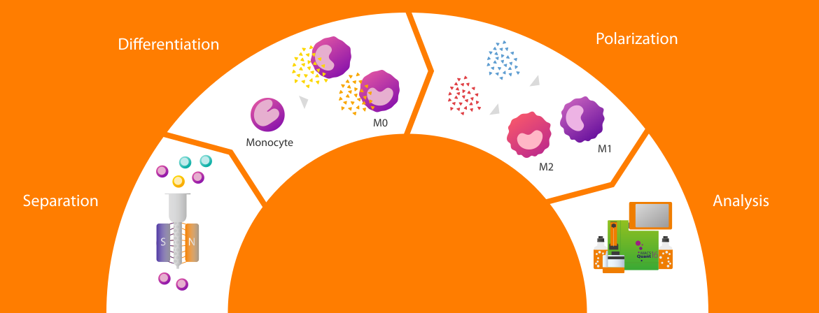
We offer a complete suite of tools designed to streamline every step: From monocyte isolation to macrophage polarization and phenotypic profiling including applications in preclinical stages of monocyte and macrophage cell therapy development 1. Whether you're studying inflammation, cancer, or tissue regeneration, our integrated workflow empowers your monocyte-derived macrophage (MDM) research with speed and precision.

A workflow for macrophage polarization and characterization

Magnetic isolation

Efficient magnetic cell separation yields a highly pure starting monocyte cell population for reliable downstream processing (e.g., efficient macrophage polarization).
See details
Illustration showing cell separation process for monocytes with Miltenyi Biotec columns.

Magnetic isolation

Efficient magnetic cell separation yields a highly pure starting monocyte cell population for reliable downstream processing (e.g., efficient macrophage polarization).
See details

Differentiation

MACS® Cytokines M‑CSF and GM‑CSF ensure reliable consistency through lot‑specific activity values, supporting efficient and reproducible differentiation of monocytes into M0 macrophages
See details
Illustration showing the differentiation from monocytes to macrophages using Miltenyi Biotec cytokines.

Differentiation

MACS® Cytokines M‑CSF and GM‑CSF ensure reliable consistency through lot‑specific activity values, supporting efficient and reproducible differentiation of monocytes into M0 macrophages
See details

M1/M2 Polarization

MACS® Cytokines ensure reliable consistency through lot‑specific activity values, supporting efficient and reproducible polarization of M0 into distinct M1 or M2 phenotypes.
See details
Illustration of polarization of two macrophage M1 and M2 using two different cytokines from Miltenyi Biotec.

M1/M2 Polarization

MACS® Cytokines ensure reliable consistency through lot‑specific activity values, supporting efficient and reproducible polarization of M0 into distinct M1 or M2 phenotypes.
See details

Flow analysis

Flow cytometry phenotyping of polarized macrophages, accurately distinguishing M1 or M2 phenotypes is achieved with REAfinity® Recombinant Antibodies.
See details
Illustration showing the MACS Quant Analyzer 16 for efficient flow cytometry.

Flow analysis

Flow cytometry phenotyping of polarized macrophages, accurately distinguishing M1 or M2 phenotypes is achieved with REAfinity® Recombinant Antibodies.
See details
Workflow illustration step-by-step Miltenyi Biotec instruments and reagents needed for monocyte-derived macrophage polarization and characterization.

Magnetic isolation

Efficient magnetic cell separation yields a highly pure starting monocyte cell population for reliable downstream processing (e.g., efficient macrophage polarization).
See details
Illustration showing cell separation process for monocytes with Miltenyi Biotec columns.

Magnetic isolation

Efficient magnetic cell separation yields a highly pure starting monocyte cell population for reliable downstream processing (e.g., efficient macrophage polarization).
See details

Differentiation

MACS® Cytokines M‑CSF and GM‑CSF ensure reliable consistency through lot‑specific activity values, supporting efficient and reproducible differentiation of monocytes into M0 macrophages
See details
Illustration showing the differentiation from monocytes to macrophages using Miltenyi Biotec cytokines.

Differentiation

MACS® Cytokines M‑CSF and GM‑CSF ensure reliable consistency through lot‑specific activity values, supporting efficient and reproducible differentiation of monocytes into M0 macrophages
See details

M1/M2 Polarization

MACS® Cytokines ensure reliable consistency through lot‑specific activity values, supporting efficient and reproducible polarization of M0 into distinct M1 or M2 phenotypes.
See details
Illustration of polarization of two macrophage M1 and M2 using two different cytokines from Miltenyi Biotec.

M1/M2 Polarization

MACS® Cytokines ensure reliable consistency through lot‑specific activity values, supporting efficient and reproducible polarization of M0 into distinct M1 or M2 phenotypes.
See details

Flow analysis

Flow cytometry phenotyping of polarized macrophages, accurately distinguishing M1 or M2 phenotypes is achieved with REAfinity® Recombinant Antibodies.
See details
Illustration showing the MACS Quant Analyzer 16 for efficient flow cytometry.

Flow analysis

Flow cytometry phenotyping of polarized macrophages, accurately distinguishing M1 or M2 phenotypes is achieved with REAfinity® Recombinant Antibodies.
See details

1. Monocyte isolation

We offer a variety of solutions that enable the isolation of monocytes with very high purity, recovery, and viability. Whether you are isolating monocytes from PBMCs or directly from blood, we have the right solution for you. We provide the appropriate kit to cover your specific needs, whether you are interested in pan monocytes, CD14+ or CD16+ monocytes.

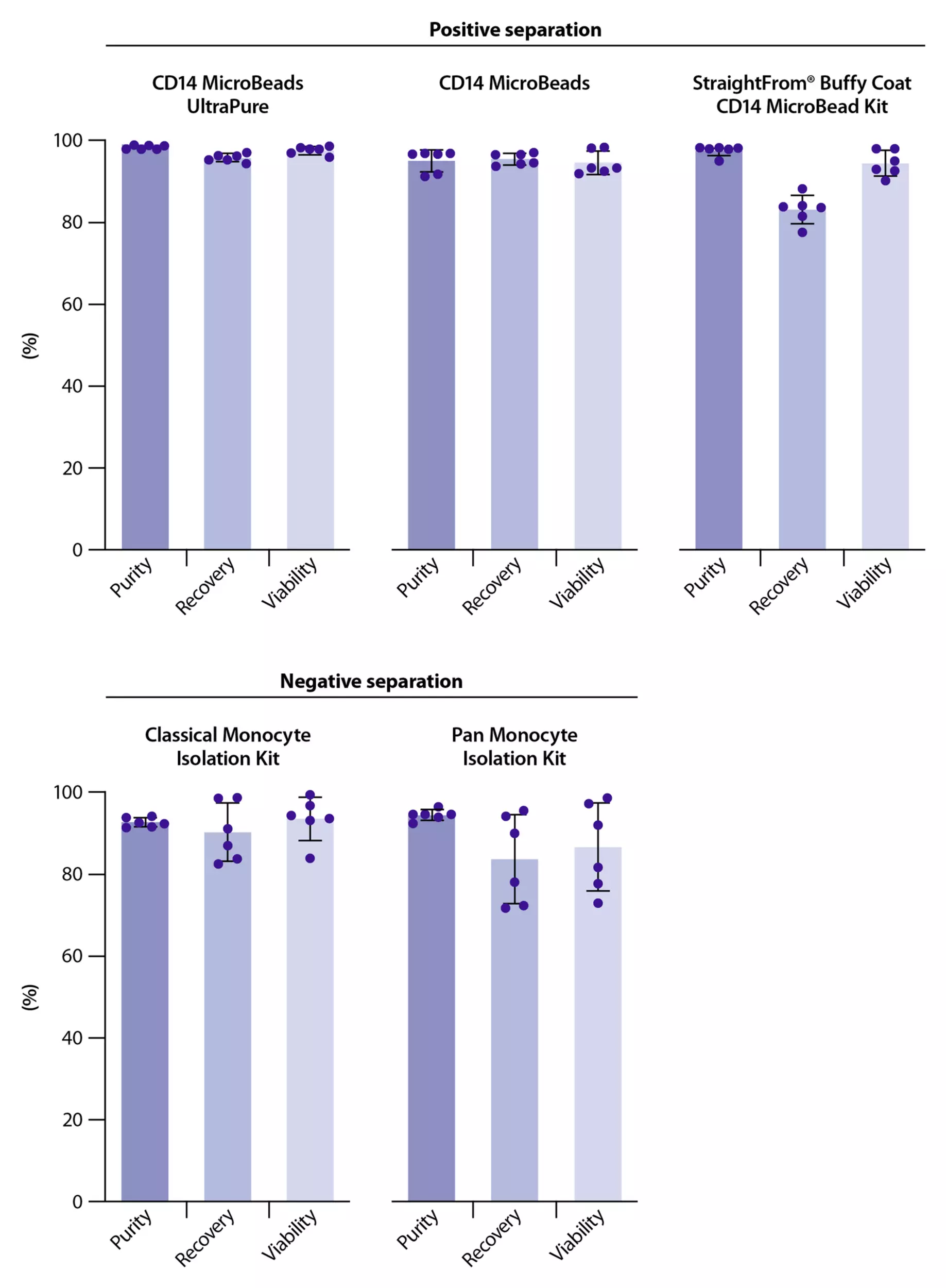
Barchart illustration showing performance of monocyte with multiple separation solutions from Miltenyi Biotec.
High cell purity, recovery, and viability is retained with each monocyte cell separation kit.

Monocytes can be isolated via positive selection or negative isolation from PBMCs using our MicroBeads or Isolation Kits respectively. Also, monocytes can be directly isolated from whole blood, buffy coat, and Leukopak® using our StraightFrom® Technology ensuring excellent purity, recovery, and viability. If you need assistance choosing the right separation kit, you can find an overview of all our solutions in the table below.

The magnetic isolation process can be achieved manually using the appropriate MACS® Columns or in a semi-automated process by utilizing the MultiMACS™ Cell24 Separator Plus. For a fully automated process the autoMACS® NEO Separator is the appropriate solution.

Cell separation methods for isolation of human monocytes

Product nameIsolationStarting materialTarget cellsCell capacity
CD14 MicroBeads, human and
CD14 MicroBeads, Ultrapure, human
positivePBMCsCD14+ monocytes1 × 109
CD16 MicroBeads, humanpositivePBMCsCD16+ monocytes2 × 109
CD11b MicroBeads, human and mousepositivePBMCs, mouse cells
Myeloid cells2 × 109
CD163 MicroBead Kit, humanpositivePBMCsMacrophages1 × 109
Pan Monocyte Isolation Kit, humanuntouchedPBMCsCD14+ and CD16+ monocytes1 × 109
Classical Monocyte Isolation Kit, humanuntouchedPBMCsCD14+ monocytes1 × 109
CD16+ Monocyte Isolation Kit, humanuntouchedPBMCsCD16+ monocytes2 × 109
StraightFrom® Whole Blood CD14 MicroBeads, humanpositiveWhole bloodCD14+ monocytes40 mL whole blood
StraightFrom® Buffy Coat CD14 MicroBead Kit, humanpositiveBuffy CoatCD14+ monocytes1 Buffy coat
StraightFrom® LRSC CD14 MicroBead Kit, humanpositiveLRSCCD14+ monocytes1 LRSC
StraightFrom® Leukopak® CD14 MicroBead Kit, humanpositiveLeukopakCD14+ monocytes½ Leukopak

2. Macrophage differentiation and polarization into M1 or M2 phenotypes

We provide ready-to-use solutions for macrophage differentiation, including M1 and M2 polarization. These options are backed by high-quality recombinant cytokines. In an in vitro setting, monocytes can be differentiated into macrophages (M0) by culturing with specific cytokines. Commonly used differentiation protocols include2:

  • GM-CSF (Granulocyte-Macrophage Colony-Stimulating Factor) which tends to drive cells toward a proinflammatory phenotype.
  • M-CSF (Macrophage Colony-Stimulating Factor) which supports the development of macrophages with anti-inflammatory and tissue-supportive characteristics.

After initial differentiation (typically over five to seven days), macrophages can be polarized into distinct functional phenotypes, as described in the table below.


Cell typeRole in the immune systemDifferentiation to M0 phenotype (five days)Polarization to M1 or M2 phenotype (two days)
M1Pathogen killing, inflammation, anti-tumor immunity
GM-CSFIFN-γ + TNF-α or IFN-γ + LPS
M2Tissue repair, immunoregulation, angiogenenesisM-CSFIL-4 + IL-13 (for M2a) or IL-10 + TGF-β (for M2c)

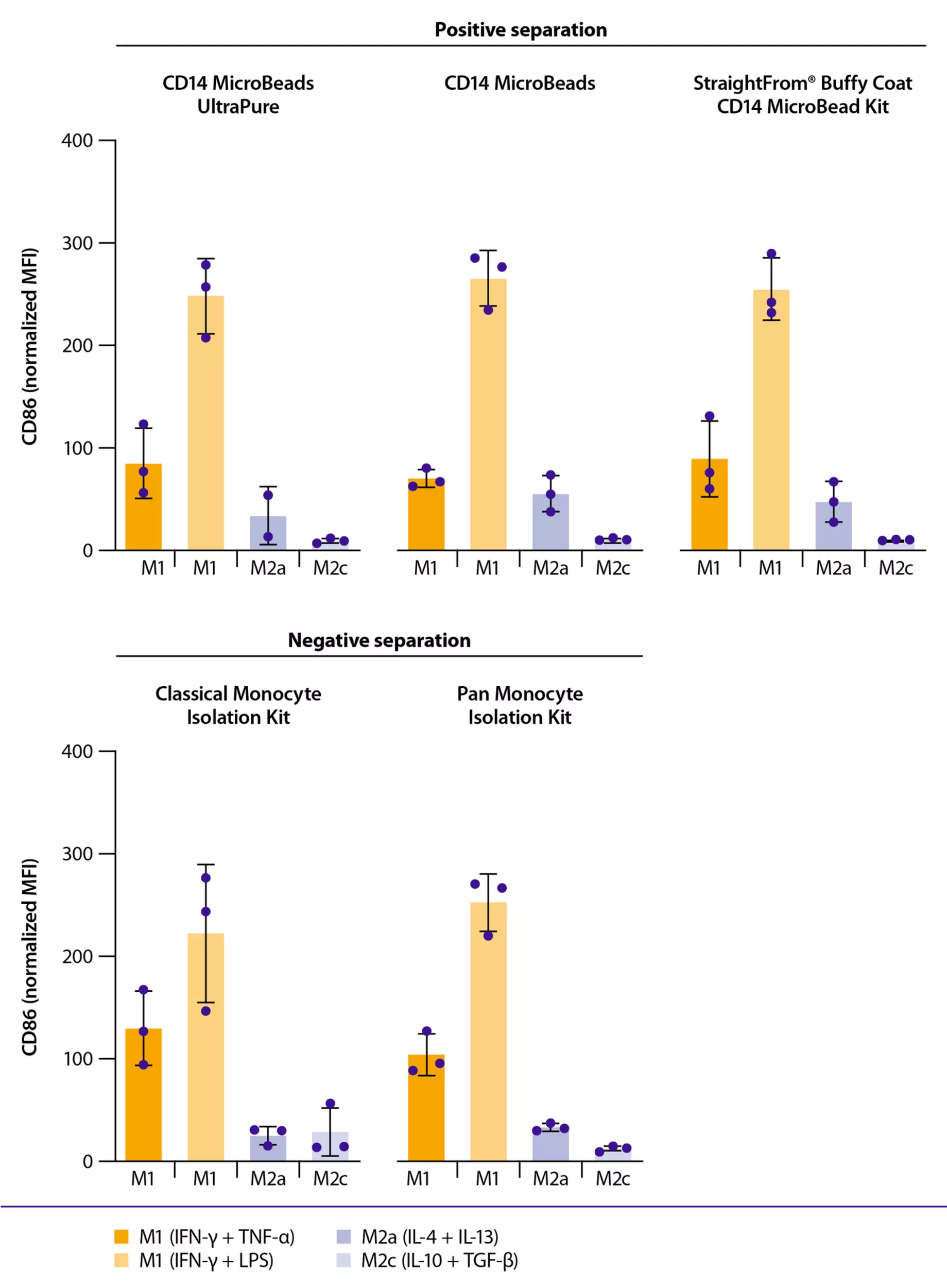
Barchart illustration showing M1
 macrophage polarization upon monocyte isolation with Miltenyi Biotec's cell separation kits.
M1 macrophage polarization upon monocyte isolation with several cell separation kits.

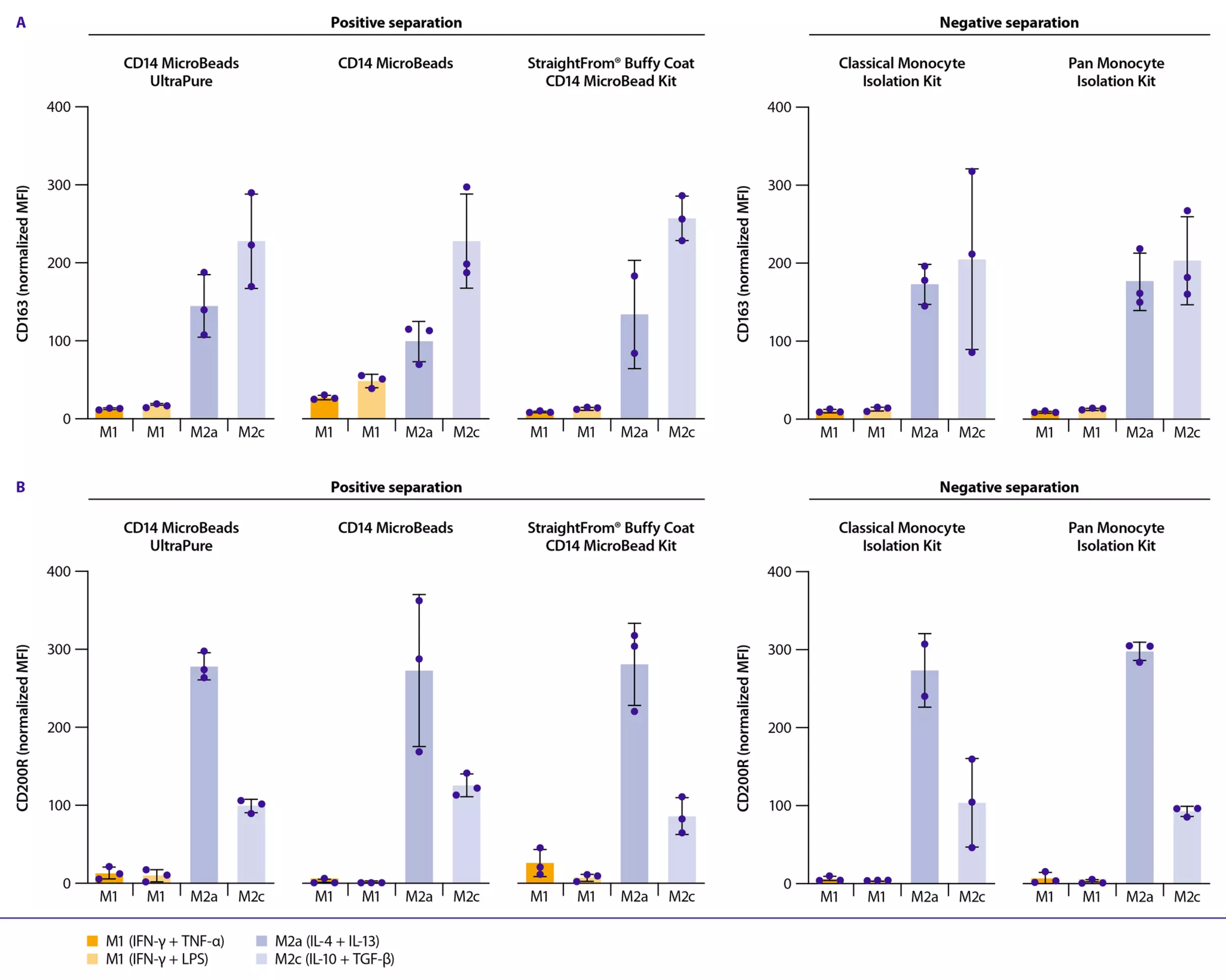
Using several of our separation kits, we demonstrate excellent M1 and M2 macrophage differentiation and polarization according to the appropriate markers expressed: CD86 expressed in M1 macrophages and CD163, CD200R expressed in M2 macrophages.

Bar chart showing M2 macrophage polarization marker in monocyte-derived macrophages, generated from monocytes isolated using different Miltenyi Biotec separation kits.
M2 macrophage polarization upon monocyte isolation with several cell separation kits.

The magnetic isolation process can be achieved manually using the appropriate MACS® Columns or in a semi-automated process by utilizing the MultiMACS Cell24 Separator Plus. For a fully automated process the autoMACS® NEO Separator is the appropriate solution.

3. Phenotypic characterization

Phenotypic characterization is essential to confirm successful macrophage polarization. This is typically performed using flow cytometry, microscopy, or gene expression analysis to assess surface marker expression. We support these workflows with high-quality recombinant REAfinity® Antibodies and the MACSQuant® Flow Cytometer, so you can achieve precise and reproducible immunophenotyping.

Macrophage polarization results in distinct marker profiles corresponding to their functional phenotypes3:

  • M1 macrophages are typically characterized by CD80, CD86, and HLA-DR.
  • M2 macrophages marker profiles include:
    • M2a macrophages: express CD209, CD200R, CD163, and CD206.
    • M2c macrophages: express high levels of CD163 and variable CD206.

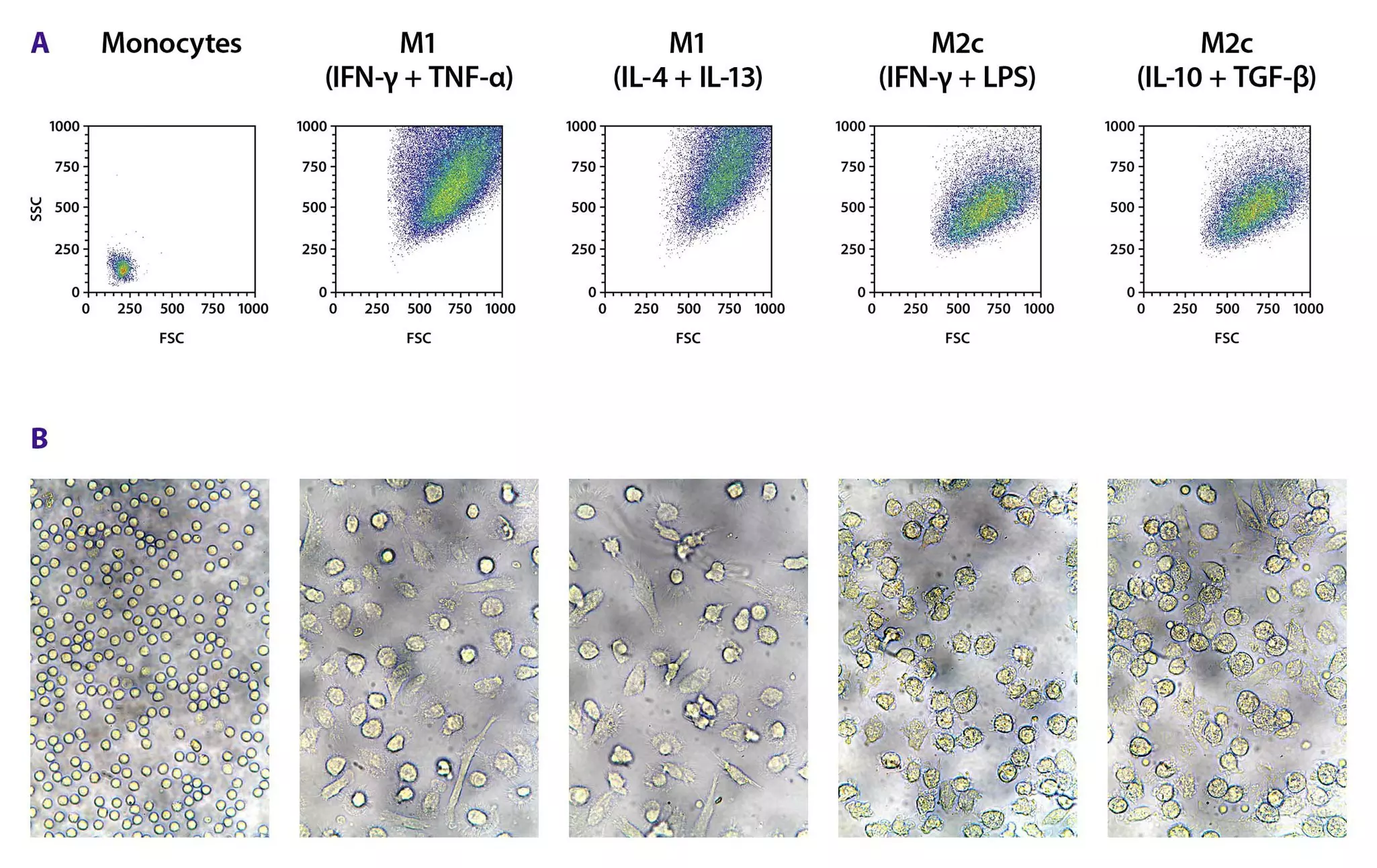
Flow cytometry and light sheet microscopy analysis of cell size and granularity showing morphological changes during monocyte-to-macrophage differentiation and polarization.
Flow cytometry and light microscopy analysis of cell size and granularity.

The different phenotypes of macrophages are evaluated according to cell size and granularity based on forward scatter (FSC) and side scatter (SSC). These methods are used after monocyte isolation with CD14 MicroBeads Ultrapure, differentiation, and polarization. A progressive increase in both parameters is observed. M1 macrophages show increased spreading and irregular shapes, while M2 macrophages exhibit a more round morphology with pronounced cytoplasmic extensions.

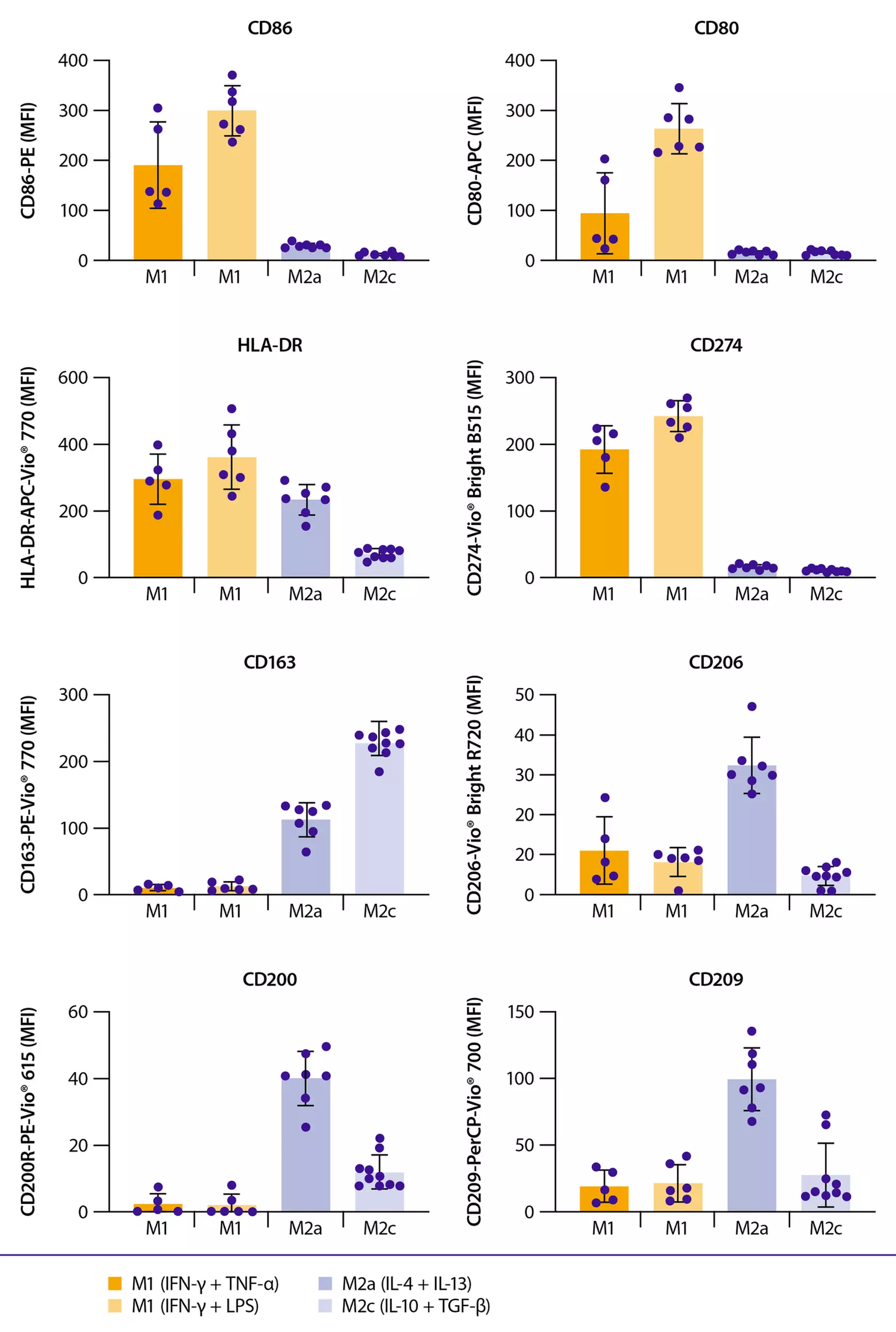
Surface marker expression after monocyte-to-macrophage differentiation and polarization using Miltenyi Biotec Antibodies and Cytokines.
Surface marker expression after monocyte-to-macrophage differentiation and polarization.

The different types of macrophages are assesed for characteristic surface marker expression with a panel of REAfinity Antibodies of CD86, CD80, HLA-DR, CD274, CD163, CD206, CD200R, and CD209. Flow cytometric analysis is performed on a MACSQuant Analyzer 16. The results demonstrate that M1 macrophages predominantly express CD86, CD80, HLA-DR, and CD274, whereas M2 macrophages show increased expression of CD163. Furthermore, the M2a subset is characterized by additional expression of CD206, CD200R, and CD209. These findings confirm the distinct phenotypic profiles of M1 and M2 macrophage subsets which can be obtained utilizing Miltenyi Biotec’s cell separation, cell culture, and cell analysis solutions.

.

4. Functional assays for macrophage activity

Phagocytosis is one of the defining functions of macrophages, including those derived from monocytes. It’s a receptor-mediated process by which macrophages engulf and degrade foreign particles, pathogens, apoptotic cells, and debris. This is essential for both innate immunity and tissue homeostasis 4.

Efferocytosis is a specialized form of phagocytosis in which phagocytic cells, primarily macrophages, recognize, engulf, and remove apoptotic cells in a controlled, non-inflammatory manner.

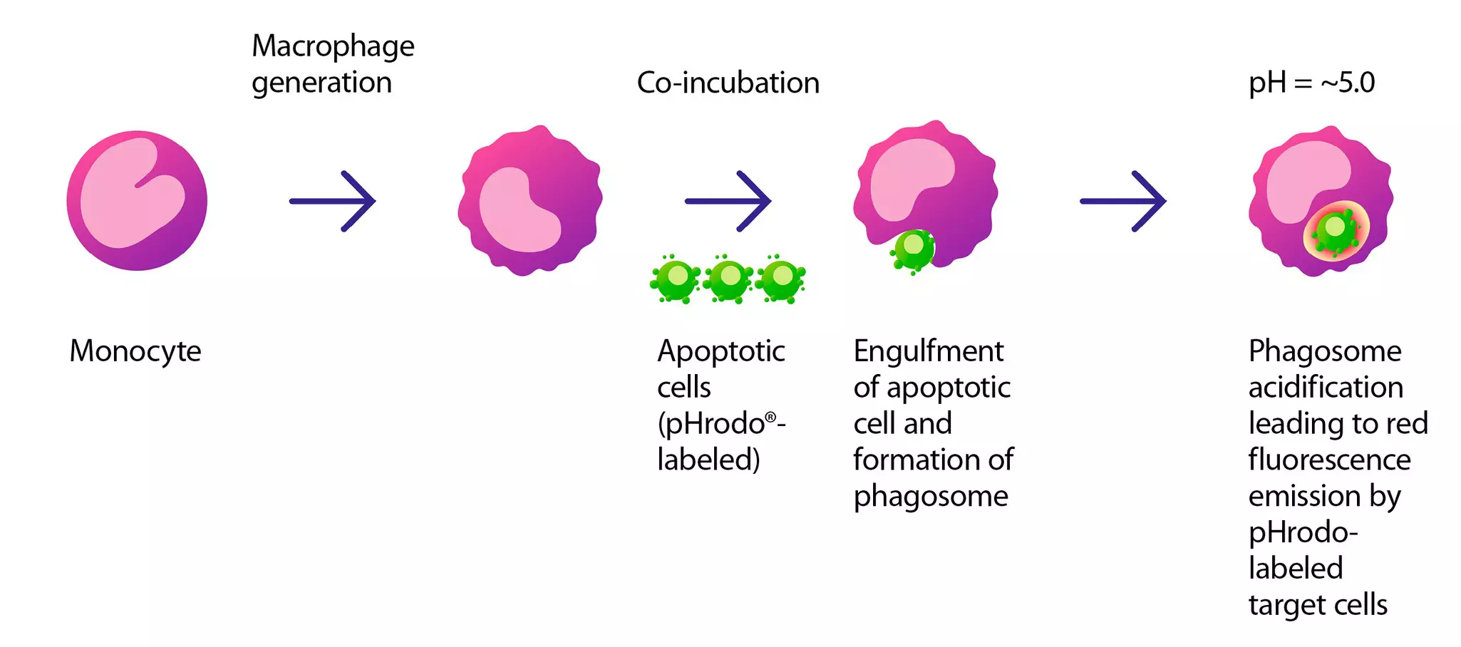
Illustration of a schematic efferocytosis assay, showing surface marker expression after monocyte-to-macrophage differentiation and polarization.
Principle of efferocytosis assay. Macrophages engulf pHrodo Red–labeled apoptotic cells. Subsequent acidification of the phagosome leads to red fluorescence emission, which can be detected by fluorescence microscopy.

This process can be monitored using pH-sensitive fluorescent dyes such as pHrodo® Red. Upon recognition of apoptotic targets, macrophages extend actin-rich pseudopodia to engulf the cells, forming phagosomes that subsequently fuse with lysosomes, triggering pHrodo fluorescence indicative of efferocytosis as shown below with exemplary data.

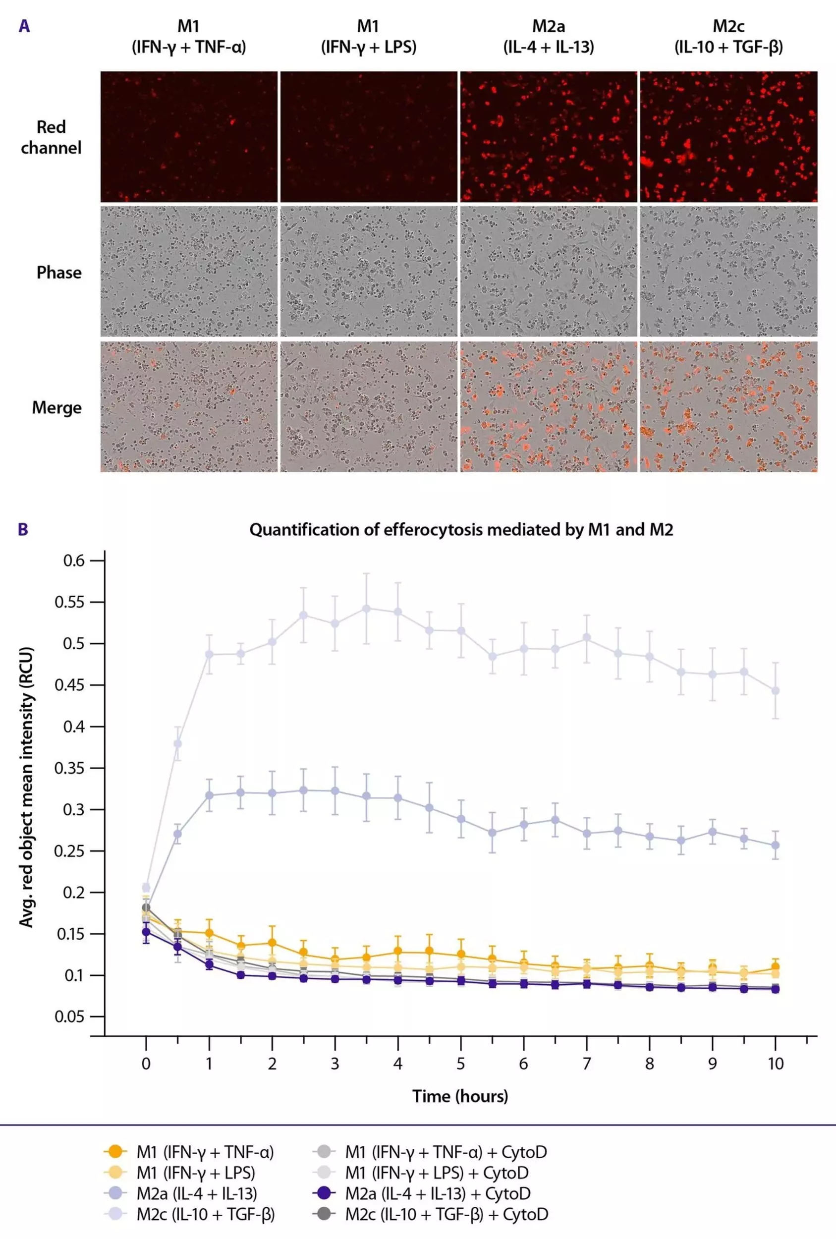
Immunofluorescene and histogram assessment of efferocytic capacity of M2c macrophages.
Efferocytic capacity of M1 and M2 macrophages.

Monocytes were isolated from PBMCs using CD14 MicroBeads Ultrapure, and differentiated and polarized into M1 and M2 macrophages. Their efferocytic capacity was subsequently assessed as a functional readout. The results demonstrate a markedly enhanced efferocytic capacity of M2 macrophage subsets compared to M1 macrophages. This is consistent with their established anti-inflammatory and tissue-repair phenotypes.

Illustration of a schematic antibody-dependent cellular phagocytosis of M2c macrophages.
Illustration of macrophage ADCP activity.

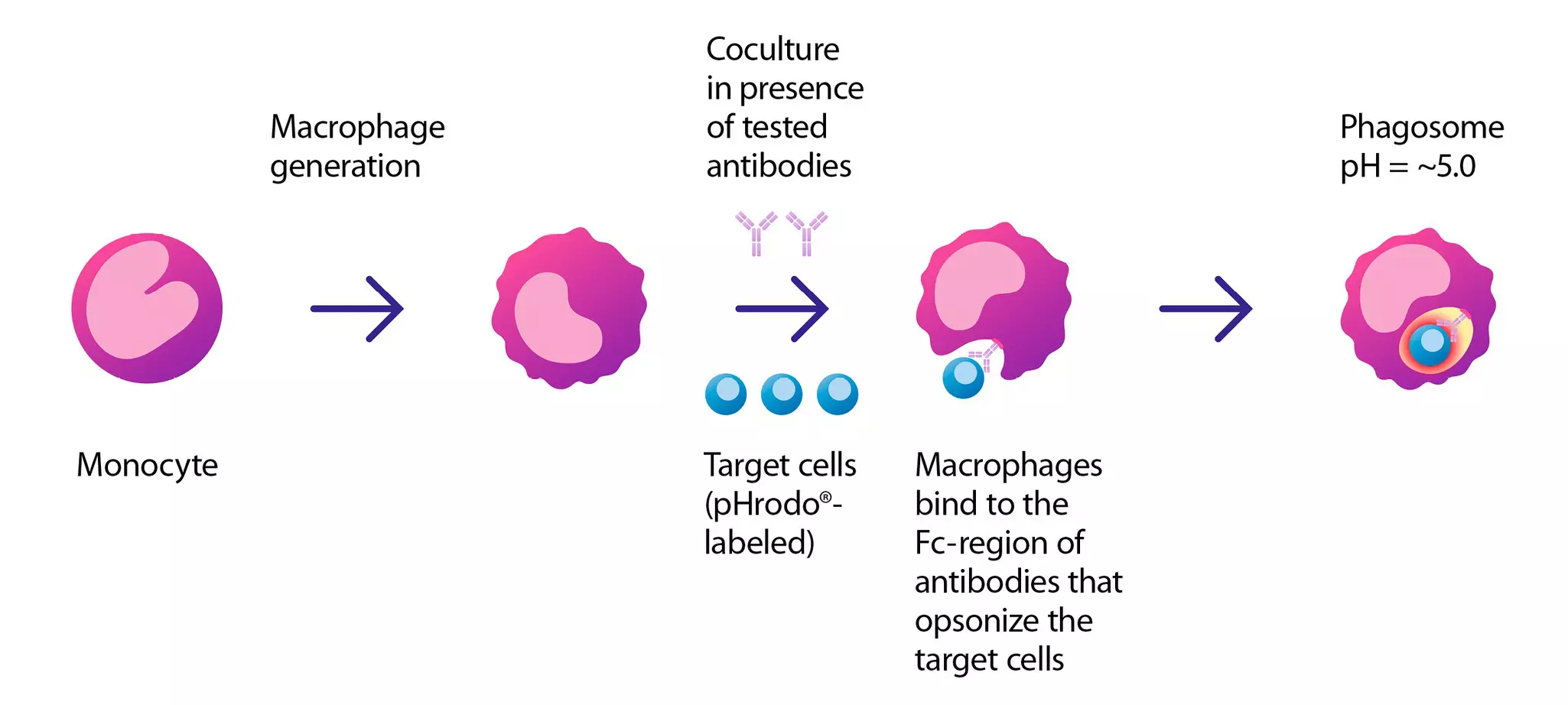
Antibody-dependent cellular phagocytosis (ADCP) is also an indicator of M2c macrophage functionality. Labeled target cells are opsonized with specific antibodies, enabling their recognition by Fc receptors on macrophages. Following antibody-dependent binding, macrophages internalize the targets via phagocytosis, forming phagosomes that progressively acidify (pH ≈ 5.0) upon fusion with lysosomes. This acidification activates pHrodo® fluorescence, generating a strong red signal indicative of successful internalization and phagosomal maturation.

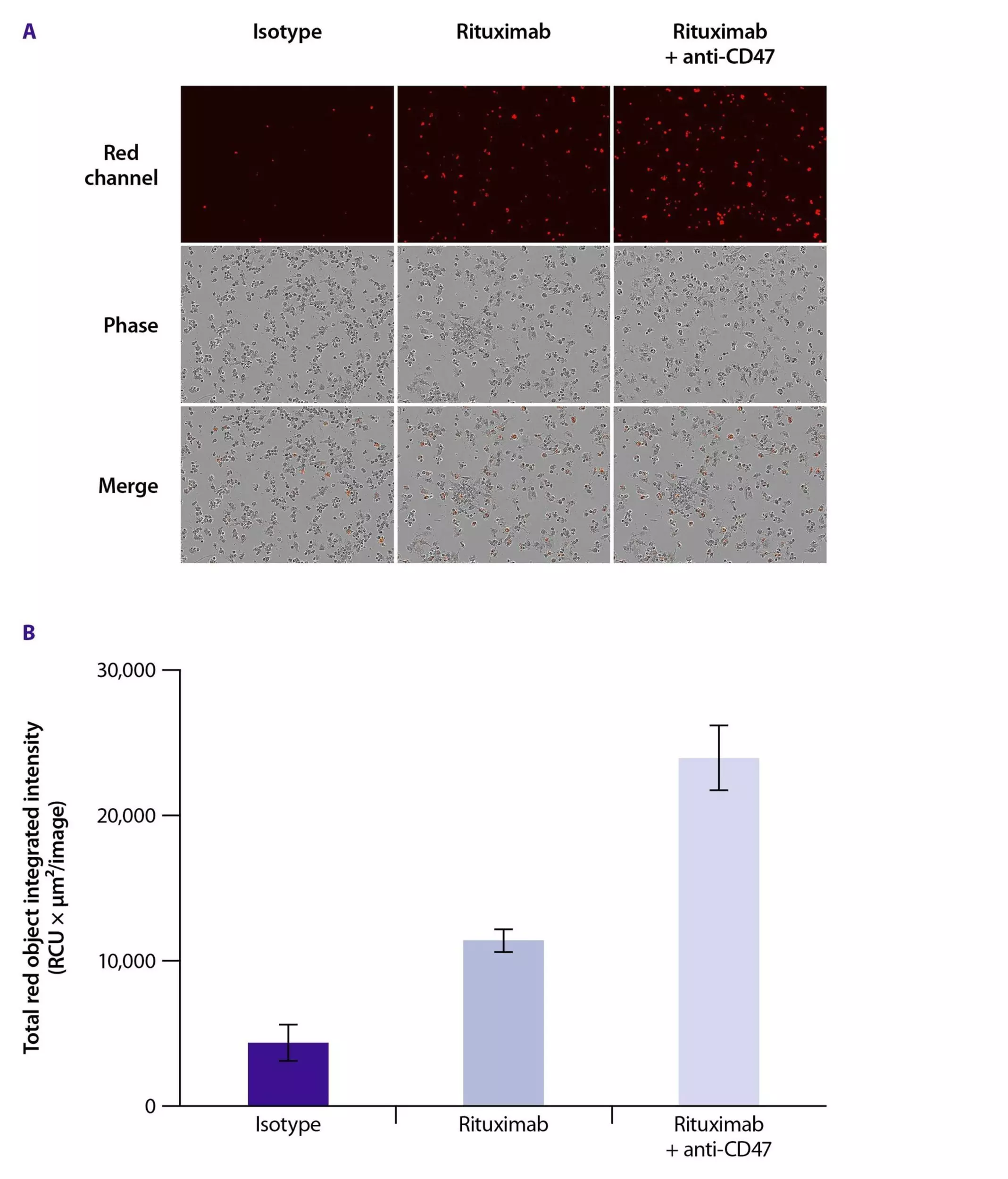
ADCP capacity of M2c macrophages. Immunofluorescene and barchart of antibody-dependent cellular phagocytosis capacity of M2c macrophages.
ADCP capacity of M2c macrophages.

M2c macrophage–mediated uptake of pHrodo® Red–labeled Jeko-1 target cells in the presence of the following antibodies: Isotype control, Rituximab, and Rituximab + anti-CD47. The top row shows the red fluorescence channel, visualizing pHrodo signal that becomes activated upon cell phagocytosis and phagosome acidification. The middle row displays phase-contrast images of macrophages and target cells. The bottom row presents merged overlays of both channels. Enhanced red fluorescence intensity correlates with higher levels of antibody-dependent target internalization and phagosome maturation, reflecting increased ADCP activity as it is shown in the coculture conditions containing Rituximab and the antagonistic anti-CD47 antibody.

Additional step-by-step protocol

Generation of Mo-DCs

This application protocol describes the generation of monocyte-derived dendritic cells (Mo-DCs). Using high-quality reagents that ensure cell viability, the resulting Mo-DCs can be used for downstream phenotypic and functional characterization.

See the details

Shop all products for this application

20 products found

|

You have previously chosen International as your location.
What do you want to do?