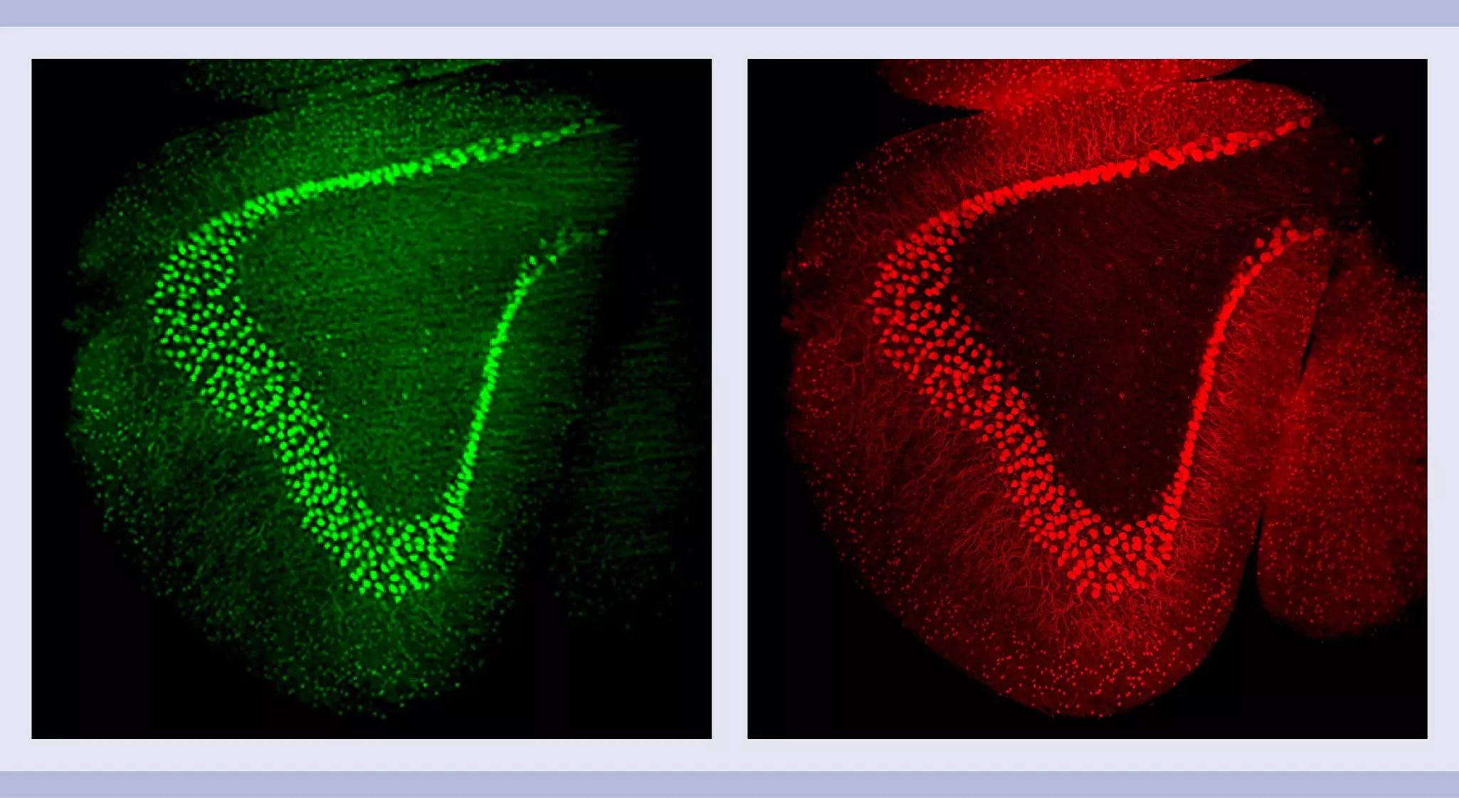
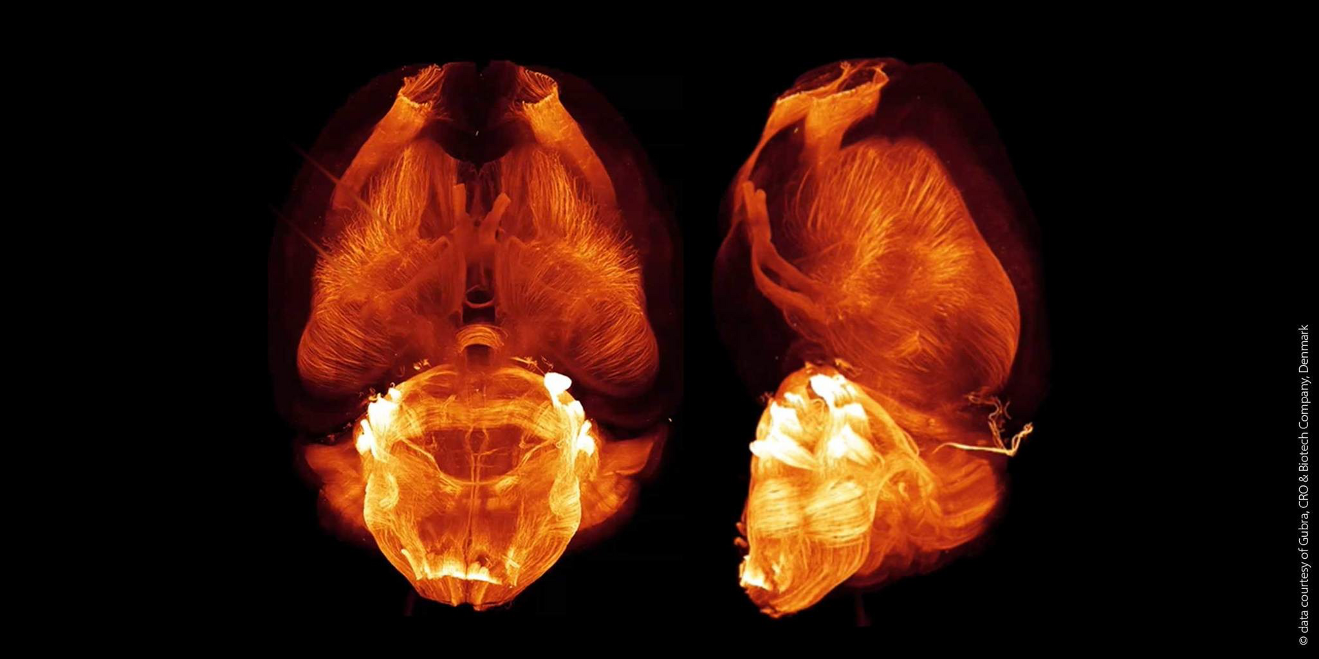
While tissues hold a wealth of information, their natural opacity makes it difficult to see beyond the surface. This is where tissue clearing comes in. Tissue clearing refers to a group of techniques that make biological tissues transparent to analyze them using microscopy techniques like confocal or light sheet microscopy.
Why do we need cleared tissue?
Thick tissues are opaque because light scatters as it travels through the different components like carbohydrates, proteins, lipids, and other pigmented molecules. Tissue clearing tackles this problem by making the entire tissue transparent. This allows scientists to see structures and interactions throughout the whole sample, not just a thin slice.
Tissue clearing vs. slicing
Traditionally, scientists would slice tissues into thin sections for study under a microscope. However, this method can damage the delicate structures and distort the natural 3D arrangement of cells, resulting in data compromise and inaccurate 3D construction of the tissue morphology. Tissue clearing avoids these issues by allowing researchers to image the entire tissue intact.
Is tissue clearing complicated?
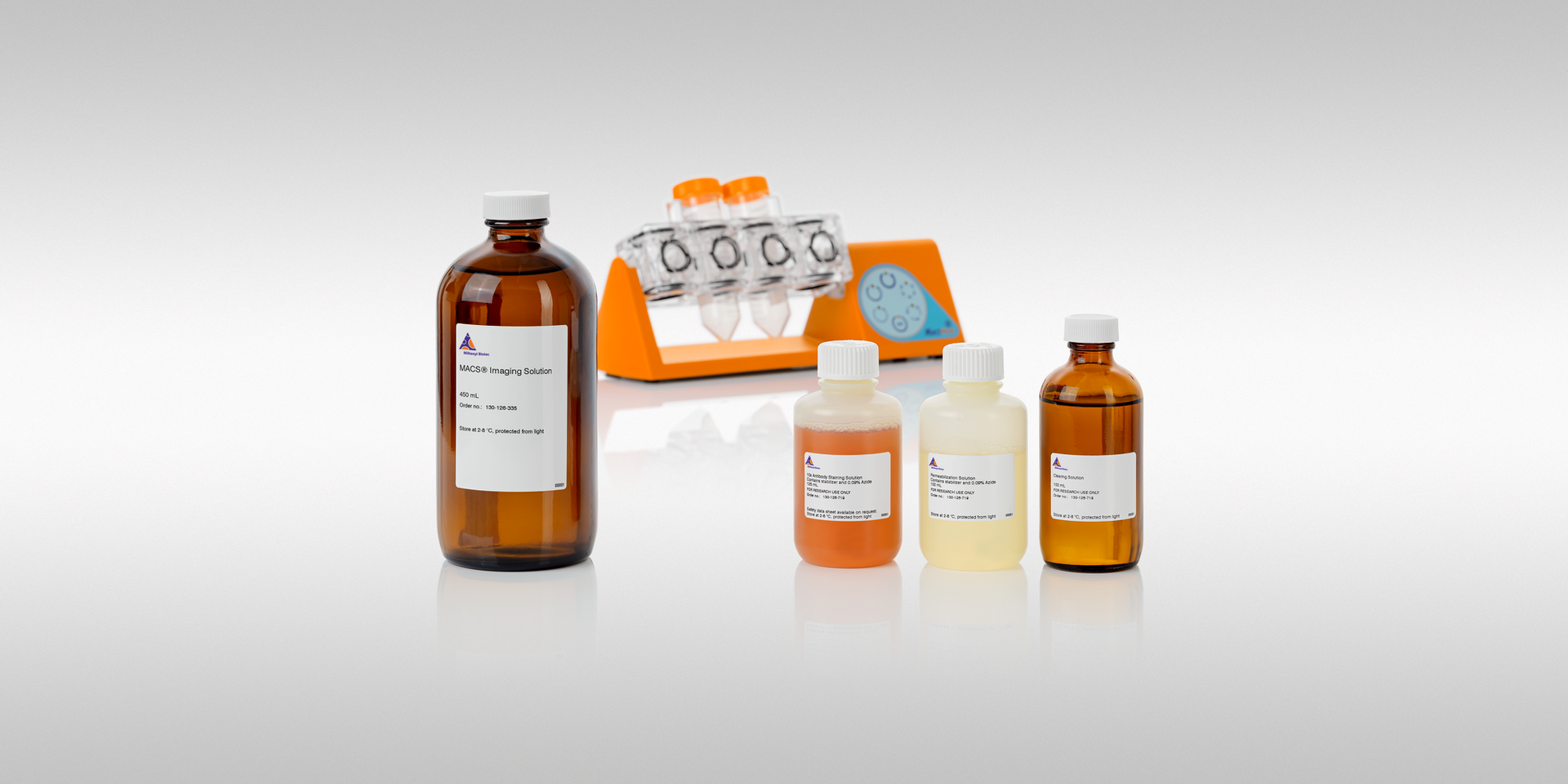
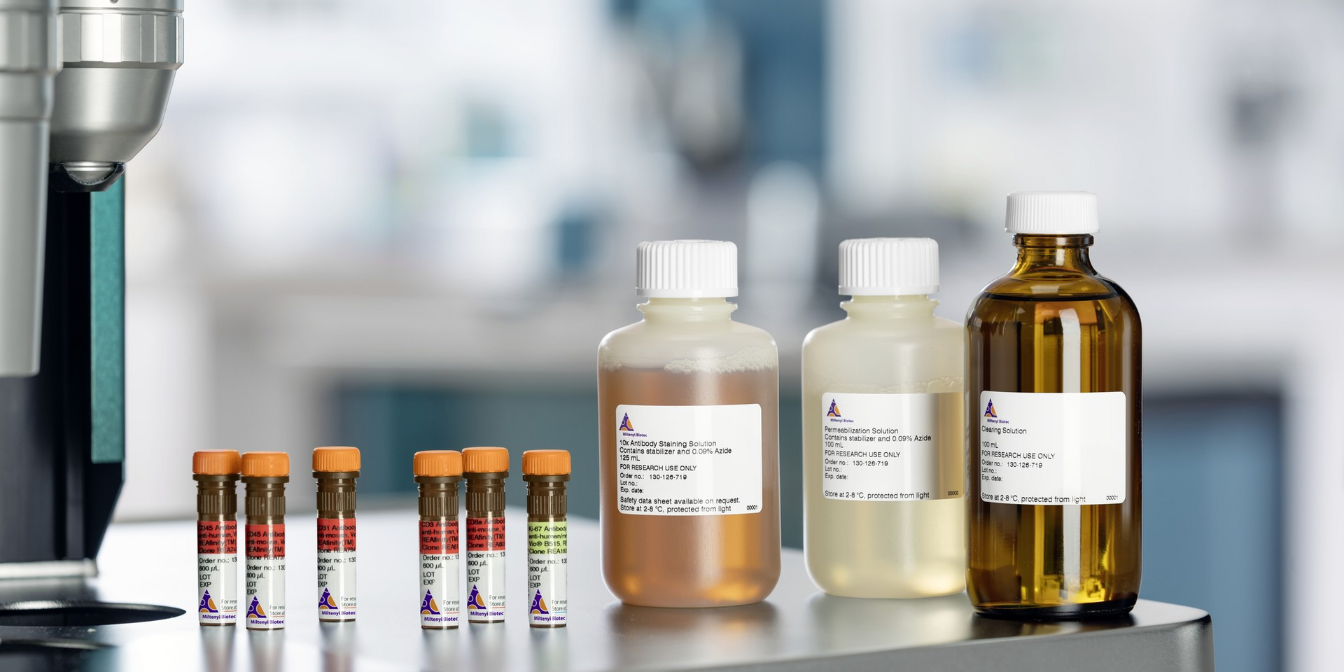
Maybe you're picturing complex procedures, but tissue clearing isn’t rocket science! There are simple methods and easy-to-follow protocols available. While setting up the process in a lab might take some initial effort, the techniques themselves are accessible to researchers of all experience levels. The key is to have the right protocol and materials.