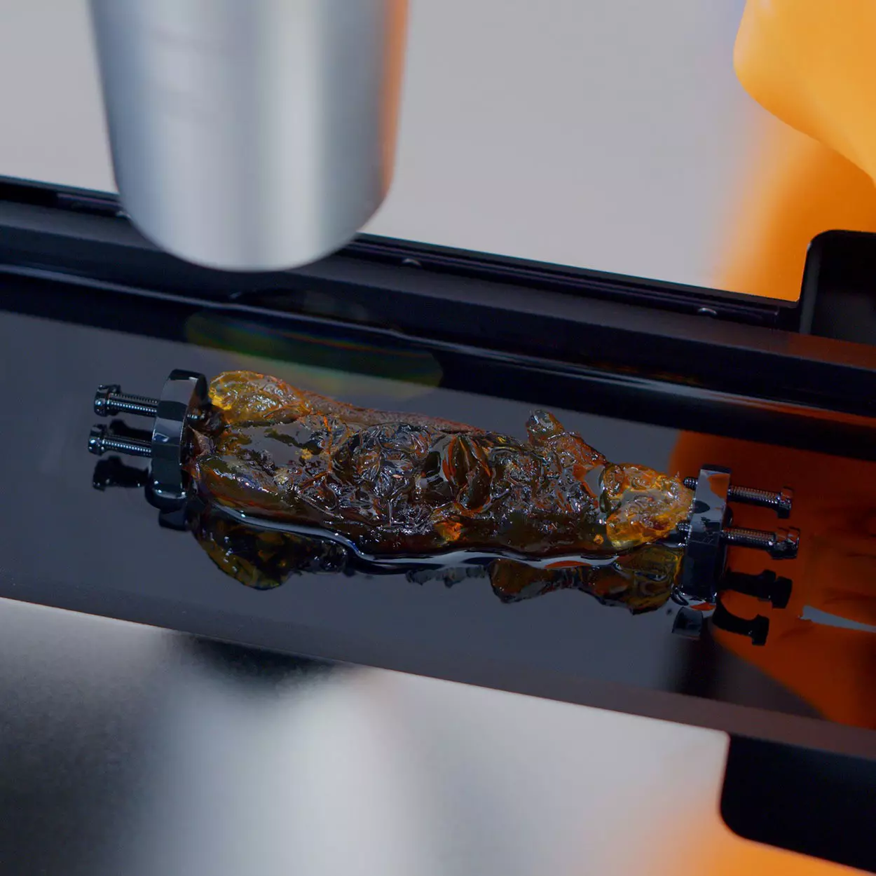
The UltraMicroscope Choros™ is our light sheet microscope designed for users who want to push the boundaries of 3D imaging of large samples. With a dedicated standalone stage, the UltraMicroscope Choros offers twice the imaging volume of the UltraMicroscope Blaze™ and its standard cuvette, while still offering the same outstanding benefits. This allows for effortless imaging of whole cleared mice and entire human organs or biopsy tissues, without the need to section or trim precious samples to fit into the imaging cuvette.
For high-throughput applications, the UltraMicroscope Choros can be paired with the MACS® UltraMount workflows, enabling efficient imaging of multiple samples in a single acquisition.
The UltraMicroscope Choros is compatible with LightSpeed Mode and MACS iQ View – 3D Large Volume, providing a complete 3D workflow solution from sample preparation to image processing.
With its extended stage travel range and dedicated large imaging cuvette, the UltraMicroscope Choros offers the widest imaging range available on the UltraMicroscope platform.
The highest throughput
When paired with the MACS® UltraMount workflows, the UltraMicroscope Choros is ideal for high-throughput screening applications.
High speed imaging
With LightSpeed Mode, the UltraMicroscope Choros unlocks unrivaled imaging speed while preserving the high resolution that the UltraMicroscope systems are renowned for.
High resolution
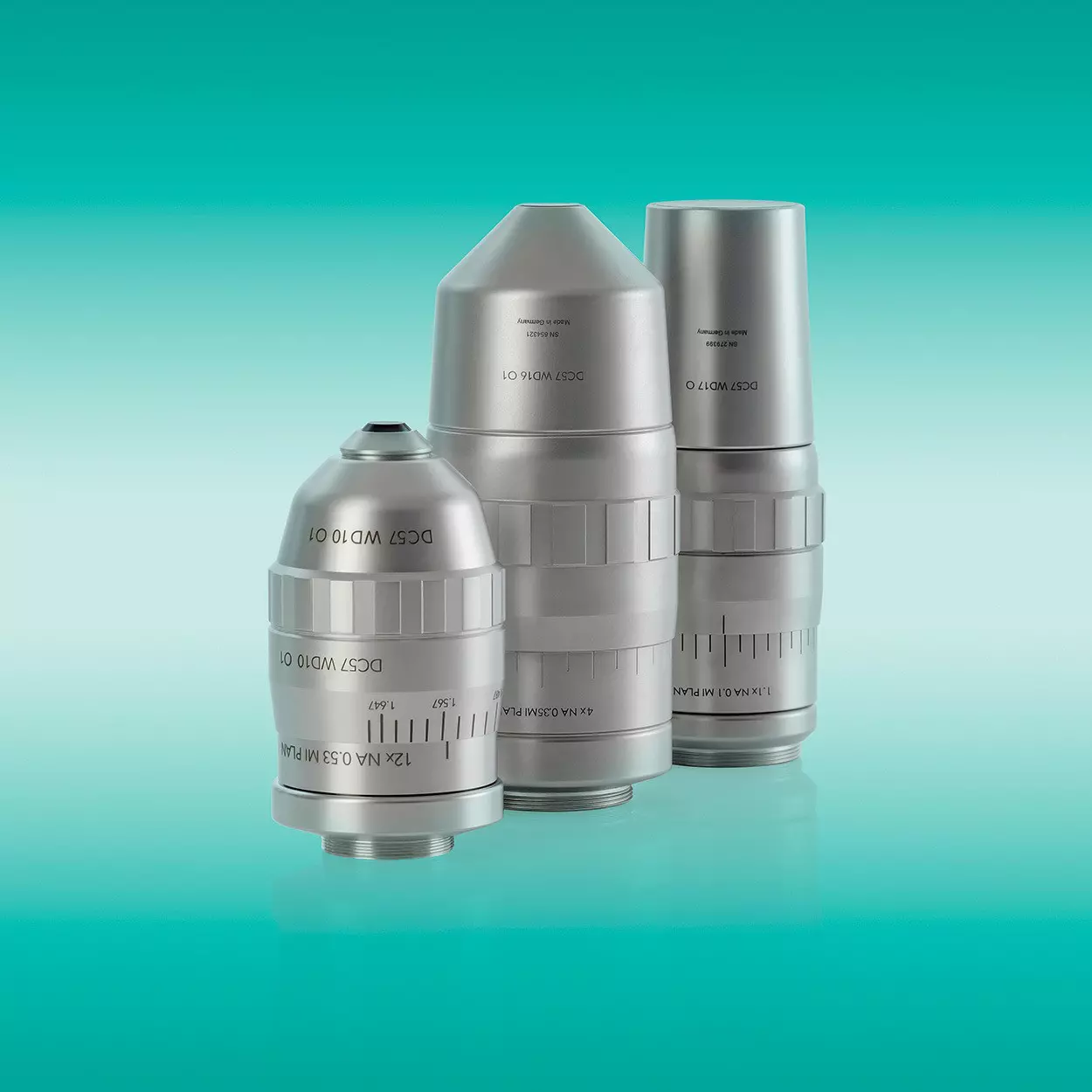
The combination of the MI Plan objective lenses and the cutting-edge light sheet illumination technology ensures unprecedented image quality in all clearing media – from water to organic solvents.
From whole organisms to high-throughput studies at cellular resolution
Purpose-built for imaging large samples and supporting high-throughput studies, the UltraMicroscope Choros offers twice the imaging volume of the UltraMicroscope Blaze. Complementary sample-handling solutions, including a large mouse holder for larger specimens and MACS UltraMount workflows for high-throughput imaging, further enhance its versatility.
Applications
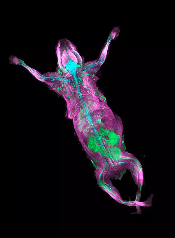
Whole mouse body shown in a 3D fly-through animation
Take a virtual flight through an entire mouse body, imaged in 3D using UltraMicroscope technology combined with tissue clearing. Microscopy data were acquired by Dr. P. Wartenberg (Prof. Dr. U. Boehm’s group, PZMS, Saarland University, Germany).
Imaging single cancer cells disseminated in whole mouse bodies
What if we could identify tiny metastases throughout the body that remain invisible to conventional methods? The group of Ali Ertürk from the Institute for Tissue Engineering and Regenerative Medicine (iTERM) at Helmholtz Zentrum München, Germany used UltraMicroscope technology to address this challenge. In a study published in Cell, they identified hundreds of small metastases in mouse models and determined precisely which metastasis was targeted by a drug candidate and which was missed. According to the researchers, this method could significantly increase the success rate of cancer drug candidates.
Pan, C. et al. (2019) Cell 179: 1661–1676.e19. (PMID:31835038).
Imaging whole human organs
Human anatomy has long been explored through conventional imaging of thin two-dimensional sections, limiting our ability to fully capture and understand large-scale anatomical structures. In this work, the integration of tissue clearing and light sheet microscopy using the UltraMicroscope provides a transformative solution, enabling three-dimensional visualization of human organs at microscopic resolution and allowing anatomical investigations across multiple spatial scales.
The video shows 3D volume of a human lung and colon. Policet-Bétend, H. et al. (2026) bioRxiv 2026.07.16.738626. (doi:10.64898/2026.07.16.738626).
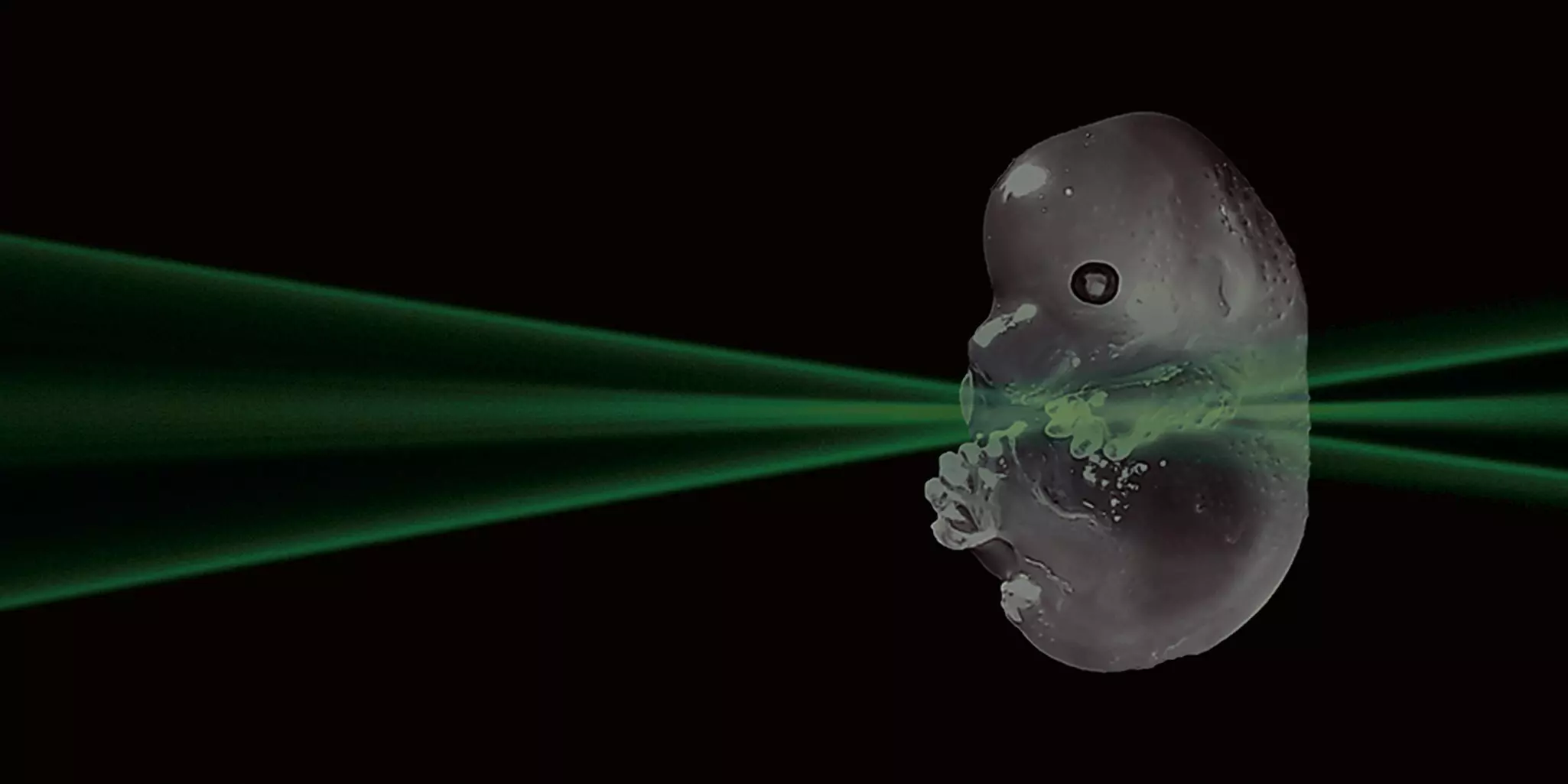
As the generated light sheet diverges, it approximates a plane only over a given horizontal range. This is where the light sheet is thinnest and where fluorescence detection takes place. To achieve an appropriate illumination for a particular sample, the planar range of the light sheet has to be matched with the sample size and the desired field of view (FOV). The light sheet of the UltraMicroscope Choros is tailored to the sample size by using adjustable light sheet parameters.
A low numerical aperture (NA) of illumination results in a broad FOV at the expense of a low Z-resolution when imaging large samples (A). In contrast, a high NA results in a high Z-resolution and a narrow FOV suitable for imaging small samples (B), with a full range of gradations in between. Where both high Z-resolution and a large FOV are essential, for example when imaging larger samples, the UltraMicroscope Choros offers dynamic horizontal focusing. To that end, a sequential series of high-resolution images are taken across the desired FOV and automatically stitched together into a single high-quality image by our ImSpector image acquisition software (C).
MI Plan objectives optimized for high-resolution light sheet imaging
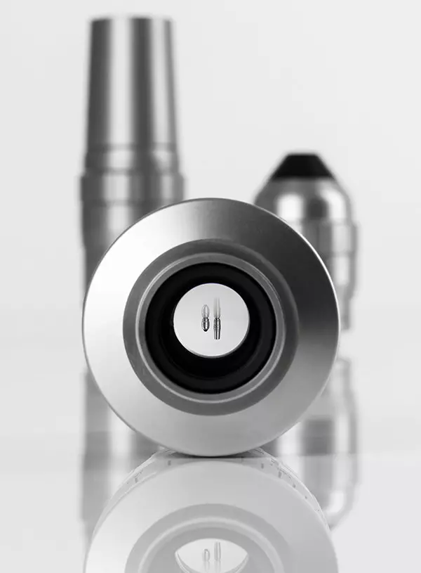
Objective lenses can produce artifacts that increase in severity with the size of FOV. However, the flat-field correction in the MI Plan objectives generate images that are in focus throughout the area of interest. The MI Plan objective series is designed and optimized for high-resolution light sheet imaging. The combination of flat-field correction and long working distances makes these objectives well suited for imaging large samples. They can be coupled to a wide variety of dipping caps that allow high-resolution and distortion-free imaging in all clearing solutions, whether based on aqueous or organic solvents (refractive indices between 1.33 and 1.56).
Multiple light sheets optimized for uniform illumination
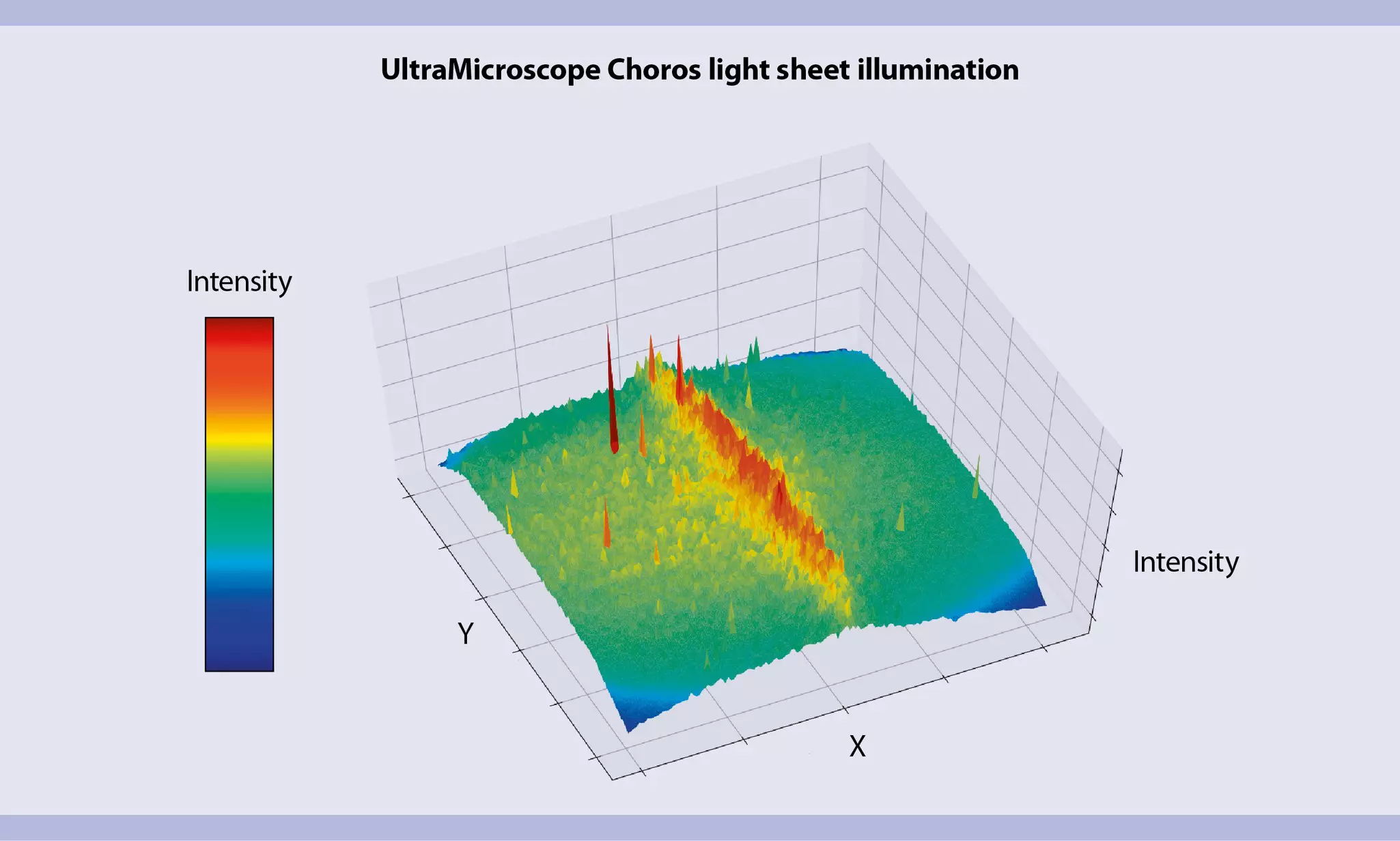
The cutting-edge illumination optics of the UltraMicroscope generates multiple light sheets that overlap in the desired FOV and excite the sample from both sides. This results in a uniform illumination profile, improved optical sectioning, and thereby a higher image quality.
Most clearing reagents differ from protocol to protocol, and so do their optical properties. Light can travel differently in different imaging media, which can create distortions and thereby lower the image quality. To compensate for this, the UltraMicroscope's onboard software allows users to choose between various predefined clearing and imaging solutions. This provides the perfect setting for every imaging media.
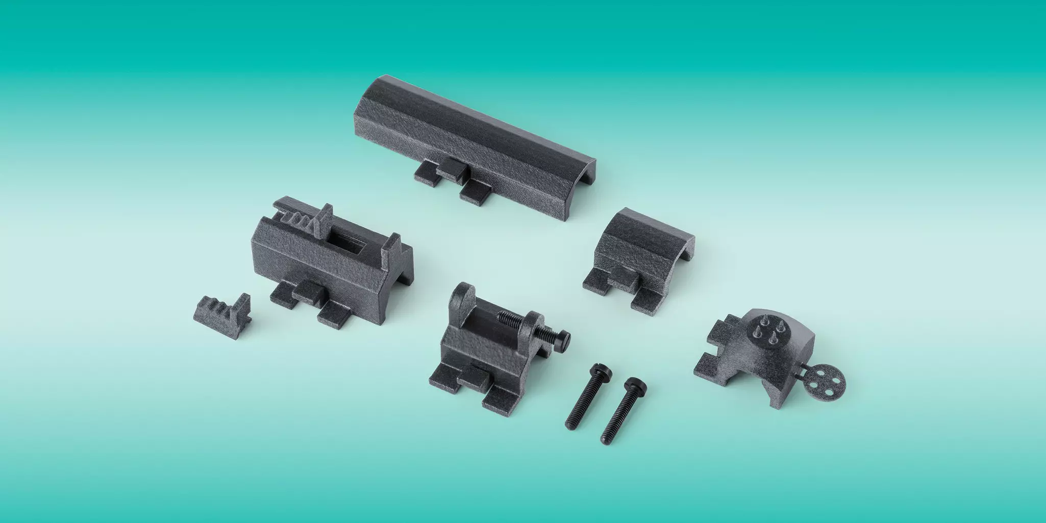
The UltraMicroscope Choros includes a standard set of sample holders for common 3D imaging needs. Additionally, application-specific holders are available, like those designed for high-throughput organoid/spheroid imaging or multiple brains/organs. Some holders come with preconfigured software templates to streamline your workflow and minimize hands-on time.
UltraMicroscope technology allows us to see every single cancer metastasis in the whole bodies of transparent mice and we can also see if drugs are targeting all those tiny micro-metastases. UltraMicroscope technology will be a powerful tool for drug development in oncology.
Dr. Ali Ertürk
CEO of Deep Piction
Neuherberg, Germany
Slide 2 of 2
LightSpeed mode has the potential to dramatically reduce acquisition time, allowing me to collect very large datasets in a fraction of the time that was previously feasible.
Dr. Rory Cooper
Department of Genetics and Evolution
Université de Genève
LightSpeed Mode
Fast-track your experiments with this new feature!
The LightSpeed Mode for the UltraMicroscope Choros unlocks unprecedented imaging speed while preserving the high resolution the UltraMicroscope is known for. Capture an entire adult mouse in less than 35 minutes – a process that would take hours with other light sheet microscopes. Complete the workflow with MACS iQ View – 3D Large Volume to stitch large datasets in just 7 minutes for fast, seamless whole-body volumetric imaging.
Upgrade an existing UltraMicroscope Choros or get a new one with LightSpeed Mode.
Experience unparalleled image quality at 60× faster imaging speeds.
Take high-throughput 3D imaging to a whole new level.
Unlock a new level of quality for UltraMicroscope Choros data with MACS iQ View – 3D Large Volume. This user-friendly software streamlines processing massive 3D datasets, stitching images, and enhancing image quality to reveal intricate details within your sample's structures. The intuitive interface makes it accessible to researchers of all experience levels.






 View details