Research solutions for extracellular vesicle (EV) isolation and analysis
As intercellular communication vehicles, EVs, such as exosomes, carry molecules and cellular components that reflect their cell of origin and that may influence their function. Analyzing EVs can thus provide valuable insights into disease-associated cellular processes, e.g. in cancer.
Our solutions cover all steps of EV analysis, offering a reliable isolation method that does not require ultracentrifugation, and a flow cytometry-based assay for fast screening of numerous EV surface epitopes associated with specific application fields, such as immuno-oncology research.
EV isolation
EV isolation and characterization have proven challenging due to their small size and heterogeneity in composition. Our solutions enable you to carry out EV isolation by positive selection using MicroBeads recognizing the tetraspanin proteins CD9, CD63, and CD81. Our EV Isolation Kit saves time, capital equipment, and requires low sample volume – a true alternative for conventional ultracentrifugation.

Isolation of EVs using MACS® Technology
Using our EV Isolation Kit, enrichment of EVs has never been easier.
- Selection of EVs according to specific surface markers i.e. CD9, CD63, or CD81
- Low starting sample volume (0.5 to 2 mL)
- Isolation from multiple sample types e.g. cell culture supernatant, urine, plasma, cerebrospinal fluid (CSF), serum, ascites etc
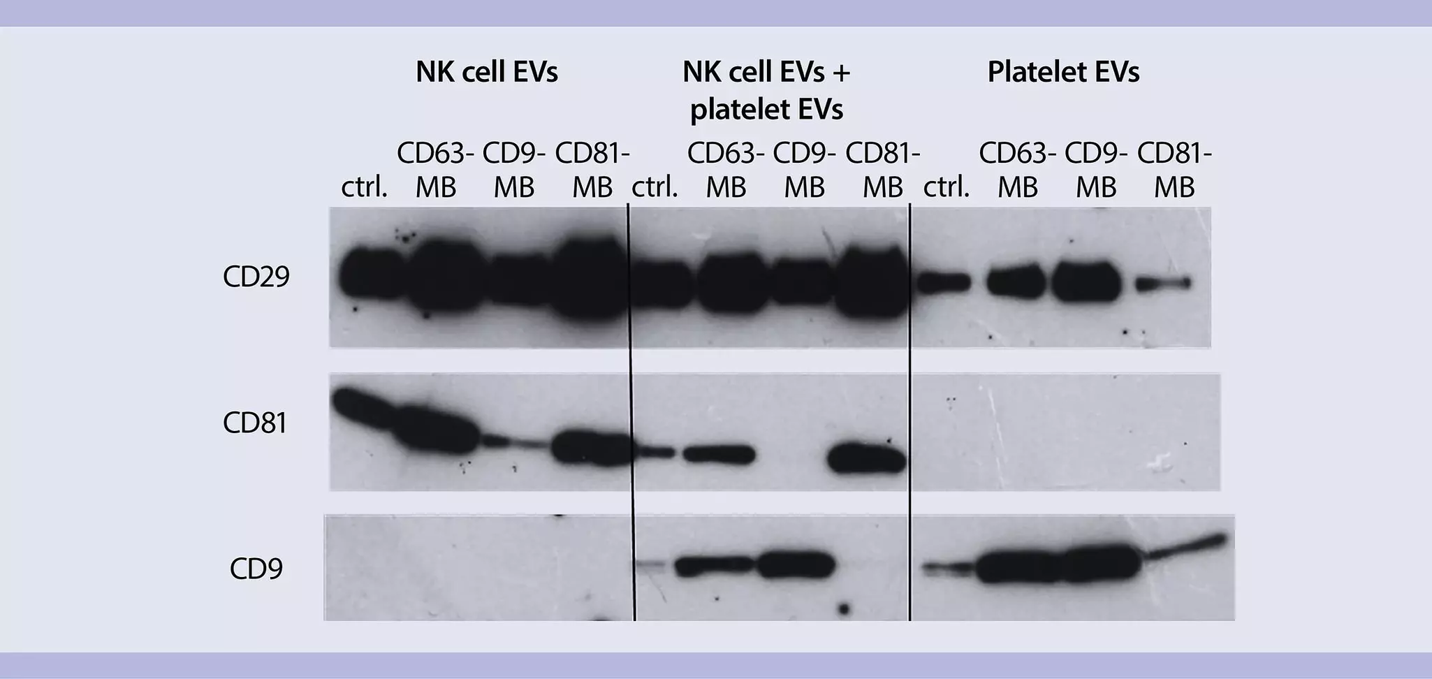
Separation of EV subpopulations
Subpopulations of EVs can be separated using superparamagnetic MACS® MicroBeads. For example, NK cell EVs and platelet EVs can be separated by positive selection using MicroBeads that recognize EV-specific tetraspanins, such as CD81 or CD9.
EV analysis
For further downstream analysis of EVs we have established the MACSPlex EV Kit. This multiplex bead-based assay consists of a cocktail of differently fluorescently labeled capture bead populations, which can be distinguished by flow cytometry.

Advantages of the MACSPlex EV Assay
Comprehensive – Fast and easy analysis of 37 EV surface markers plus 2 isotype controls simultaneously
Customizable – Additional antibodies can be added for multi-parameter analysis
Flexible – Compatible with different EV sample types and diverse sample preparation protocols
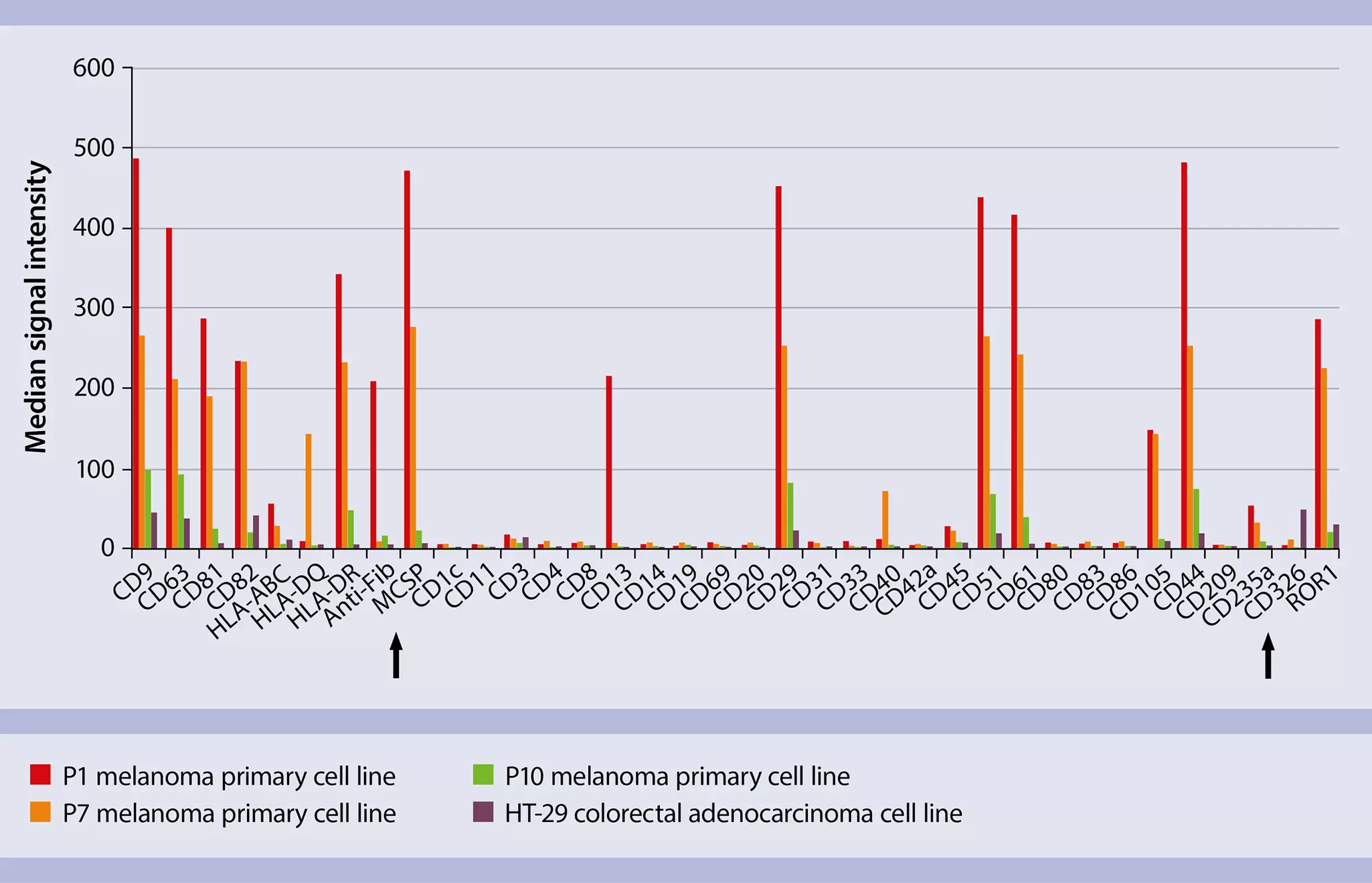
Discrimination of tumor-specific EV populations with MACSPlex EV Kit IO
EVs isolated from distinct cancer cell lines can be discriminated based on their surface markers that are specific to their originary tumor cell. EVs from melanoma cell line for instance show different surface markers then EVs from colon cancer cells, as demonstrated by binding to the respective MACSPlex EV Kit IO capture beads.
Video
Extracellular vesicle analysis by flow cytometry using the MACSPlex EV Kits
This video provides step-by-step instructions on how to use the MACSPlex EV Kit, human. It explains how to prepare the extracellular vesicle (EV) sample, couple the EVs to the capture beads, and stain the EV sample with the detection reagents. The video also provides practical tips for improved sample handling.
Step-by-step protocols for EV research
Get acquainted with our protocols for the isolation and analysis of EVs in cancer research.
Search:
2 results
You have previously chosen International as your location.
What do you want to do?